
intensive care unit admission, discharge, and triage.

- Recommendations

Recommendations  

Reference

General considerations


We recommend that critically ill patients be transferred from the emergency department ,ward bed or Intermediate Care Units (IMCUs)  to an available Intensive Care Unit (ICU) bed as rapidly as possible, ideally in less than or equal to 1 hour from the time the ICU bed is confirmed available and the decision to transfer is finalized. This is to minimize delays in accessing specialized ICU monitoring and interventions and to reduce potential risks associated with prolonged holding in non-ICU environments (Strong recommendation, low-quality evidence).

1,2

We recommend that in circumstances where a critically ill patient requires ICU services that are unavailable within the current hospital or cannot be provided in a timely manner, clinicians immediately utilize officially established national or regional referral pathways to facilitate rapid assessment and transfer to an appropriate ICU. In Egypt, this includes contacting the National Project of Critical Care, Neonatal, and Emergency Services "Rayat Misr" through its designated electronic platform or emergency hotline (137) as per national protocols (Strong recommendation, low -quality evidence).

1,2,3,4,5,6,7

We recommend that individual hospitals and their Intensive Care Unit (ICU) leadership formulate and regularly review explicit ICU admission and triage policies follow this guidline. These policies should be tailored to their specific institutional context, including the scope of clinical services provided (e.g., trauma, burn, neurocritical care, hepatic care), the demographic characteristics of their patient population, current ICU capacity, and available therapeutic resources (Strong recommendation, moderate-quality evidence).

1,8,9,10

We suggest that Triage decisions must be made transparently and without any prejudice. Factors like ethnicity, race, sex, social standing, or financial resources are wholly irrelevant. (Good practice statement, low-quality evidence).

1,8,9,10

We recommend that hospitals and their ICU leadership develop, implement, and regularly review explicit, transparent, and fair policies for the triage and allocation of ICU beds. These policies should provide a clear framework for balancing scheduled postoperative admissions with emergency admissions. This includes defining a process for temporarily reserving beds for imminent high-risk surgical arrivals while also having a dynamic override mechanism, managed by a designated triage officer or clinical leader (e.g., ICU director), to ensure any patient with an immediate life-threatening condition can access a necessary bed with minimal delay. Unjustified or prolonged reservation of an empty ICU bed in the face of a critically ill patient needing admission is inconsistent with ethical principles of justice and beneficence (Strong recommendation, moderate-quality evidence).

The core issue is one of triage and resource allocation. An ICU bed is a scarce, high-cost resource. A structured, transparent policy is essential to:

  1. Ensure Patient Safety: By minimizing delays for any patient who develops a critical need for ICU care.
  2. Maintain Access for Planned, High-Risk Procedures: Life-saving or life-improving surgeries (e.g., cardiac surgery, transplants, major cancer surgery) cannot be safely performed without a guaranteed postoperative ICU bed. Canceling these surgeries also causes harm.
  3. Promote Fairness and Equity: A clear policy prevents ad-hoc or inequitable decisions.
  4. Optimize Hospital Flow and Resource Use: Predictable ICU bed availability is essential for operating room scheduling and managing patient flow from the emergency department.

1,2, 11

We recommend that hospitals, where feasible and appropriate based on patient population needs, establish Intermediate Care Units (IMCUs). These units should serve as a bridge between the general ward and the Intensive Care Unit to provide enhanced monitoring and a higher level of nursing care for two primary patient groups: those who no longer require intensive care but are too unstable for the general ward (step-down), and those who are deteriorating on the ward but may not require immediate ICU-level organ support (step-up). The implementation of IMCUs is intended to improve patient safety, reduce ICU readmissions, optimize the use of critical care beds, and potentially lower overall costs for specific high-risk patient populations. (Strong recommendation, moderate-quality evidence).

The optimization of hospital resources while ensuring patient safety represents a significant challenge in modern healthcare. A critical gap often exists between the high-intensity, resource-heavy environment of the Intensive Care Unit (ICU) and the standard monitoring available on a general medical or surgical ward. Intermediate Care Units (IMCUs), also known as Step-Down or High-Dependency Units, are designed to bridge this gap, offering a level of care for patients who are too ill or unstable for the general ward but do not require the advanced organ support of an ICU.

12,13,14,15, 390

We recommend that hospitals develop and implement a Rapid Response System to facilitate early identification, bedside assessment, and appropriate triage of clinically deteriorating patients on general wards. This system should include a clear set of vital sign-based trigger criteria (such as: Modified Early Warning Score )  Fig. 1 and a designated team with critical care skills to respond promptly Rapid Response Team (RRT). A primary function of this team should be to stabilize the patient and facilitate timely and appropriate admission to an Intensive Care Unit (ICU) or intermediate care unit (IMCU) when indicated (Strong recommendation, High-quality evidence).

A key element of contemporary patient safety programs is the Rapid Response System (RRS). Its purpose is to recognize and intervene with patients whose condition is worsening, aiming to prevent serious adverse events like cardiac arrest. This forward-thinking strategy has demonstrated a reduction in hospital deaths and an enhancement of patient results. Putting a successful RRS into practice involves several key aspects: straightforward activation standards, a specialized and capable response team, thorough training for all staff, and a dedication to ongoing quality enhancement.

This guide details the fundamental elements and stages for creating and rolling out a successful Rapid response System in a hospital environment.

The Four Cornerstones of a Rapid Response System

An effective RRS is founded on four interrelated cornerstones:

  • The "Trigger" Component (Afferent Limb): This refers to the method used to spot a patient who is at risk. It consists of a defined set of indicators that, when observed, trigger a call to the Rapid Response Team.
  • The "Response" Component (Efferent Limb): This is the Rapid Response Team (RRT) itself. This team is composed of clinicians with advanced skills who deliver critical care knowledge directly to the patient's location.
  • The Administrative Component: This element supplies the structural support, leadership, and necessary resources that allow the RRS to operate smoothly.
  • The Quality Improvement Component: This involves the continuous gathering and examination of data to evaluate the RRS's performance and pinpoint opportunities for refinement.

16, 17, 18 ,19 ,20, 21, 22, 23,26, 27, 353, 391

We recommend that RRSs be utilized for early review of acutely ill non-ICU patients to identify patients who need or would benefit from ICU admission and treatment and to prevent unnecessary ICU admissions (Strong recommendation, moderate-quality evidence).

When a patient on a general ward triggers the RRS—often due to concerning changes in vital signs, respiratory distress, or altered mental status—a team of critical care experts, typically including an ICU nurse, a respiratory therapist, and sometimes a critical care physician or advanced practice provider, is dispatched to the patient's bedside.

The RRS team conducts a rapid but comprehensive assessment that goes beyond a simple review of vital signs. This evaluation is tailored to the patient's specific presentation and may include:

  • A thorough physical examination: Focusing on the cardiovascular, respiratory, and neurological systems to determine the severity and cause of the deterioration.
  • Review of the patient's chart and recent trends: To understand the patient's baseline health, recent treatments, and the trajectory of their illness.
  • Point-of-care testing: This may involve immediate blood gas analysis, lactate measurement, or other diagnostics to quickly assess physiological derangements.
  • Focused assessments for specific conditions: Such as a swallow evaluation if aspiration is suspected, or a detailed neurological exam in cases of altered consciousness.

The decision to transfer a patient to the ICU is not based on a single parameter but is a complex clinical judgment made by the RRS team in collaboration with the primary medical team. The core of this decision-making process revolves around three key questions:

1.  Does the patient require advanced organ support? This is a primary driver for ICU admission and includes the need for mechanical ventilation, vasopressors to support blood pressure, or renal replacement therapy. The RRS team's critical care expertise is vital in identifying the subtle signs that a patient is heading towards requiring such interventions.

2.  Would the patient benefit from the level of monitoring available only in the ICU? Some patients may not immediately require invasive interventions but are at high risk of rapid deterioration. The continuous monitoring of vital signs, cardiac rhythms, and other physiological parameters in the ICU can be life-saving for these individuals.

A key function of the RRS is to act as an "ICU without walls," bringing critical care expertise to the patient's bedside and often preventing the need for a physical transfer to the ICU. The RRS can achieve this through several interventions:

  • Stabilization on the ward: The team can initiate immediate treatments such as administering intravenous fluids, adjusting medications, providing non-invasive ventilation, or optimizing oxygen therapy, which may be sufficient to stabilize the patient and reverse the acute deterioration.
  • Enhanced care on the ward: The RRS can recommend and help implement a higher level of care on the general ward, such as more frequent vital sign monitoring, specific nursing interventions, or closer follow-up by the primary team.
  • Facilitating goals of care discussions: By engaging patients and families in conversations about their wishes and the realities of their prognosis, the RRS can help to ensure that ICU admission aligns with the patient's values. This is a powerful tool in preventing admissions that are not desired by the patient or are medically futile.

23, 24, 25, 26, 27, 391

We recommend implementing ICU consult teams to support ward/IMCU staff with deteriorating patients, facilitate transfer to ICU, and reduce ICU readmission rates. (Good practice statement, low-quality evidence).

1, 26, 27, 28, 29, 30

Admission & Triage


We suggest using the following tools for bed allocation during the admission and triage processes(Good practice statement, low-quality evidence). :

Guide to resource allocation of intensive monitoring and care including levels of monitoring, care, and nursing ratios (table 2)

ICU Admission Prioritization framework(table 3)

1, 17, 18, 31, 32

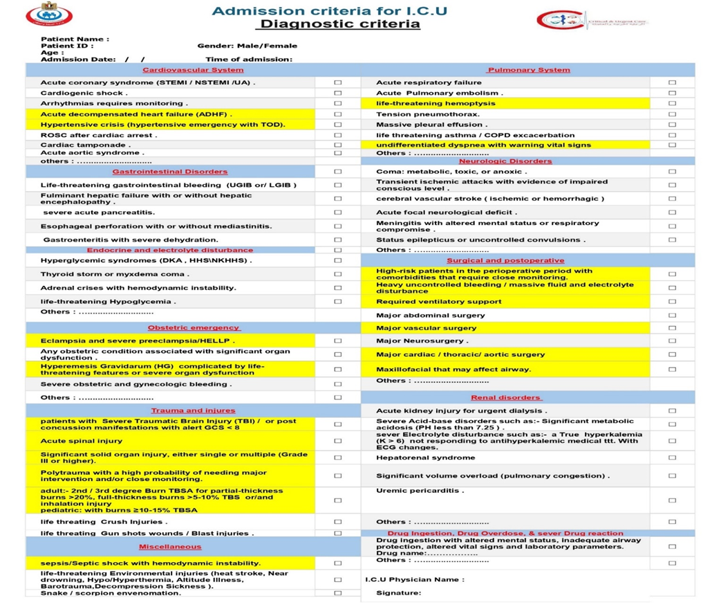
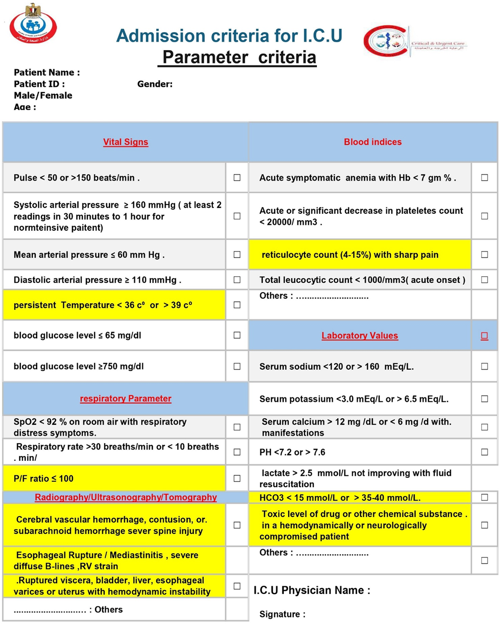
We suggest using the following tools as core of addmsion and discharge criteria checklist(Good practice statement, low-quality evidence):

1- addmsion checklist (table 4,5)


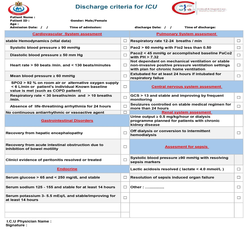
2- discharge criteria checklist(table 6):


1, 13, 27, 33, 35, 418

We recommend that the Acute Physiology and Chronic Health Evaluation (APACHE II) score, or a similar validated illness severity scoring system, be used as an adjunct to clinical assessment for patients being considered for Intensive Care Unit (ICU) admission. The calculation of an APACHE score upon presentation to the emergency department or for deteriorating ED/ward patients can provide valuable, objective information regarding the severity of illness and risk of mortality. A high or significantly rising score should be considered a strong indicator for the need for ICU-level care, facilitating timely and appropriate triage, especially for patients with conditions known to have a high mortality risk such as sepsis, acute respiratory distress syndrome (ARDS), and major trauma. It is crucial to emphasize that the APACHE score should not be used in isolation to determine ICU admission or discharge. It is a decision-support tool that, when integrated with a comprehensive clinical evaluation, patient preferences, and the overall clinical context, can lead to more informed and effective patient care.(Good practice statement, low-quality evidence).

34 ,36, 37, 38

We recommended that patients with invasive mechanical ventilation or complex life-threatening conditions, including sepsis, receive care in an ICU. Additionally, weaning from mechanical ventilation should be restricted to an ICU or In necessities step down to a intermediate care unit (IMCU) , and not take place on a general ward. (Strong recommendation, moderate-quality evidence).

1, 33, 39

We suggest avoiding admitting to a specialized ICU patients with a primary diagnosis not associated with that specialty (i.e., boarding) (Strong recommendation, moderate-quality evidence).

1, 40, 285

Triage


We suggest that hospitals and ICU leaders develop and implement admission and triage policies that are designed to minimize under-triage and overtriage is more acceptable (Good practice statement, low-quality evidence).

Rationale for the Principle of Preferring Over-Triage:

This principle is a cornerstone of patient safety in emergency and critical care triage. It acknowledges the different consequences of the two types of triage errors:

  • Under-triage: This occurs when a patient who requires ICU-level care is not admitted to the ICU and is instead managed in a lower-acuity setting (e.g., a general ward). This error can lead to a "failure to rescue" from clinical deterioration, resulting in preventable cardiac arrest, irreversible organ damage, or death for that individual patient. The harm is direct and potentially catastrophic.
  • Over-triage: This occurs when a patient who could have been safely managed in a lower-acuity setting is admitted to the ICU. The primary consequence is inefficient use of a scarce, high-cost resource (the ICU bed). While this can affect system capacity and potentially delay care for another patient, the direct harm to the admitted individual is minimal compared to the harm of under-triage (provided the ICU stay itself does not cause iatrogenic harm).

1, 2, 17, 24, 41

Diagnostic criteria


We recommend that all patients presenting with ST-Elevation Myocardial Infarction (STEMI), ST-Elevation Myocardial Infarction (STEMI), Non-ST-Elevation Myocardial Infarction (NSTEMI)  and other patients with Acute Coronary Syndromes (ACS) identified as high-risk (based on clinical features, ECG findings, cardiac biomarkers, hemodynamic instability, life-threatening arrhythmias, ongoing ischemia, acute heart failure and validated risk scores) be admitted to a Coronary Care Unit (CCU) an Intensive Care Unit (ICU), or an intermediate care unit (IMCU) with. This setting is necessary for continuous cardiac and hemodynamic monitoring, prompt management of life-threatening complications facilitation of timely reperfusion or invasive strategies, and optimization of medical therapy that require Intensive care, such as heart failure, cardiogenic shock, or serious arrhythmias, ventricular septal rupture, even if their initial condition appears stable, facilitation of timely reperfusion/revascularization strategies, and optimization of medical therapy (Strong recommendation, moderate-quality evidence).

The primary purpose of admitting high-risk ACS patients to an ICU/CCU is to provide intensive monitoring and immediate intervention for life-threatening complications that are most common in the early hours and days after the acute event.

  1. For ST-Elevation Myocardial Infarction (STEMI):
    • STEMI results from an acute, total occlusion of a coronary artery, leading to a high immediate risk of:
      • Life-Threatening Arrhythmias: Ventricular tachycardia (VT) and ventricular fibrillation (VF) are most frequent in the initial hours and are a leading cause of pre-hospital and early in-hospital death. Continuous ECG monitoring with immediate defibrillation capability is life-saving.
      • Hemodynamic Instability: Acute myocardial dysfunction can lead to acute heart failure, cardiogenic shock, or severe bradyarrhythmias.
      • Mechanical Complications: Such as ventricular septal rupture, papillary muscle rupture leading to acute mitral regurgitation, or free wall rupture.
    • ICU/CCU admission is considered standard for all STEMI patients to facilitate continuous monitoring, manage complications, and provide care before, during, and after emergent reperfusion therapy (primary PCI or fibrinolysis).
  2. For Non-ST-Elevation ACS (NSTEMI and Unstable Angina):
    • This is a heterogeneous group, and admission to an ICU/CCU is based on risk stratification. Patients are deemed high-risk if they have:
      • Hemodynamic Instability: Hypotension or cardiogenic shock.
      • Life-Threatening Arrhythmias.
      • Recurrent or Ongoing Ischemia: Refractory chest pain or dynamic ST-segment changes.
      • Acute Heart Failure.
      • Mechanical Complications.
      • High-Risk Features on non-invasive testing or high-risk scores (e.g., GRACE score >140).
    • ICU/CCU admission for these high-risk patients allows for intensive monitoring and stabilization prior to an early invasive strategy (coronary angiography and revascularization)

33, 42, 43, 44, 45, 46, 47, 48

We recommend that all patients diagnosed with cardiogenic shock characterized by persistent hypotension despite adequate fluid status and signs of end-organ hypoperfusion. be immediately admitted to an to a Coronary Care Unit (CCU) or Intensive Care Unit (ICU) or a specialized cardiac/cardiovascular ICU with equivalent capabilities This is to facilitate immediate and continuous hemodynamic monitoring, aggressive management with intravenous vasoactive medications, advanced respiratory support, rapid investigation and treatment of the underlying cause, and consideration for mechanical circulatory support (Strong recommendation, moderate-quality evidence).

Admition is considered essential due to the need for:

  • Advanced Hemodynamic Monitoring: Continuous invasive arterial blood pressure monitoring, central venous pressure monitoring, and often advanced cardiac output monitoring to guide therapy.
  • Pharmacological Hemodynamic Support: Titration of intravenous vasopressors (e.g., norepinephrine) and inotropes (e.g., dobutamine, milrinone) to maintain organ perfusion.
  • Mechanical Ventilation: For respiratory failure secondary to pulmonary edema, to reduce the work of breathing, or for airway protection in obtunded patients.
  • Identification and Treatment of Reversible Causes: Urgent investigation (e.g., echocardiography, coronary angiography) and intervention (e.g., revascularization for acute myocardial infarction, valve surgery, arrhythmia management).
  • Mechanical Circulatory Support (MCS): Consideration and potential deployment of devices like intra-aortic balloon pumps (IABP), percutaneous ventricular assist devices (e.g., Impella, TandemHeart), or Extracorporeal Membrane Oxygenation (ECMO) in refractory shock.
  • Management of Multi-Organ Dysfunction: Support for associated renal failure (renal replacement therapy), hepatic dysfunction, coagulopathy, etc.
  • Continuous Expert Nursing Care and Rapid Response Capability.

33, 42, 43, 44, 45, 46, 47,49, 50, 51, 52, 53, 54

We recommend that patients presenting with arrhythmias be admitted to an a specialized Coronary Care Unit (CCU)  or Intensive Care Unit (ICU) if the arrhythmia is life-threatening (e.g., sustained ventricular tachycardia, ventricular fibrillation, symptomatic high-grade atrioventricular block), causes hemodynamic instability (e.g., hypotension, shock, acute heart failure), is associated with severe symptoms directly attributable to the arrhythmia (e.g., syncope with a high-risk rhythm), or requires urgent electrical interventions (cardioversion, defibrillation, pacing) or pharmacological interventions with intravenous agents that necessitate continuous cardiorespiratory monitoring not feasible or safe in a lower-acuity setting (Strong recommendation, moderate-quality evidence).

Patients with specific arrhythmias are admitted to an ICU/CCU due to:

  • Hemodynamic Instability: Arrhythmias causing hypotension, shock, acute heart failure, or signs of end-organ hypoperfusion.
  • Life-Threatening Nature: Rhythms that can degenerate into cardiac arrest (e.g., sustained ventricular tachycardia, certain bradyarrhythmias with high risk of asystole).
  • Need for Urgent Intervention: Requirement for immediate electrical cardioversion, defibrillation, or temporary cardiac pacing.
  • Pharmacological Management Requiring Close Monitoring: Initiation or titration of intravenous antiarrhythmic drugs that have a narrow therapeutic window or potential for proarrhythmia or significant hemodynamic side effects.
  • Continuous ECG and Hemodynamic Monitoring: To assess arrhythmia burden, response to treatment, and detect early signs of deterioration.
  • Management of Underlying Cause: Arrhythmias are often a manifestation of an acute underlying condition (e.g., acute myocardial infarction, electrolyte imbalance, sepsis, drug toxicity) that itself requires intensive care.

33, 55, 56, 57, 58, 358

We recommend that patients with acute decompensated heart failure (ADHF) who exhibit signs of hemodynamic instability (e.g., hypotension, cardiogenic shock), severe respiratory distress or failure (requiring non-invasive or invasive mechanical ventilation), life-threatening arrhythmias, or who require intravenous vasoactive medications or invasive hemodynamic monitoring, be admitted to an Intensive Care Unit (ICU) or a Intensive Coronary Care Unit (CCU) for intensive management and monitoring (Strong recommendation, moderate-quality evidence).

ADHF represents a spectrum of clinical severity. While many patients can be managed on general medical wards, those with severe ADHF require ICU/CCU admission due to:

  • Hemodynamic Instability: Hypotension, cardiogenic shock, or the need for intravenous vasoactive medications (inotropes, vasopressors, or potent vasodilators) requiring continuous hemodynamic monitoring (often invasive).
  • Severe Respiratory Distress/Failure: Acute pulmonary edema requiring non-invasive ventilation (NIV) or invasive mechanical ventilation (IMV), or significant hypoxemia unresponsive to standard oxygen therapy.
  • Life-Threatening Arrhythmias: Ventricular or supraventricular arrhythmias causing hemodynamic compromise or requiring urgent intervention and close monitoring.
  • Need for Advanced Monitoring: Continuous ECG, invasive arterial blood pressure, central venous pressure, or pulmonary artery catheter monitoring in select cases.
  • Failure of Initial Therapies: Patients not responding to initial diuretics and vasodilators who may require escalation of care.
  • Presence of Critical Precipitating Factors or Complications: Such as acute coronary syndrome, severe valvular dysfunction, hypertensive emergency, or significant electrolyte disturbances requiring intensive correction and monitoring.
  • Potential Need for Mechanical Circulatory Support (MCS): In refractory cardiogenic shock.

33, 48, 49, 59, 60, 358

We recommend that patients presenting with a hypertensive emergency (defined as severely elevated blood pressure accompanied by acute, ongoing target organ damage) be admitted to an Intensive Care Unit (ICU) or a Intensive Coronary Care Unit (CCU) or a comparable high-acuity monitored setting. This is to facilitate immediate and controlled parenteral antihypertensive therapy, continuous (often invasive) blood pressure monitoring, and close observation and management of specific target organ dysfunction (Strong recommendation, moderate-quality evidence).

It is vital to differentiate:

  1. Hypertensive Emergency: Severely elevated blood pressure (BP) – often SBP >180 mmHg or DBP >120 mmHg, though the absolute level is less critical than the presence of organ damage – accompanied by evidence of acute, ongoing target organ damage (TOD). Examples include hypertensive encephalopathy, acute myocardial infarction/ischemia, acute pulmonary edema, eclampsia/severe pre-eclampsia, acute aortic dissection, stroke (ischemic or hemorrhagic), or acute kidney injury. These require immediate, controlled BP reduction with parenteral medication and intensive monitoring, typically in an ICU.
  2. Hypertensive Urgency: Severely elevated BP in a similar range, but without evidence of acute TOD. These patients usually do not require ICU admission and can be managed with gradual BP reduction using oral medications, often on an outpatient or short-stay basis.

Patients with hypertensive emergencies require ICU admission due to the need for:

  • Immediate and controlled reduction of BP using titratable intravenous (IV) antihypertensive agents to limit or reverse target organ damage.
  • Continuous, often invasive, arterial blood pressure monitoring to guide therapy and avoid overly rapid or excessive BP lowering, which can be harmful (e.g., precipitate cerebral or myocardial ischemia).
  • Close monitoring for progression or resolution of target organ damage (neurological status, cardiac function, renal function, etc.).
  • Management of specific target organ damage (e.g., ventilatory support for pulmonary edema, specific neurological interventions).
  • Identification and management of underlying causes or precipitating factors.

33, 61, 62, 63, 64, 65

We recommend that adult patients who achieve Return of Spontaneous Circulation (ROSC) after cardiac arrest and remain comatose or hemodynamically unstable, or who require ongoing ventilatory support, be admitted to an Intensive Care Unit (ICU) or a Coronary Care Unit (CCU) with equivalent ICU capabilities for comprehensive post-resuscitation care (Strong recommendation, moderate-quality evidence).

Patients who achieve ROSC after cardiac arrest are critically ill and suffer from post-cardiac arrest syndrome. This complex syndrome includes:

  1. Post-cardiac arrest brain injury: Due to initial hypoxia/ischemia and subsequent reperfusion injury, leading to a spectrum of neurological deficits, cerebral edema, and potential seizures.
  2. Post-cardiac arrest myocardial dysfunction: Stunning of the heart muscle, even if the arrest was not primarily cardiac, leading to hemodynamic instability.
  3. Systemic ischemia/reperfusion response: A generalized inflammatory response affecting multiple organs.
  4. Persistent precipitating pathology: The underlying cause of the cardiac arrest (e.g., acute coronary syndrome, pulmonary embolism, sepsis, respiratory failure, electrolyte abnormalities, toxidrome) often requires ongoing management.

Management of these patients in an ICU is essential for:

  • Targeted Temperature Management (TTM): For many comatose adult survivors to improve neurological outcome.
  • Optimization of ventilation and oxygenation: Often requiring mechanical ventilation with lung-protective strategies.
  • Hemodynamic monitoring and support: Including management of blood pressure, cardiac output, and tissue perfusion, often with vasoactive medications.
  • Continuous neurological monitoring: Including EEG for seizure detection, and management of cerebral edema or intracranial hypertension.
  • Identification and treatment of the cause of cardiac arrest.
  • Metabolic control: E.g., glycemic control, correction of electrolyte imbalances.
  • Prevention and treatment of complications: Such as infections, further organ injury, and seizures.

33, 66, 67, 68, 69

We recommend that patients diagnosed with cardiac tamponade, especially those exhibiting hemodynamic instability or requiring urgent pericardial drainage, be admitted to an Intensive Care Unit (ICU) or a Coronary Care Unit (CCU) with equivalent capabilities for continuous cardiorespiratory monitoring, management of potential complications, and further diagnostic evaluation and treatment of the underlying cause (Strong recommendation, moderate-quality evidence)

Admission to an ICU/CCU is generally indicated for patients with cardiac tamponade, particularly if they exhibit hemodynamic compromise, due to the need for:

  • Urgent Diagnostic Confirmation: Often requiring echocardiography at the bedside.
  • Continuous Hemodynamic Monitoring: Including invasive arterial blood pressure monitoring, and potentially central venous pressure, to assess the severity and response to intervention.
  • Emergency Pericardial Drainage: Pericardiocentesis (needle drainage) or surgical drainage, which are procedures that require a controlled environment with resuscitation capabilities and skilled personnel.
  • Management of Hemodynamic Instability: Including fluid resuscitation, and potentially vasopressor or inotropic support before, during, and after drainage.
  • Post-Procedure Monitoring: Close observation for complications of the procedure (e.g., arrhythmias, pneumothorax, myocardial puncture, bleeding) and for re-accumulation of pericardial fluid.
  • Identification and Management of the Underlying Cause: The etiology of the pericardial effusion and tamponade often requires further investigation and specific treatment, which may necessitate ongoing intensive care.

33, 70, 71, 72

We recommend that all patients diagnosed with or strongly suspected of having an Acute Aortic Syndrome (including aortic dissection, intramural hematoma, or penetrating atherosclerotic ulcer) be immediately admitted to an Intensive Care Unit (ICU) or a specialized cardiovascular ICU capable of providing comprehensive medical and peri-interventional management (Strong recommendation, moderate-quality evidence).

AAS encompasses a group of life-threatening conditions including aortic dissection (AD), intramural hematoma (IMH), and penetrating atherosclerotic ulcer (PAU). These conditions are medical emergencies due to the high risk of aortic rupture, end-organ malperfusion, and death.

Patients diagnosed with or strongly suspected of having AAS require immediate admission to an ICU or a specialized cardiovascular ICU. This is critical due to the need for:

  1. Aggressive Hemodynamic Control: Immediate and meticulous control of blood pressure (target SBP often 100-120 mmHg) and heart rate (target <60 bpm) using titratable intravenous medications (e.g., beta-blockers, vasodilators – known as anti-impulse therapy) to reduce aortic wall stress and limit progression of the aortic injury.
  2. Continuous Invasive Monitoring: Arterial line for continuous blood pressure monitoring is essential. Central venous access may also be required.
  3. Pain Management: Severe pain is common and requires potent analgesia, which can also help in hemodynamic control.
  4. Serial Neurological and Vascular Assessments: To detect and manage potential malperfusion syndromes affecting the brain, spinal cord, limbs, or visceral organs.
  5. Urgent Diagnostic Imaging and Consultation: Rapid access to advanced imaging (CT angiography, transesophageal echocardiography) and immediate consultation with cardiac and vascular surgical teams are crucial for definitive management (surgery for Type A dissections and complicated Type B AAS; medical management for uncomplicated Type B AAS often still initiated in ICU).
  6. Management of Complications: Such as cardiac tamponade, acute aortic regurgitation, stroke, or visceral ischemia.

33, 73, 74, 75, 76, 77, 78

We recommend that adult patients presenting with sever valvular heart disease such as severe acute mitral regurgitation, particularly when characterized by signs of respiratory failure (e.g., pulmonary edema requiring advanced respiratory support) or hemodynamic instability (e.g., hypotension, shock), be promptly admitted to an Intensive Care Unit (ICU) or a specialized Cardiovascular/Coronary Care Unit (CVICU/CCU). This is to facilitate immediate hemodynamic stabilization with intravenous vasoactive medications (vasodilators and/or inotropes), advanced respiratory support (non-invasive or invasive ventilation), intensive monitoring, and optimization as a bridge to urgent surgical intervention, which is the definitive therapy (Strong recommendation, moderate-quality evidence).
Acute mitral regurgitation is a medical and surgical emergency. Unlike chronic MR where the heart has time to adapt, AMR imposes a sudden, large volume load on an unprepared left atrium and ventricle. This leads to a rapid rise in left atrial and pulmonary pressures and a sharp drop in effective forward cardiac output.

ICU/CCU admission is critical for patients with hemodynamically significant AMR due to the need for:

  1. Management of Acute Pulmonary Edema and Respiratory Failure: The rapid backflow of blood into the lungs causes fulminant pulmonary edema and severe hypoxemic respiratory failure. This requires aggressive diuresis and advanced respiratory support, including non-invasive ventilation (NIV) or endotracheal intubation and invasive mechanical ventilation (IMV).
  2. Management of Cardiogenic Shock: The abrupt decrease in forward stroke volume leads to hypotension, end-organ hypoperfusion, and cardiogenic shock. This requires intensive hemodynamic monitoring (often with an invasive arterial line and sometimes a pulmonary artery catheter) and support with intravenous vasoactive medications.
  3. Specialized Pharmacotherapy:
    • Afterload Reduction: Intravenous vasodilators (e.g., sodium nitroprusside) are crucial to decrease systemic vascular resistance, which promotes forward blood flow and reduces the regurgitant volume. Their use requires continuous, close blood pressure monitoring.
    • Inotropic Support: Inotropes (e.g., dobutamine) may be needed to improve cardiac contractility in the setting of cardiogenic shock.
  4. Mechanical Circulatory Support: An intra-aortic balloon pump (IABP) may be used as a bridge to surgery to decrease afterload and augment coronary perfusion.
  5. Urgent Diagnosis and Surgical Planning: Transesophageal echocardiography (TEE) is often required for detailed anatomical assessment and is frequently performed in the ICU or operating room on unstable patients. ICU care is essential to stabilize the patient as a "bridge to surgery," as emergency mitral valve repair or replacement is the definitive treatment for severe AMR.
  6. Management of the Underlying Cause: The cause of AMR (e.g., acute myocardial infarction with papillary muscle rupture, infective endocarditis with leaflet destruction) is often a critical illness that independently requires ICU management.

33,53, 54, 420, 421, 422, 423, 424, 425

Gastrointestinal Disorders


We recommend that patients presenting with life-threatening gastrointestinal bleeding (UGIB or LGIB), characterized by hemodynamic instability (e.g., shock, persistent hypotension despite initial resuscitation), ongoing massive hemorrhage requiring significant transfusion, or validated high-risk scores indicating severe bleeding or high re-bleeding risk, be admitted to an Intensive Care Unit (ICU) or intermediate care unit (IMCU) with equivalent capabilities. This is to facilitate aggressive resuscitation, continuous hemodynamic monitoring, urgent diagnostic and therapeutic interventions (often endoscopic), specialized pharmacotherapy, and management of potential complications (Strong recommendation, moderate-quality evidence).

Admission to an ICU (or a high-dependency unit with equivalent capabilities) is crucial for such patients due to the need for:

  • risk scoring systems (e.g., Glasgow-Blatchford, Rockall for UGIB; Oakland score for LGIB) that help identify patients at high risk for adverse outcomes, including re-bleeding and mortality. Patients scoring in the high-risk categories often have features (like hemodynamic instability, significant comorbidities) that necessitate intensive care.
  • Aggressive Resuscitation: Rapid administration of intravenous fluids and blood products to restore and maintain hemodynamic stability.
  • Continuous Hemodynamic Monitoring: Often including invasive arterial blood pressure monitoring and central venous access for medication delivery and volume status assessment.
  • Vasopressor/Inotropic Support: For patients with refractory shock despite fluid resuscitation.
  • Airway Management: Potential need for endotracheal intubation and mechanical ventilation, especially in patients with massive hematemesis, altered mental status, or severe respiratory distress.
  • Urgent Diagnostic and Therapeutic Endoscopy: Which may be complex in unstable patients and require advanced airway management or anaesthetic support.
  • Management of Coagulopathy: Correction of underlying or acquired coagulopathies.
  • Specialized Pharmacotherapy: Such as intravenous proton pump inhibitors for UGIB, or vasoactive drugs (e.g., octreotide, terlipressin) and prophylactic antibiotics for suspected acute variceal bleeding.
  • Close Monitoring for Re-bleeding and Complications: Including cardiovascular events, aspiration, and adverse effects of interventions.
  • Facilitation of Further Interventions: Such as interventional radiology (angiography and embolization) or emergency surgery if endoscopic hemostasis fails or is not possible.

33, 79, 80, 81, 82, 83, 84, 85, 86, 87

We recommend that all patients diagnosed with Acute Liver Failure (ALF) / Fulminant Liver Failure (FLF) – characterized by severe acute liver injury with coagulopathy (INR ≥1.5) and any degree of hepatic encephalopathy (or those without initial encephalopathy but with rapid deterioration of liver function and coagulopathy indicating high risk of imminent ALF/FLF) – be promptly admitted to an Intensive Care Unit (ICU). Ideally, this should be a specialized liver unit or transplant center operating under a high-intensity ICU model, characterized by the intensivist being primarily responsible for or having mandatory daily co-management in the day-to-day care of the patient, ensuring expert critical care input alongside hepatology or/and transplant surgery expertise (Strong recommendation, moderate-quality evidence).

Acute Liver Failure is a critical medical emergency characterized by rapid and severe liver injury, impaired liver synthetic function (coagulopathy with INR ≥1.5), and the development of hepatic encephalopathy (any degree of altered mental status) in individuals without pre-existing cirrhosis. The condition is associated with a high risk of life-threatening complications, including:

  • Cerebral Edema and Intracranial Hypertension (ICH): A major cause of mortality.
  • Multi-Organ Failure (MOF): Including acute kidney injury (AKI), cardiovascular instability (vasodilatory shock, myocardial dysfunction), and respiratory failure (ARDS).
  • Severe Coagulopathy and Bleeding.
  • High Susceptibility to Infections and Sepsis.
  • Profound Metabolic Disturbances: Such as hypoglycemia and severe electrolyte imbalances.

33, 88, 89, 90, 91, 92, 93

We recommend that all patients diagnosed with severe acute pancreatitis, defined by the presence of persistent organ failure (>48 hours), be admitted to an Intensive Care Unit (ICU) or a intermediate care unit (IMCU) with equivalent capabilities for comprehensive critical care management. This includes, but is not limited to, aggressive goal-directed fluid therapy, management of respiratory and cardiovascular failure, renal replacement therapy if indicated, specialized nutritional support, advanced pain control, and monitoring and management of local and systemic complications (Strong recommendation, moderate-quality evidence).

Severe acute pancreatitis, as defined by the Revised Atlanta Classification and other international guidelines, is characterized by persistent organ failure (lasting >48 hours), which may involve one or multiple organ systems (respiratory, cardiovascular, renal). It is often associated with pancreatic necrosis and a profound systemic inflammatory response syndrome (SIRS), leading to multi-organ dysfunction syndrome (MODS). This life-threatening condition necessitates ICU admission for:

  • Management of Organ Failure: Including mechanical ventilation for Acute Respiratory Distress Syndrome (ARDS), vasopressor and inotropic support for cardiovascular collapse/shock, and renal replacement therapy (RRT) for acute kidney injury (AKI).
  • Aggressive Hemodynamic Monitoring and Fluid Resuscitation: Goal-directed fluid therapy, often guided by invasive monitoring, is crucial in the early phase.
  • Specialized Nutritional Support: Early enteral nutrition is preferred, which may require post-pyloric feeding tube placement and careful management.
  • Advanced Pain Control: Severe pain often requires multimodal analgesia, including parenteral opioids.
  • Monitoring and Management of Local Complications: Such as infected pancreatic necrosis, abscesses, or pseudocysts, which may require percutaneous drainage or surgical intervention.
  • Management of Systemic Complications: Including SIRS, MODS, electrolyte disturbances, and metabolic derangements.
  • Close Monitoring for Deterioration: And prompt intervention for any new or worsening organ dysfunction.

33, 94, 95, 96, 97, 98, 99, 100, 101

We recommend that patients diagnosed with or strongly suspected of having esophageal perforation, with or without initially evident mediastinitis, be promptly admitted to an Intensive Care Unit (ICU). (Strong recommendation, moderate-quality evidence).

Esophageal perforation is a highly lethal surgical emergency. The breach in esophageal integrity allows immediate and continued contamination of the mediastinum and/or pleural or peritoneal cavities with oropharyngeal secretions, gastric contents, and bacteria. This leads to:

  • Rapid development of severe chemical and bacterial mediastinitis, pleuritis, or peritonitis.
  • Systemic Inflammatory Response Syndrome (SIRS) and progression to sepsis and septic shock.
  • Multi-organ dysfunction syndrome (MODS), including acute respiratory distress syndrome (ARDS), acute kidney injury (AKI), and cardiovascular collapse.
  • High mortality rates, significantly increased by any delay in diagnosis or appropriate intervention.

Given these life-threatening sequelae, patients with esophageal perforation almost invariably require admission to an ICU or a specialized high-acuity surgical unit. This is essential for:

  • Aggressive Resuscitation: Immediate and ongoing intravenous fluid resuscitation, and often vasopressor support for septic shock.
  • Continuous Hemodynamic and Respiratory Monitoring: Including invasive arterial blood pressure monitoring, central venous access, and continuous cardiorespiratory monitoring.
  • Airway Management and Mechanical Ventilation: Frequently required due to sepsis-induced ARDS, pleural effusions, or altered consciousness.
  • Early Administration of Broad-Spectrum Intravenous Antibiotics.
  • Urgent Diagnostic Workup: To confirm the perforation, localize it, and assess the extent of contamination (usually CT scan with oral contrast).
  • Prompt Source Control: This is paramount and may involve:
    • Emergency surgery (primary repair, esophageal diversion, resection, debridement, and wide drainage).
    • Endoscopic interventions (stent placement, clipping, vacuum-assisted closure).
    • Interventional radiology for drainage of collections. All these interventions require significant peri-procedural critical care.
  • Management of Sepsis and Organ Support: Adherence to sepsis bundles, support of failing organs.
  • Advanced Pain Management.
  • Specialized Nutritional Support: Patients are typically kept nil per os (NPO) initially, requiring early consideration of enteral (e.g., feeding jejunostomy) or parenteral nutrition.

33, 102, 103, 104, 105, 106, 107

We recommend that patients presenting with acute gastroenteritis complicated by severe dehydration leading to hemodynamic instability (e.g., shock, manifested by hypovolemic shock ,persistent hypotension despite initial fluid challenge, significant end-organ hypoperfusion (e.g., oliguria/anuria, altered mental status)), or critical electrolyte or acid-base or acute kidney injury requiring intensive management  – be admitted to an Intensive Care Unit (ICU) or a intermediate care unit (IMCU) with equivalent capabilities. This is to facilitate aggressive and goal-directed fluid resuscitation, continuous hemodynamic and laboratory monitoring, potential vasopressor support, correction of critical electrolyte and acid-base abnormalities, and management of organ dysfunction (Strong recommendation, moderate-quality evidence).

gastroenteritis are self-limiting, severe dehydration is a life-threatening complication that can lead to:

  • Hypovolemic Shock: Characterized by tachycardia, hypotension, poor peripheral perfusion, cool extremities, delayed capillary refill, oliguria/anuria, and altered mental status.
  • Severe Electrolyte Imbalances: Such as critical hypokalemia, hyperkalemia, hyponatremia, or hypernatremia, which can lead to cardiac arrhythmias, muscle weakness, or neurological complications.
  • Severe Acid-Base Disturbances: Commonly metabolic acidosis (due to bicarbonate loss in diarrhea or lactic acidosis from shock).
  • Acute Kidney Injury (AKI): Secondary to profound hypovolemia and reduced renal perfusion.
  • End-Organ Dysfunction: Due to prolonged hypoperfusion.

Admission to an ICU is typically indicated when severe dehydration leads to these complications, requiring:

  • Rapid and Aggressive Intravenous Fluid Resuscitation: Often involving large volumes of crystalloids to restore circulating volume.
  • Continuous Hemodynamic Monitoring: Including frequent vital signs, urine output, and often invasive monitoring (e.g., arterial line, central venous access) in cases of shock.
  • Management of Shock: Including potential use of vasopressors if hypotension is refractory to initial fluid resuscitation.
  • Correction of Severe Electrolyte and Acid-Base Disturbances: Requiring frequent laboratory monitoring and careful intravenous replacement or corrective therapies.
  • Monitoring and Support of Organ Function: Including renal support if severe AKI develops.
  • Identification of High-Risk Patients: Elderly patients, infants, or those with significant comorbidities are at higher risk for rapid decompensation.

33, 108, 109, 110

Endocrine and electrolyte disturbance


We recommend that adult patients presenting with severe Diabetic Ketoacidosis (DKA) – characterized by criteria such as arterial pH <7.1, serum bicarbonate <10 mEq/L, significant ketonemia/ketonuria with altered mental status (stupor/coma), or hemodynamic instability – and all patients with Hyperosmolar Hyperglycemic State (HHS) – characterized by marked hyperglycemia (often >600 mg/dL or >33.3 mmol/L), high effective serum osmolality (often >320 mOsm/kg), profound dehydration, and altered mental status – be admitted to an Intensive Care Unit (ICU) or a intermediate care unit (IMCU) with equivalent capabilities. This admission is essential for aggressive intravenous fluid and electrolyte management, continuous intravenous insulin therapy with frequent and precise monitoring, close observation for neurological and cardiovascular complications, and identification and management of precipitating factors or concurrent organ dysfunction (Strong recommendation, moderate-quality evidence).

severe DKA and virtually all cases of HHS are life-threatening metabolic emergencies. Admission to an ICU/HDU is generally indicated for these severe presentations due to:

  1. Profound Dehydration and Hyperosmolality: Particularly prominent in HHS, requiring meticulous and aggressive intravenous fluid resuscitation, with careful monitoring of fluid balance, electrolytes, neurological status, and hemodynamic parameters to prevent complications like cerebral edema (especially in DKA) or central pontine myelinolysis.
  2. Severe Metabolic Acidosis (primarily in DKA): Characterized by a significant anion gap, requiring continuous intravenous insulin infusion, potential bicarbonate administration in extremely select cases of severe acidemia, and frequent monitoring of arterial/venous blood gases, serum ketones, and electrolytes.
  3. Critical Electrolyte Disturbances: Especially potassium (total body depletion despite potential initial serum hyperkalemia, followed by a rapid fall to hypokalemia with insulin therapy and fluid resuscitation), but also sodium, phosphate, and magnesium. These require frequent (often hourly to bi-hourly) monitoring and precise intravenous replacement.
  4. Continuous Intravenous Insulin Infusion: Essential for gradually lowering blood glucose, correcting ketosis (in DKA), and resolving acidosis. This requires frequent blood glucose monitoring (often hourly) and careful titration to avoid hypoglycemia and overly rapid correction of metabolic derangements.
  5. Altered Mental Status or Coma: Common in severe DKA (especially with severe acidosis) and almost universal in HHS (due to extreme hyperosmolality). This necessitates close neurological observation, potential airway protection (if GCS is low or airway reflexes are compromised), and management of cerebral edema if it occurs.
  6. Hemodynamic Instability: Shock (hypovolemic or distributive if complicated by sepsis) can occur, requiring invasive hemodynamic monitoring and potentially vasopressor support.
  7. Identification and Management of Precipitating Factors: Such as infections (a common trigger), myocardial infarction, stroke, pancreatitis, or other acute illnesses that may themselves require intensive care.
  8. Monitoring for and Management of Complications: Including cerebral edema, severe dyskalemias, hypoglycemia, thromboembolic events, rhabdomyolysis (particularly in HHS), and acute respiratory distress syndrome (ARDS).

33, 111, 112, 113, 114, 115, 116

We recommend that all patients diagnosed with or highly suspected of having thyroid storm be promptly admitted to an Intensive Care Unit (ICU) or intermediate care unit (IMCU) with equivalent capabilities for comprehensive critical care management. Given the complexity and potential for rapid deterioration, we suggest their care be managed within ICU model, characterized by the intensivist being responsible for or having a mandatory and active role in the day-to-day co-management of the patient, in close collaboration with endocrinology specialists (Strong recommendation, moderate-quality evidence).

Thyroid storm is a rare but life-threatening exacerbation of hyperthyroidism, characterized by acute and severe multisystem decompensation. It carries a significant mortality rate (10-30%) even with treatment. Admission to an ICU is considered essential due to the need for:

  1. Intensive Monitoring: Continuous monitoring of vital signs (heart rate, blood pressure, temperature, respiratory rate, oxygen saturation), cardiac rhythm (ECG), neurological status, and fluid balance is critical. Invasive hemodynamic monitoring (e.g., arterial line) may be required in cases of shock or severe cardiovascular instability.
  2. Aggressive Management of Hyperthermia: High fever is a hallmark and requires active cooling measures.
  3. Control of Severe Tachycardia and Arrhythmias: Beta-blockers (often intravenous, like esmolol or propranolol) are crucial. Life-threatening arrhythmias (e.g., atrial fibrillation with rapid ventricular response, ventricular tachycardia) may require cardioversion or specific antiarrhythmic therapy.
  4. Management of Cardiovascular Dysfunction: This can range from high-output heart failure to cardiogenic shock, requiring specific therapies including diuretics, vasodilators, or inotropes/vasopressors.
  5. Support for Central Nervous System Dysfunction: Agitation, delirium, psychosis, seizures, or coma may occur, necessitating sedation, airway protection, and neurological support.
  6. Multi-modal Pharmacotherapy:
    • Inhibition of new thyroid hormone synthesis (thionamides: propylthiouracil (PTU) or methimazole).
    • Inhibition of thyroid hormone release (iodine solutions, given at least 1 hour after thionamide administration).
    • Peripheral blockade of thyroid hormone effects (beta-blockers).
    • Prevention of peripheral conversion of T4 to T3 (corticosteroids, PTU, propranolol).
    • Corticosteroids (e.g., hydrocortisone, dexamethasone) also address potential relative adrenal insufficiency.
  7. Management of Gastrointestinal-Hepatic Dysfunction: Nausea, vomiting, diarrhea, and potentially jaundice or liver failure require supportive care.
  8. Treatment of Precipitating Factors: Identifying and aggressively treating any underlying trigger (e.g., infection, diabetic ketoacidosis, surgery, trauma, radioactive iodine therapy, withdrawal from antithyroid drugs).
  9. Support for Other Organ Systems: Such as respiratory support (oxygen, mechanical ventilation if needed) or management of electrolyte imbalances.

33, 117, 118, 119, 120, 121

We recommend that all patients diagnosed with or highly suspected of having myxedema coma be promptly admitted to an Intensive Care Unit (ICU) or a intermediate care unit (IMCU) with equivalent capabilities for comprehensive critical care management. Given the complexity and potential for rapid deterioration, we suggest their care be managed within ICU model, characterized by the intensivist being responsible for or having a mandatory and active role in the day-to-day co-management of the patient, in close collaboration with endocrinology specialists(Strong recommendation, moderate-quality evidence).

Myxedema coma is a rare, life-threatening, extreme manifestation of decompensated hypothyroidism. It is a medical emergency with a high mortality rate (historically 25-60%, though improving with early recognition and aggressive management). Admission to an ICU is considered essential due to the profound multi-system derangements and the need for intensive monitoring and support, including:

  1. Altered Mental Status: Ranging from lethargy and confusion to profound coma, often requiring airway protection and mechanical ventilation.
  2. Hypothermia: Core body temperature can be significantly low, requiring careful rewarming strategies and monitoring for associated complications.
  3. Respiratory Failure: Hypoventilation due to decreased respiratory drive, respiratory muscle weakness, macroglossia, or obstructive sleep apnea is common, leading to hypercapnia and hypoxia, often necessitating mechanical ventilation.
  4. Cardiovascular Instability: Bradycardia, hypotension, reduced cardiac output, pericardial effusion (sometimes with tamponade physiology), and increased susceptibility to arrhythmias. Vasopressor or inotropic support may be required.
  5. Severe Metabolic Disturbances: Hyponatremia (often dilutional due to impaired water excretion), hypoglycemia, and potential adrenal insufficiency (requiring stress-dose glucocorticoids until coexisting adrenal failure is excluded).
  6. Parenteral Thyroid Hormone Replacement: Intravenous levothyroxine (and sometimes liothyronine) is required for initial loading and maintenance, with careful monitoring of cardiac and neurological response.
  7. Management of Precipitating Factors: Identifying and treating underlying triggers such as infection (sepsis often presents atypically), cold exposure, medications (e.g., sedatives, opioids, amiodarone), stroke, or trauma.
  8. General Supportive Critical Care: Including fluid and electrolyte management, nutritional support, and prevention of complications like pressure sores, DVT, and infections.

33, 122, 123, 124, 125, 126, 127

We recommend that all adult patients presenting with adrenal crisis complicated by hemodynamic instability (e.g., hypotension refractory to initial fluid challenges, signs of shock) be promptly admitted to an Intensive Care Unit (ICU) or a intermediate care unit (IMCU) with equivalent capabilities for comprehensive critical care management.  Given the complexity of managing shock, electrolyte disturbances, and potential multi-organ involvement, we suggest their care managed within ICU model, characterized by the intensivist being responsible for or having a mandatory and active role in the day-to-day co-management of the patient, in close collaboration with endocrinology specialists(Strong recommendation, moderate-quality evidence).

Adrenal crisis (acute adrenal insufficiency) is a life-threatening endocrine emergency characterized by an acute deficiency of cortisol, often accompanied by aldosterone deficiency in primary adrenal insufficiency. When complicated by hemodynamic instability (i.e., hypotension progressing to shock, often refractory to initial fluid resuscitation alone), it signifies a state of severe physiological decompensation with high mortality if not recognized and treated promptly and aggressively.

ICU admission is essential for such patients due to the need for:

  1. Rapid and Aggressive Hemodynamic Resuscitation: Large-volume intravenous (IV) crystalloid (typically isotonic saline) administration to correct profound volume depletion.
  2. Immediate Parenteral Glucocorticoid Administration: High-dose IV hydrocortisone is life-saving and a cornerstone of treatment. Dexamethasone may be used initially if diagnostic testing (ACTH stimulation test) is planned before confirming the diagnosis.
  3. Vasopressor Support: For patients with shock that does not respond adequately to initial fluid and glucocorticoid therapy. The hemodynamic effects of catecholamines are often blunted in cortisol deficiency, and glucocorticoids help restore vascular responsiveness.
  4. Continuous Hemodynamic Monitoring: Including invasive arterial blood pressure monitoring and potentially central venous access for fluid/vasopressor administration and CVP monitoring.
  5. Frequent Monitoring and Correction of Electrolyte and Glucose Abnormalities: Hyponatremia, hyperkalemia (especially in primary AI), and hypoglycemia are common and can be life-threatening, requiring careful correction.
  6. Identification and Treatment of Precipitating Factors: Adrenal crisis is often triggered by infection (sepsis), surgery, trauma, or other stressors in patients with known or undiagnosed adrenal insufficiency. The underlying trigger often requires intensive management itself.
  7. General Supportive Critical Care: Management of any associated organ dysfunction (e.g., respiratory support, renal support).

33, 128, 129, 130, 131

We recommend that adult patients presenting with life-threatening hypoglycemia – characterized by severe neuroglycopenia (e.g., coma, seizures, significantly altered mental status requiring external assistance for recovery) or those requiring continuous intravenous glucose infusions with very frequent monitoring to prevent relapse and maintain euglycemia – be promptly admitted to an Intensive Care Unit (ICU) or a intermediate care unit (IMCU) with equivalent capabilities for comprehensive critical care management.  (Strong recommendation, moderate-quality evidence).

Admission to an ICU is typically indicated for such patients due to the need for:

  1. Immediate and Controlled Glucose Correction: Administration of intravenous (IV) dextrose (bolus followed by continuous infusion) to restore and maintain euglycemia.
  2. Intensive Glucose Monitoring: Frequent (often every 15-60 minutes initially) or continuous glucose monitoring to guide therapy, prevent overcorrection (hyperglycemia), and detect recurrent hypoglycemia.
  3. Management of Severe Neurological Complications: Airway protection (including endotracheal intubation and mechanical ventilation if comatose or unable to protect airway), management of seizures with anticonvulsants.
  4. Identification and Treatment of the Underlying Cause: This is crucial to prevent recurrence and may involve investigating for insulinoma, sulfonylurea overdose, adrenal insufficiency, severe liver or kidney disease, sepsis, or errors in diabetes management. Some causes (e.g., long-acting sulfonylurea overdose) require prolonged observation and glucose support.
  5. Administration of Specific Antidotes or Therapies: Such as glucagon (if IV access is delayed or in specific scenarios) or octreotide (for sulfonylurea-induced hypoglycemia).
  6. Hemodynamic and Respiratory Support: If hypoglycemia has led to cardiovascular instability or respiratory compromise.
  7. Nutritional Support: To provide a sustained source of glucose once the acute episode is controlled.
  8. Management of Associated Conditions or Complications.

33, 132, 133, 134, 135, 136, 137, 138

Obstetric emergency


We recommend that all patients presenting with eclampsia, severe pre-eclampsia with severe features (including but not limited to severe hypertension, evidence of significant end-organ dysfunction), or HELLP syndrome be promptly admitted to an Intensive Care Unit (ICU) or a intermediate care unit (IMCU) with equivalent capabilities for comprehensive critical care management with obstetric expertise. Given the complexity and potential for rapid deterioration, we suggest their care be managed within a high-intensity ICU/ IMCU model, characterized by experienced multidisciplinary input including obstetricians, anesthesiologists, intensivists, and neonatologists, with clear protocols for escalation and management This includes continuous hemodynamic monitoring, seizure prophylaxis with magnesium sulfate, antihypertensive therapy, and preparation for potential delivery. (Strong recommendation, moderate-quality evidence).

These conditions represent the most severe end of the spectrum of hypertensive disorders of pregnancy and are major causes of maternal and perinatal morbidity and mortality. Admission to an ICU/ IMCU is crucial due to the need for:

  1. Management and Prevention of Seizures: Eclampsia involves active seizures. Severe pre-eclampsia carries a high risk of progressing to eclampsia. Intravenous magnesium sulfate (MgSO4) is the cornerstone for both treatment and prophylaxis, requiring continuous infusion and vigilant monitoring for therapeutic efficacy and signs of toxicity (respiratory depression, loss of reflexes, cardiac arrest).
  2. Airway Management and Respiratory Support: Patients may require airway protection during/after seizures, or mechanical ventilation due to coma, recurrent seizures, pulmonary edema (a common complication of severe pre-eclampsia/HELLP), or ARDS.
  3. Aggressive Control of Severe Hypertension: Severely elevated blood pressure (e.g., SBP ≥160 mmHg or DBP ≥110 mmHg) requires immediate and controlled reduction with intravenous antihypertensive agents (e.g., labetalol, hydralazine, nifedipine) to prevent maternal stroke, cardiac decompensation, and placental abruption. This necessitates continuous or very frequent blood pressure monitoring, often with an invasive arterial line.
  4. Intensive Neurological Monitoring: Assessing level of consciousness, monitoring for signs of cerebral edema, hemorrhage, or visual disturbances (features of severe pre-eclampsia/eclampsia).
  5. Management of HELLP Syndrome Complications: (Hemolysis, Elevated Liver enzymes, Low Platelets). This includes monitoring for and managing severe thrombocytopenia, coagulopathy (including DIC), potential liver hematoma or rupture, and acute kidney injury. Blood product transfusion is often necessary.
  6. Fluid Management and Monitoring for End-Organ Dysfunction: Careful fluid balance is critical due to endothelial dysfunction, capillary leak, and the risk of both pulmonary edema and oliguric AKI. Close monitoring of renal function, liver function, and hematological parameters is essential.
  7. Fetal Monitoring and Timely Delivery: Delivery of the fetus and placenta is the definitive treatment. Maternal stabilization in an ICU/HDU is often required before, during, and immediately after delivery.
  8. Close Postpartum Monitoring: The risk of eclampsia, HELLP syndrome progression, and other complications can persist or even peak in the postpartum period, necessitating continued intensive observation.

33, 139, 140, 141, 142, 143, 144, 145, 146, 147, 148

We recommend that all obstetric patients (pregnant or postpartum) who develop significant acute organ dysfunction (e.g., sever postpartum hemaorage, cardiovascular collapse/shock, severe respiratory failure requiring advanced support, acute kidney injury requiring renal replacement therapy, severe coagulopathy, or profound neurological impairment, sever trauma) be promptly admitted to an an Intensive Care Unit (ICU) or a intermediate care unit (IMCU) with equivalent capabilities for comprehensive critical care management with obstetric expertise. Given the unique physiological changes of pregnancy and the need for coordinated multidisciplinary care (obstetrics, critical care, anesthesia, neonatology), we suggest their care be managed within a high-intensity ICU/ IMCU model includes continuous hemodynamic monitoring, respiratory support, renal replacement therapy, and other advanced interventions as necessary. (Strong recommendation, moderate-quality evidence).

Obstetric patients can develop significant organ dysfunction due to a variety of pregnancy-specific conditions (e.g., obstetric hemorrhage, amniotic fluid embolism, acute fatty liver of pregnancy, peripartum cardiomyopathy) or non-obstetric conditions occurring during or aggravated by pregnancy (e.g., sepsis, pneumonia/ARDS, cardiac disease, trauma). "Significant organ dysfunction" implies failure of one or more vital organ systems (cardiovascular, respiratory, renal, hepatic, neurological, hematological) that threatens the mother's life and often the fetus's well-being.

Admission to an ICU/ IMCU is crucial in such cases for:

  1. Advanced Monitoring: Continuous invasive and non-invasive monitoring of maternal hemodynamics, respiratory function, neurological status, renal function, and fetal status (if applicable and viable).
  2. Organ Support: Provision of life-sustaining therapies such as mechanical ventilation (invasive or non-invasive), vasopressor/inotropic support for shock, renal replacement therapy (RRT), massive transfusion protocols for hemorrhage, and management of severe coagulopathy.
  3. Specialized Obstetric and Critical Care Expertise: Coordinated care by a multidisciplinary team including obstetricians, intensivists, anesthesiologists, maternal-fetal medicine specialists, neonatologists, and specialized nurses.
  4. Management of Specific Obstetric Emergencies: Such as control of massive obstetric hemorrhage, management of severe hypertensive disorders, treatment of amniotic fluid embolism, or urgent peripartum/postpartum interventions.
  5. Prevention and Management of Complications: Including sepsis, thromboembolism, and further organ deterioration.
  6. Facilitation of Timely Delivery if Indicated: While stabilizing the mother is paramount, the ICU/ IMCU setting allows for optimized maternal status before, during, and after delivery in critically ill patients.

33, 139, 140, 141, 149, 150, 151, 152, 153

We recommend that pregnant patients with Hyperemesis Gravidarum (HG) who develop severe, life-threatening complications. Such complications include, but are not limited to, profound dehydration leading to hypovolemic shock or severe acute kidney injury (AKI), severe refractory electrolyte imbalances (e.g., critically low potassium or sodium) causing cardiac or neurological symptoms, Wernicke's encephalopathy, or esophageal complications. be admitted to an Intensive Care Unit (ICU) or a intermediate care unit (IMCU) with equivalent capabilities for comprehensive critical care management with obstetric expertise. Given the unique physiological changes of pregnancy and the need for coordinated multidisciplinary care (obstetrics, critical care, anesthesia, neonatology), we suggest their care be managed within a high-intensity ICU/ IMCU model includes continuous hemodynamic monitoring, respiratory support, renal replacement therapy, and other advanced interventions as necessary. (Strong recommendation, moderate-quality evidence).

While most cases of HG are managed on general inpatient wards or as outpatients, a small but critical subset can develop severe, life-threatening complications requiring ICU admission. These complications include:

  1. Profound Dehydration and Hypovolemic Shock: Leading to cardiovascular collapse, refractory hypotension, or severe acute kidney injury (AKI) requiring intensive fluid resuscitation, hemodynamic monitoring (potentially invasive), and vasopressor support.
  2. Severe and Refractory Electrolyte Disturbances: Such as critically low potassium (hypokalemia) leading to cardiac arrhythmias, muscle weakness, or paralysis; severe hyponatremia or hypernatremia causing neurological symptoms (seizures, altered mental status); or severe acid-base derangements. These require meticulous and frequent monitoring with careful intravenous correction.
  3. Wernicke's Encephalopathy (WE): A severe, potentially irreversible neurological complication due to thiamine (vitamin B1) deficiency, caused by prolonged vomiting and malnutrition. It presents with ataxia, ophthalmoplegia, and altered mental status (confusion, stupor, coma). WE is a medical emergency requiring immediate thiamine replacement and intensive neurological support.
  4. Esophageal Injury: Such as Mallory-Weiss tears or, rarely, esophageal rupture, due to severe and persistent vomiting, leading to gastrointestinal bleeding or mediastinitis.
  5. Acute Liver Dysfunction/Failure: Significant elevation of liver enzymes, jaundice, or rarely, hepatic encephalopathy.
  6. Severe Malnutrition and Metabolic Derangements: Beyond electrolyte imbalances, leading to cachexia or other organ dysfunctions.
  7. Failure to Respond to Aggressive Ward Management: Patients whose vomiting, dehydration, or electrolyte imbalances remain intractable despite maximal therapy on a general ward.
  8. Associated Critical Illness: If HG is complicated by severe infection, sepsis, or other critical conditions.

33, 139, 140, 154, 155, 156, 157

We recommend that adult patients presenting with severe obstetric or gynecologic bleeding, characterized by hemodynamic instability (e.g., shock, persistent hypotension despite initial fluid resuscitation), ongoing massive hemorrhage requiring significant transfusion (e.g., activation of massive transfusion protocol), or evidence of significant end-organ hypoperfusion or coagulopathy, be admitted to an Intensive Care Unit (ICU) or an intermediate care unit (IMCU) with equivalent capabilities. This is to facilitate aggressive resuscitation including balanced blood product replacement, continuous hemodynamic monitoring, urgent interventions for source control (medical, radiological, or surgical), specialized pharmacotherapy (e.g., tranexamic acid, uterotonics), and management of potential complications such as coagulopathy, ARDS, or AKI (Strong recommendation, moderate-quality evidence).

every obstetric bleeding (e.g., postpartum hemorrhage (PPH), placental abruption, ruptured ectopic pregnancy, placenta accreta spectrum) and severe acute gynecologic bleeding (e.g., from ruptured ovarian cysts, advanced gynecologic cancers, severe abnormal uterine bleeding, postoperative hemorrhage) are major causes of maternal/female morbidity and mortality. "Severe" bleeding implies hemodynamic instability (hypovolemic shock), the need for massive transfusion, or evidence of end-organ hypoperfusion and coagulopathy.

Admission to an ICU or a unit with intermediate care capabilities is crucial for such patients due to the need for:

  1. Rapid and Aggressive Hemodynamic Resuscitation: Large-volume intravenous crystalloid administration and rapid transfusion of blood products (red blood cells, fresh frozen plasma, platelets, cryoprecipitate/fibrinogen) often guided by massive transfusion protocols (MTPs).
  2. Continuous Hemodynamic Monitoring: Including invasive arterial blood pressure monitoring, central venous access for infusions and monitoring, and potentially advanced cardiac output monitoring to guide resuscitation.
  3. Vasopressor and Inotropic Support: For patients with persistent shock despite adequate volume replacement.
  4. Urgent Source Control: This is paramount and often performed concurrently with resuscitation. It may involve:
    • Obstetric: Uterotonic medications, uterine massage, intrauterine balloon tamponade, uterine compression sutures, uterine artery embolization by interventional radiology, or emergency surgical interventions (e.g., exploratory laparotomy, hysterectomy).
    • Gynecologic: Surgical exploration, hemostatic procedures, embolization, or resection.
  5. Management of Coagulopathy: Including administration of tranexamic acid, reversal of anticoagulants if applicable, and targeted replacement of coagulation factors based on laboratory testing (e.g., prothrombin time/INR, aPTT, fibrinogen levels, viscoelastic tests like TEG/ROTEM).
  6. Airway Management and Respiratory Support: Mechanical ventilation may be required for patients in severe shock, with altered mental status, significant metabolic acidosis, or developing ARDS/TRALI.
  7. Temperature Management: Prevention and treatment of hypothermia, which exacerbates coagulopathy.
  8. Intensive Monitoring and Management of Multi-Organ Dysfunction: Including acute kidney injury (AKI), acute respiratory distress syndrome (ARDS), and disseminated intravascular coagulation (DIC).

33, 139, 140, 141, 149, 158, 159, 160, 161, 162, 163, 164

Trauma and injures


We recommend that all adult patients presenting with severe traumatic brain injury or post concussion manifestations, defined by a Glasgow Coma Scale (GCS) score of ≤ 8 after initial resuscitation, be promptly admitted to an Intensive Care Unit (ICU) capable of providing comprehensive neurocritical care. This includes airway protection, mechanical ventilation, intracranial pressure monitoring, hemodynamic support, and multidisciplinary care. Given the complexity of managing severe TBI and the potential for rapid neurological deterioration, we suggest their care be managed within a high-intensity neuro-ICU model, characterized by the intensivist (often a neurointensivist or an intensivist with neurocritical care expertise) being primarily responsible for or having a mandatory and active role in the day-to-day co-management of the patient, in close collaboration with neurosurgery and other relevant specialists (Strong recommendation for ICU admission, moderate-quality evidence Conditional recommendation for specific ICU model,low quality evidence)

Patients with severe TBI (GCS ≤ 8) are at high risk of mortality and severe long-term disability. ICU admission is critical for:

  1. Airway Management and Mechanical Ventilation: Patients with GCS ≤ 8 are unable to protect their airway and require endotracheal intubation and mechanical ventilation to ensure adequate oxygenation, ventilation (control of PaCO2 which influences intracranial pressure), and prevention of aspiration.
  2. Neurological Monitoring: Frequent and detailed neurological examinations, continuous monitoring for signs of neurological deterioration or herniation syndromes.
  3. Intracranial Pressure (ICP) Monitoring and Management: For many patients with severe TBI, ICP monitoring is indicated to detect and treat intracranial hypertension, a major cause of secondary brain injury. Management involves tiered therapies (sedation, osmotic agents, CSF drainage, etc.).
  4. Cerebral Perfusion Pressure (CPP) Optimization: Maintaining adequate CPP (Mean Arterial Pressure - ICP) is crucial to ensure cerebral blood flow. This often requires invasive arterial blood pressure monitoring and careful hemodynamic management.
  5. Prevention and Management of Secondary Brain Injury: This includes avoiding hypoxia, hypotension, hyperthermia, hyperglycemia/hypoglycemia, and electrolyte disturbances, all of which can exacerbate the initial brain injury.
  6. Seizure Prophylaxis and Treatment: Post-traumatic seizures are common and can worsen outcome.
  7. Hemodynamic Management: Addressing potential hypotension due to associated injuries, neurogenic shock, or effects of sedation.
  8. Management of Associated Injuries: Severe TBI often occurs in the context of polytrauma.
  9. General Critical Care Support: Including nutritional support, DVT prophylaxis, stress ulcer prophylaxis, and management of infections.

33, 165, 166, 167, 168, 169

We recommend that adult patients with an acute spinal cord injury resulting in significant neurological deficit (e.g., quadriplegia, paraplegia) that causes or poses an imminent risk of respiratory failure (typically injuries at or above the C5 level) or hemodynamic instability (neurogenic shock, typically from injuries at or above the T6 level) be promptly admitted to  preferably a specialized Neuro-ICU or Intensive Care Unit (ICU), preferably one with expertise in neurocritical care and trauma. This is to facilitate advanced airway and respiratory support (including mechanical ventilation), intensive hemodynamic monitoring and management to maintain spinal cord perfusion, prevention and treatment of systemic complications, and expert multidisciplinary care in conjunction with neurosurgery and spine surgery teams (Strong recommendation, moderate-quality evidence).

acute SCI, particularly injuries affecting the cervical or high thoracic spinal cord, often leads to life-threatening physiological derangements that necessitate admission to an ICU. The primary drivers for ICU admission are:

  1. Acute Respiratory Failure: This is the leading cause of morbidity and mortality in acute SCI.
    • High Cervical Injuries (above C5): Result in paralysis of the diaphragm and intercostal muscles, leading to immediate ventilatory failure requiring endotracheal intubation and mechanical ventilation.
    • Lower Cervical and High Thoracic Injuries: Paralyze intercostal and abdominal muscles, leading to a severely impaired cough, inability to clear secretions, progressive atelectasis, and a high risk of pneumonia and hypoxemic/hypercapnic respiratory failure. Intensive respiratory therapy and monitoring, and often mechanical ventilation, are required.
  2. Neurogenic Shock: A form of distributive shock seen in injuries at or above the T6 level, caused by loss of sympathetic nervous system tone. This results in vasodilation (leading to hypotension) and unopposed vagal tone to the heart (leading to bradycardia). Management requires intensive monitoring, intravenous fluids, and vasopressors (often those with both alpha- and beta-adrenergic effects like norepinephrine).
  3. Prevention of Secondary Injury: A key goal of early management is to prevent secondary injury to the spinal cord from ischemia. This involves maintaining adequate spinal cord perfusion, which often requires maintaining a target Mean Arterial Pressure (MAP) (e.g., 85-90 mmHg), necessitating invasive arterial blood pressure monitoring and vasopressor infusions.
  4. Cardiovascular Instability: Besides neurogenic shock, severe bradycardia is common and can require treatment with atropine or even temporary cardiac pacing.
  5. Spinal Immobilization and Surgical Stabilization: Patients require meticulous spinal precautions and handling. Those undergoing surgical decompression and stabilization need intensive pre-operative optimization and post-operative critical care.
  6. Management of Systemic Complications: Including temperature dysregulation, paralytic ileus, stress ulcer prophylaxis, deep vein thrombosis (DVT) prophylaxis, and management of associated traumatic injuries (SCI often occurs in the context of polytrauma).

33, 165, 170, 171, 172, 173, 174, 175

We recommend that adult patients presenting with significant solid organ injury, classified as American Association for the Surgery of Trauma (AAST) Grade III or higher (either single or multiple organs), be admitted to an Intensive Care Unit (ICU) or an intermediate care unit (IMCU) with equivalent capabilities for intensive monitoring and management. This is to facilitate continuous hemodynamic and hematological assessment, early detection of bleeding or failure of non-operative management, readiness for urgent angiographic or surgical intervention, aggressive pain control, and management of associated injuries or systemic complications. For patients who are hemodynamically unstable or have evidence of ongoing bleeding despite initial resuscitation, direct ICU admission is strongly recommended (Strong recommendation, moderate-quality evidence).

Patients sustaining solid organ injuries (e.g., to the liver, spleen, kidneys, pancreas, urogenital ) classified as American Association for the Surgery of Trauma (AAST) Grade III or higher are at substantial risk for serious complications, even if initially hemodynamically stable and selected for non-operative management (NOM). Admission to an ICU or a closely monitored environment like an IMCU is often crucial for:

  1. Close Hemodynamic and Hematological Monitoring: Continuous monitoring of vital signs, urine output, and frequent serial assessments of hemoglobin/hematocrit are essential to detect early signs of ongoing or delayed hemorrhage. Invasive hemodynamic monitoring (e.g., arterial line) may be indicated in patients with borderline stability or high-risk injuries.
  2. Monitoring for Failure of Non-Operative Management (NOM): Higher-grade injuries managed non-operatively have a risk of failure (e.g., persistent bleeding, development of pseudoaneurysm, organ-specific complications) requiring prompt recognition and intervention.
  3. Readiness for Urgent Intervention: Rapid access to angiography with embolization or surgical exploration is critical if hemodynamic instability develops, NOM fails, or complications arise. This level of readiness is best supported by an ICU/HDU environment.
  4. Pain Management: Significant solid organ injuries can cause severe pain requiring aggressive multimodal analgesia, including parenteral opioids, which necessitates careful respiratory and neurological monitoring.
  5. Management of Associated Injuries: Patients with Grade III+ solid organ injuries often have multiple associated injuries (polytrauma), each potentially requiring specific monitoring and management within a critical care setting.
  6. Detection and Management of Organ-Specific Complications: Such as bile leaks or bilomas (liver injury), pancreatic fistulae or pseudocysts (pancreatic injury), urinomas or delayed renal hemorrhage (kidney injury), or delayed splenic rupture.
  7. Systemic Inflammatory Response Syndrome (SIRS) and Sepsis: Particularly with pancreatic injuries or if open injuries/contamination occurs.
  8. Blood Product Transfusion: Higher-grade injuries are more likely to require blood transfusions, and massive transfusion protocols may need to be initiated if severe hemorrhage occurs.

33, 165, 176, 178, 179, 180, 181, 182, 183

We recommend that all adult patients presenting with polytrauma who have a high probability of needing major intervention (e.g., surgery, angiography) and/or require intensive physiological monitoring and support due to actual or impending organ dysfunction or hemodynamic instability be promptly admitted to an Intensive Care Unit (ICU), preferably one within a designated trauma center with readily available multidisciplinary trauma expertise. (Strong recommendation, moderate-quality evidence).

Admission to an ICU is crucial for such patients due to the need for:

  Management of Hemorrhagic Shock and Trauma-Induced Coagulopathy (TIC): Aggressive resuscitation with blood products according to massive transfusion protocols (MTPs), control of ongoing hemorrhage (surgical, angiographic), and specific management of TIC.

  Airway Management and Advanced Respiratory Support: Many polytrauma patients require endotracheal intubation and mechanical ventilation due to traumatic brain injury (TBI with GCS ≤ 8), thoracic trauma (e.g., flail chest, pulmonary contusions, hemopneumothorax), altered mental status from shock, or for operative procedures. ARDS is a common complication requiring specialized ventilator strategies.

  Intensive Neurological Monitoring and Management: For patients with associated TBI, including monitoring for and management of intracranial hypertension (ICHtn) and optimization of cerebral perfusion pressure (CPP).

  Support for Multiple Organ Dysfunction Syndrome (MODS): Polytrauma frequently leads to SIRS and MODS, affecting cardiovascular, respiratory, renal, hepatic, and coagulation systems, requiring integrated organ support.

  Continuous Hemodynamic Monitoring: Often invasive (arterial line, central venous catheter, advanced cardiac output monitoring) to guide resuscitation and vasoactive drug therapy.

  Post-Operative Critical Care: Following damage control surgery or other major operative interventions.

  Pain Management: Complex pain management strategies are often required for multiple injuries.

  Monitoring for Missed Injuries and Complications: Such as compartment syndromes, sepsis, venous thromboembolism (VTE), fat embolism syndrome.

  Coordination of Multidisciplinary Care: Involving trauma surgeons, intensivists, orthopedic surgeons, neurosurgeons, anesthesiologists, interventional radiologists, and other specialists.

33, 165, 176, 178, 179, 180, 184, 185, 186, 187

We recommend that decisions regarding ICU admission for adult burn patients should carefully consider factors such as the type of burn, percentage of Total Body Surface Area (%TBSA) affected, presence of inhalation injury, location of burns, associated trauma, patient age, pre-existing comorbidities, and the number of victims in large-scale mass casualty incidents. Specifically, under normal circumstances, we advise that adult patients presenting with severe second- or third-degree burns—including partial-thickness burns exceeding  15–20% TBSA, full-thickness burns exceeding  5–10% TBSA, require mechanical ventilation (eg, significant inhalation injuries, massive fluid resuscitation), electrical or major chemical burns, burns involving critical areas (face, hands, feet, genitalia, perineum, or major joints), or burns in individuals with significant pre-existing medical conditions or accompanying traumatic injuries—be ideally admitted to a dedicated Burn Intensive Care Unit (ICU) within a specialized Burn Center. Given the complex and highly specialized nature of burn care, we strongly suggest that these patients be managed by an experienced multidisciplinary burn team, comprising intensivists, burn surgeons, specialized nurses, respiratory therapists, and nutritionists within an ICU setting. (Strong recommendation, moderate-quality evidence).

Not all 2nd or 3rd-degree burns necessitate ICU admission. The decision is critically dependent on Burn Center care for severe injuries inherently involves ICU-level management for the acute phase. Criteria typically include:

  • Partial-thickness burns greater than 20% TBSA.
  • Burns that involve the face, hands, feet, genitalia, perineum, or major joints.
  • Third-degree burns in any age group (often specifying >10% TBSA for mandatory burn center care).
  • Electrical burns, including lightning injury.
  • Chemical burns.
  • Inhalation injury.
  • Burn injury in patients with pre-existing medical disorders that could complicate management, prolong recovery, or affect mortality.
  • Any patients with burns and concomitant trauma (such as fractures) in which the burn injury poses the greatest risk of morbidity or mortality.
  • Burned children in hospitals without qualified personnel or equipment for the care of children.
  • Burn injury in patients who will require special social, emotional, or rehabilitative intervention. Patients meeting these criteria, especially those with large TBSA burns or inhalation injury, are typically managed in a Burn ICU.

Severe burns are profound injuries leading to complex pathophysiological changes that require intensive, multidisciplinary care. ICU admission is crucial for:

  1. Aggressive Fluid Resuscitation: Large TBSA burns cause massive fluid shifts and "burn shock," necessitating large-volume intravenous fluid resuscitation (e.g., using Parkland, Brooke, or other formulas) with meticulous monitoring of hemodynamic response (urine output, MAP, lactate, base deficit) to prevent under- or over-resuscitation.
  2. Airway Management and Respiratory Support:
    • Inhalation Injury: Common with burns in enclosed spaces or facial burns, leading to airway edema, bronchospasm, impaired gas exchange, and ARDS. Early intubation and mechanical ventilation are often required.
    • Circumferential Chest Burns: Can restrict chest wall movement, requiring escharotomies and ventilatory support.
    • ARDS: Can develop from inhalation injury or systemic sepsis.
  3. Specialized Wound Care and Surgical Management: While definitive wound care (debridement, grafting) may occur in the OR, perioperative management, care of extensive wounds, prevention of infection, and management of escharotomies/fasciotomies for compartment syndromes often require ICU-level support.
  4. Infection Control and Sepsis Management: Burn patients are highly susceptible to severe infections and sepsis due to loss of the skin barrier and immune suppression. ICU care allows for aggressive surveillance, early antibiotic therapy, and management of septic shock.
  5. Hypermetabolism and Nutritional Support: Severe burns induce a profound hypermetabolic state requiring aggressive and specialized nutritional support (typically early enteral nutrition) to prevent catabolism and support wound healing.
  6. Pain Management: Severe pain requires multimodal analgesia, often including parenteral opioids, regional anesthesia techniques, and anxiolytics/sedatives, which need careful monitoring in an ICU.
  7. Temperature Regulation: Patients with large burns lose the ability to regulate body temperature and are prone to hypothermia, requiring a controlled thermal environment.
  8. Management of Multi-Organ Dysfunction Syndrome (MODS): Including acute kidney injury (AKI), cardiac dysfunction, hepatic dysfunction, and coagulopathy.

33, 188, 189, 190, 191, 192, 193, 194

We recommend that adult patients presenting with severe burns defined by criteria be admitted to an Intensive Care Unit (ICU). While admission to a specialized Burn ICU within a designated Burn Center is the optimal standard of care, a general ICU can and should provide the necessary life-saving critical care for these patients when a specialized Burn ICU is not immediately available or in/During mass casualty scenarios.( Good practice statement, low-quality evidence).

33, 191, 192, 193, 194

We recommend that adult patients presenting with life-threatening crush injuries – characterized by extensive muscle damage, prolonged compression, evidence of or high risk for Crush Syndrome (e.g., significant rhabdomyolysis, hyperkalemia, early AKI), severe compartment syndrome, or associated hemodynamic instability – be promptly admitted to an Intensive Care Unit (ICU) or an intermediate care unit (IMCU) with equivalent capabilities for comprehensive critical care management. This is to facilitate aggressive resuscitation including Correction of Metabolic Acidosis and Other Electrolyte Disturbances, balanced blood product replacement, continuous hemodynamic monitoring, urgent interventions for source control (medical, or surgical), Monitoring and Management of Compartment Syndrome , specialized pharmacotherapy (e.g., tranexamic acid, uterotonics), and management of potential complications such as coagulopathy, ARDS, or AKI (Strong recommendation, moderate-quality evidence).

Life-threatening crush injuries, particularly those leading to or at high risk of Crush Syndrome or severe Compartment Syndrome, are medical emergencies with significant morbidity and mortality. Crush Syndrome is a systemic manifestation of muscle cell damage resulting from prolonged pressure, leading to rhabdomyolysis (release of myoglobin, potassium, phosphate, uric acid), which can cause:

  • Acute Kidney Injury (AKI): Often severe, requiring renal replacement therapy (RRT).
  • Life-Threatening Hyperkalemia: Leading to cardiac arrhythmias and arrest.
  • Severe Metabolic Acidosis.
  • Hypocalcemia.
  • Hypovolemic Shock: Due to fluid sequestration into damaged tissues.
  • Disseminated Intravascular Coagulation (DIC).

Compartment syndrome, if not promptly treated by fasciotomy, leads to irreversible muscle and nerve damage and can contribute to systemic complications.

ICU admission is crucial for such patients due to the need for:

  1. Aggressive Fluid Resuscitation: Early, high-volume intravenous crystalloid administration (often before extrication if possible) is critical to maintain renal perfusion, prevent myoglobin precipitation in renal tubules, and correct hypovolemia. Urine output targets are often higher than usual.
  2. Intensive Monitoring of Electrolytes and Renal Function: Frequent monitoring of potassium, calcium, phosphate, uric acid, creatinine kinase (CK), myoglobin, creatinine, and urine output.
  3. Management of Life-Threatening Hyperkalemia: Immediate interventions including intravenous calcium, insulin/glucose, beta-agonists, sodium bicarbonate, cation-exchange resins, and often urgent RRT.
  4. Management of Rhabdomyolysis and AKI: Including forced diuresis with mannitol and urine alkalinization (sodium bicarbonate infusion) – though the latter is controversial, it's often considered. Early initiation of RRT is frequently required for severe AKI, refractory hyperkalemia, or severe acidosis.
  5. Monitoring and Management of Compartment Syndrome: Regular neurovascular assessments, intracompartmental pressure monitoring if indicated, and urgent surgical consultation for fasciotomy.
  6. Hemodynamic Support: Management of shock with fluids and vasopressors if needed, often guided by invasive monitoring.
  7. Correction of Metabolic Acidosis and Other Electrolyte Disturbances.
  8. Advanced Pain Management.
  9. Management of Associated Traumatic Injuries: Crush injuries are often part of polytrauma.
  10. Surgical Debridement or Amputation: For devitalized tissues.
  11. General Critical Care Support: Including mechanical ventilation if required due to associated injuries, ARDS, or complications.

33, 188, 189, 190, 191, 192, 193, 194, 195, 196, 197, 198, 199

We recommend that all patients presenting with life-threatening gunshot wounds or blast injuries, characterized by hemodynamic instability, significant hemorrhage requiring or likely to require massive transfusion, compromised airway or breathing, severe traumatic brain injury (GCS ≤ 8), or evidence of significant penetrating or multi-system trauma, be promptly admitted to an Intensive Care Unit (ICU), ideally within a designated trauma center equipped for comprehensive trauma and critical care. Given the complexity and need for coordinated, multidisciplinary expert care (trauma surgery, critical care, neurosurgery, orthopedic surgery, anesthesia, interventional radiology), we suggest their care be managed within a ICU model, characterized by the intensivist and trauma surgeon having a primary or mandatory and active co-management role (Strong recommendation, moderate-quality evidence).

life-threatening gunshot wounds and blast injuries represent severe forms of trauma, often resulting in polytrauma with complex injury patterns and a high risk of mortality and significant morbidity. ICU admission is critical for these patients due to:

  1. Management of Massive Hemorrhage and Hypovolemic/Hemorrhagic Shock: These injuries frequently involve major vascular damage or severe solid organ injury leading to rapid blood loss. ICU care is essential for damage control resuscitation, including activation of massive transfusion protocols (MTPs), aggressive fluid and blood product administration, and continuous hemodynamic monitoring (often invasive).
  2. Airway Management and Advanced Respiratory Support:
    • GSWs to the head, neck, or chest, and blast injuries (especially primary blast lung injury or associated facial/airway trauma) can compromise the airway or cause severe respiratory failure (e.g., ARDS, pulmonary contusions, hemo/pneumothorax).
    • Mechanical ventilation with lung-protective strategies is often required.
  3. Management of Traumatic Brain Injury (TBI): GSWs to the head or TBI from blast effects (primary or tertiary) are common and severe, necessitating neurocritical care including intracranial pressure (ICP) monitoring, optimization of cerebral perfusion pressure (CPP), and management of cerebral edema.
  4. Surgical Intervention and Post-Operative Critical Care: Most life-threatening GSWs and many severe blast injuries require urgent surgical intervention (e.g., exploratory laparotomy, thoracotomy, craniotomy, vascular repair, orthopedic fixation). Post-operative recovery for such extensive procedures invariably occurs in an ICU. Damage control surgery often involves planned re-operations, with ICU care crucial between stages.
  5. Trauma-Induced Coagulopathy (TIC): Severe trauma, shock, and massive transfusion can lead to TIC, requiring specific diagnostic approaches (e.g., viscoelastic testing) and targeted hemostatic resuscitation in the ICU.
  6. Monitoring and Management of Multi-Organ Dysfunction Syndrome (MODS): Sepsis (due to contamination from penetrating injuries, especially hollow viscus perforation), ARDS, acute kidney injury (AKI), and other organ failures are common sequelae requiring intensive support.
  7. Pain Management: Severe pain from multiple injuries often requires complex multimodal analgesia, including regional techniques or continuous intravenous infusions, best managed with ICU monitoring.
  8. Detection and Management of Specific Blast Injury Complications: Such as blast lung, gastrointestinal barotrauma, air embolism, and compartment syndromes.

33, 165, 176, 178, 179, 181, 182, 183, 184, 186, 200, 201

Pulmonary System


We recommend that adult patients presenting with acute respiratory failure, defined by severe hypoxemia (PaO2/FiO2 ratio < 100), hypercapnia (PaCO2 > 50 mmHg with pH < 7.35), or the need for non-invasive or invasive mechanical ventilation, be promptly admitted to an Intensive Care Unit (ICU) or an intermediate care unit (IMCU) with equivalent capabilities. This is to facilitate advanced respiratory support (including mechanical ventilation and lung-protective strategies when indicated), continuous respiratory and hemodynamic monitoring, management of the underlying cause of respiratory failure, and treatment of potential complications (Strong recommendation, high-quality evidence).
Acute respiratory failure (ARF) is a life-threatening condition where the respiratory system fails to maintain adequate gas exchange (oxygenation and/or carbon dioxide removal). It can be hypoxemic (PaO2 < 60 mmHg on room air), hypercapnic (PaCO2 > 50 mmHg), or a combination. The underlying causes are diverse (e.g., pneumonia, ARDS, COPD exacerbation, asthma, neuromuscular weakness), but the common feature is the need for advanced respiratory support and monitoring.

ICU admission is crucial for patients with ARF due to:

  1. Advanced Respiratory Support:
    • Non-invasive ventilation (NIV): For some patients with hypercapnic or mild hypoxemic respiratory failure. Requires close monitoring in a setting with rapid access to intubation.
    • Invasive mechanical ventilation (IMV): For severe hypoxemia, hypercapnia, or when NIV fails. Requires intubation and management on a mechanical ventilator.
    • Advanced modes of ventilation: Including lung-protective strategies for ARDS.
    • Adjunctive therapies: Such as prone positioning in ARDS.
  2. Continuous Monitoring of Respiratory Status:
    • Pulse oximetry, arterial blood gas (ABG) analysis, capnography.
    • Assessment of respiratory mechanics.
  3. Management of Underlying Cause:
    • Antibiotics for pneumonia.
    • Bronchodilators and corticosteroids for asthma/COPD.
    • Specific therapies for ARDS.
  4. Management of Comorbidities and Complications:
    • Sepsis.
    • Multi-organ dysfunction syndrome (MODS).
    • Pneumothorax.
  5. Airway Management:
    • Endotracheal intubation.
    • Tracheostomy.
  6. Hemodynamic Monitoring and Support:
    • Often required due to the underlying illness or complications.
  7. Sedation and Analgesia:
    • For patient comfort and to facilitate mechanical ventilation.

33, 225, 226, 227, 228, 229, 230, 231, 232, 233, 234, 235

We recommend that adult patients presenting with acute pulmonary embolism be admitted to an Intensive Care Unit (ICU) or an intermediate care unit (IMCU) with equivalent capabilities if they are classified as high-risk (i.e., presenting with hemodynamic instability/shock) or intermediate-high risk (i.e., hemodynamically stable but with evidence of both right ventricular dysfunction and myocardial injury). This is to facilitate immediate hemodynamic and respiratory support, administration and monitoring of reperfusion therapies (e.g., thrombolysis), close observation for clinical deterioration allowing for timely rescue interventions, and management of potential complications (Strong recommendation, moderate-quality evidence).

Not all patients with acute PE require ICU admission. The decision is critically dependent on risk stratification, which primarily assesses the short-term risk of death or hemodynamic decompensation. This is based on hemodynamic stability, markers of right ventricular (RV) dysfunction, and myocardial injury.

Admission to an ICU/IMCU is crucial for:

  1. High-Risk (Massive) PE: Patients presenting with hemodynamic instability (i.e., obstructive shock, defined by hypotension, end-organ hypoperfusion, or need for vasopressors). These patients require:
    • Immediate Hemodynamic Support: Management of shock with vasopressors, inotropes, and cautious fluid administration.
    • Urgent Reperfusion Therapy: Immediate administration of systemic thrombolysis, or arrangement for catheter-directed therapies or surgical embolectomy. These interventions carry significant risks (especially bleeding) and require intensive monitoring.
    • Advanced Respiratory Support: Management of severe hypoxemia, often requiring mechanical ventilation.
    • Potential for Extracorporeal Support (ECMO): For refractory shock or cardiac arrest.
  2. Intermediate-Risk PE: Patients who are hemodynamically stable but have evidence of RV strain (RV dysfunction on imaging like echocardiography or CTPA) and/or myocardial injury (elevated cardiac troponin). Particularly for the intermediate-high risk subgroup, admission to a monitored setting like an ICU/IMCU/HDU is indicated for:
    • Close Monitoring for Hemodynamic Decompensation: This group is at risk of sudden deterioration into high-risk PE. Continuous cardiac and hemodynamic monitoring can detect this early.
    • Readiness for Rescue Reperfusion: If the patient deteriorates, the ability to rapidly initiate thrombolysis or other reperfusion therapies is critical.
    • Management of significant hypoxemia or RV dysfunction.

33, 236, 237, 238, 239

We recommend that patients presenting with life-threatening hemoptysis – characterized by airway compromise, hemodynamic instability, significant respiratory failure, or bleeding volume/rate considered to be high risk – be promptly admitted to an Intensive Care Unit (ICU) or an intermediate care unit (IMCU) with equivalent capabilities. This is to facilitate immediate airway management, resuscitation, continuous cardiorespiratory monitoring, and urgent diagnostic and therapeutic interventions, most notably bronchoscopy and bronchial artery embolization (BAE), in a coordinated, multidisciplinary fashion involving intensivists, pulmonologists, interventional radiologists, and thoracic surgeons (Strong recommendation, moderate-quality evidence).

Life-threatening hemoptysis (often also termed "massive hemoptysis") is a medical emergency with high mortality. While definitions vary regarding the volume of expectorated blood (e.g., >100-600 mL/24h), a more clinically relevant definition focuses on any amount of bleeding that causes physiological compromise. The primary cause of death is asphyxiation from blood filling the airways, rather than exsanguination from blood loss.

Admission to an ICU is crucial for patients with life-threatening hemoptysis due to the need for:

  1. Airway Protection and Management: This is the highest priority. Patients may require immediate endotracheal intubation with a large-bore endotracheal tube to secure the airway, prevent aspiration of blood into the non-bleeding lung, and facilitate mechanical ventilation and bronchoscopy. Lung isolation techniques (e.g., double-lumen tube, bronchial blocker) may be necessary.
  2. Advanced Respiratory Support: Mechanical ventilation is often required to support gas exchange when alveoli are filled with blood, leading to hypoxemic respiratory failure.
  3. Hemodynamic Monitoring and Resuscitation: For patients who develop hypovolemic shock from blood loss, ICU care allows for continuous hemodynamic monitoring (often with an arterial line) and aggressive resuscitation with fluids and blood products.
  4. Facilitating Urgent Diagnostic and Therapeutic Interventions:
    • Bronchoscopy (usually flexible, sometimes rigid): To localize the site of bleeding and potentially provide temporary control (e.g., with balloon tamponade, cold saline, or epinephrine instillation). This is often performed at the ICU bedside on ventilated patients.
    • Bronchial Artery Embolization (BAE): The primary, first-line definitive treatment for most cases of massive hemoptysis. It requires immediate coordination with interventional radiology. ICU care is essential for stabilizing the patient before and managing them after this procedure.
    • Surgery: Reserved for cases where BAE fails or is not feasible, or for specific underlying conditions (e.g., aspergilloma, localized tumor). This requires intensive perioperative care in an ICU.
  5. Management of Coagulopathy: Identification and reversal of any underlying or acquired coagulopathy.
  6. Continuous Monitoring: For re-bleeding, respiratory status, and hemodynamic stability.

33, 240, 241, 242, 243, 244, 245

We recommend that all patients who have been treated for a tension pneumothorax be promptly admitted to an Intensive Care Unit (ICU) or an intermediate care unit (IMCU) with equivalent capabilities for comprehensive critical care management. This admission is essential for ongoing hemodynamic resuscitation and support, management of respiratory failure (often with mechanical ventilation), monitoring and management of the chest drain and any persistent air leak, observation for complications such as re-expansion pulmonary edema, and diagnosis and treatment of the underlying precipitating cause (Strong recommendation, moderate-quality evidence).

A tension pneumothorax is an immediate, life-threatening emergency where air progressively accumulates in the pleural space, causing complete lung collapse, mediastinal shift, and compression of the great vessels. This leads to acute respiratory failure and obstructive shock, which can rapidly progress to cardiac arrest.

While the immediate life-saving intervention is rapid decompression (via needle thoracostomy followed by definitive tube thoracostomy/chest drain), subsequent admission to an ICU or Intermediate Care Unit (IMCU) is crucial. The initial event is a sign of severe underlying injury or pathology, and these patients remain critically ill. ICU admission is necessary for:

  1. Management of Post-Decompression Shock and Hemodynamic Instability: Patients often remain hypotensive or in shock due to the preceding obstructive physiology and require ongoing resuscitation with fluids, vasopressors, and continuous invasive hemodynamic monitoring.
  2. Management of Respiratory Failure: The underlying lung injury (e.g., from trauma, barotrauma on a ventilator, or a large persistent air leak) often necessitates ongoing respiratory support, including mechanical ventilation.
  3. Chest Drain Management: Monitoring the output and air leak from the chest drain is critical. Large or persistent air leaks may require specialized management, such as low-pressure suction or consultation for bronchoscopy or thoracic surgery.
  4. Monitoring for Complications: Close observation for potential complications such as re-expansion pulmonary edema, failure of the lung to re-expand, persistent air leak, or infection (empyema).
  5. Identification and Management of the Underlying Cause: Tension pneumothorax is often caused by severe blunt or penetrating chest trauma, positive pressure ventilation (especially in patients with ARDS), or a large spontaneous pneumothorax. Management of these underlying conditions requires ICU-level care.
  6. Pain Management: Chest trauma and chest tube placement are very painful and often require significant multimodal analgesia, including parenteral medications that necessitate close monitoring.
  7. Management of Associated Injuries: In the common context of trauma, patients with tension pneumothorax frequently have other life-threatening injuries (e.g., head injury, abdominal injury, major fractures) that independently require ICU admission.

33, 246, 247, 248, 249, 250, 251

We recommend that adult patients presenting with a massive pleural effusion that causes acute respiratory failure (characterized by severe dyspnea, increased work of breathing, or hypoxemia) or hemodynamic compromise be promptly admitted to an Intensive Care Unit (ICU) or an intermediate care unit (IMCU) with equivalent capabilities for comprehensive critical care management. This is to treating underlying cause pleural effusion also facilitate treating urgent, controlled therapeutic drainage, provide advanced respiratory and hemodynamic support as needed, allow for intensive monitoring for and management of potential complications (especially re-expansion pulmonary edema), and to diagnose and manage the severe underlying cause of the effusion (Strong recommendation, moderate-quality evidence).

A massive pleural effusion is typically defined radiologically as opacification of most or all of a hemithorax, often with contralateral mediastinal shift. While the underlying cause can vary (e.g., malignancy, infection/empyema, heart failure, trauma/hemothorax, liver cirrhosis), the need for ICU admission is not determined by the effusion itself, but by its acute physiological consequences or the severity of the underlying illness.

ICU or Intermediate Care Unit (IMCU) admission is crucial for patients with a massive pleural effusion who present with:

  1. Acute Respiratory Failure: The large volume of fluid severely compresses the lung, leading to atelectasis, ventilation-perfusion mismatch, and profound hypoxemia. Patients may have severe dyspnea, tachypnea, and increased work of breathing requiring advanced respiratory support (high-flow oxygen, non-invasive ventilation [NIV], or invasive mechanical ventilation [IMV]).
  2. Hemodynamic Compromise: The mediastinal shift caused by a very large effusion can compress the great vessels and heart, impairing venous return and cardiac output, leading to obstructive shock (hypotension, tachycardia).
  3. Need for Urgent Therapeutic Drainage in a Critically Ill Patient: Draining a large effusion (thoracentesis or chest tube insertion) is the definitive treatment to relieve cardiorespiratory compromise. Performing this procedure on a patient who is unstable, hypoxic, or on mechanical ventilation requires the intensive monitoring and support of an ICU setting.
  4. Monitoring for Complications of Drainage: Rapid drainage of a large, chronic effusion carries a risk of re-expansion pulmonary edema, a life-threatening condition that requires immediate recognition and intensive respiratory support. Other procedural risks include pneumothorax and bleeding.
  5. Severe Underlying Cause: The condition causing the massive effusion is often a critical illness itself (e.g., severe sepsis from empyema, decompensated heart failure, major trauma with hemothorax, advanced malignancy with complications), independently requiring ICU-level care.

33, 253, 254, 255, 256, 257, 258, 259, 260, 261, 262

We recommend that adult patients presenting with a severe or life-threatening exacerbation of asthma or COPD be promptly admitted to an Intensive Care Unit (ICU) or an intermediate care unit (IMCU) with equivalent capabilities for comprehensive critical care management. Indications for this level of care include acute respiratory failure requiring non-invasive or invasive mechanical ventilation, severe dyspnea with signs of respiratory muscle fatigue, altered mental status, persistent or worsening hypoxemia, or severe/worsening respiratory acidosis despite initial therapy. This is to facilitate advanced respiratory support, continuous cardiorespiratory monitoring, intensive pharmacotherapy, and management of potential complications (Strong recommendation, moderate-to-high quality evidence).

While most exacerbations of asthma and COPD are managed in outpatient or general ward settings, severe exacerbations can lead to life-threatening acute respiratory failure. Admission to an ICU or an intermediate (IMCU) is crucial for patients with signs of severe distress or impending respiratory arrest.

For Severe Asthma Exacerbation (Status Asthmaticus), ICU admission is indicated for:

  • Life-threatening features: Such as drowsiness/confusion, paradoxical thoraco-abdominal breathing, a "silent chest" (indicating minimal air movement), cyanosis, bradycardia, or hypotension.
  • Severe respiratory failure: Significant hypoxemia or development of hypercapnia with respiratory acidosis (a sign of respiratory muscle fatigue).
  • Failure to respond to initial aggressive therapy: Lack of improvement despite continuous nebulized short-acting beta-agonists (SABA), systemic corticosteroids, and other first-line treatments.
  • Need for advanced interventions: Including continuous SABA nebulization, intravenous magnesium sulfate or other bronchodilators, high-flow nasal cannula (HFNC), non-invasive ventilation (NIV – used cautiously in asthma), or invasive mechanical ventilation (IMV), which is particularly high-risk due to dynamic hyperinflation (air-trapping).

For Severe COPD Exacerbation, ICU admission is indicated for:

  • Acute hypercapnic respiratory failure: Characterized by worsening respiratory acidosis (e.g., pH < 7.35, PaCO2 > 45-50 mmHg). Non-invasive ventilation (NIV) is a cornerstone of therapy in this group.
  • Severe dyspnea: With features suggesting respiratory muscle fatigue or impending respiratory arrest.
  • Altered mental status: Confusion, lethargy, or coma.
  • Failure of initial NIV trial: Requiring escalation to invasive mechanical ventilation.
  • Hemodynamic instability: Presence of shock requiring vasopressor support.
  • Significant comorbidities: Such as pneumonia, cardiac failure, or pulmonary embolism contributing to the patient's instability.

33, 263, 264, 265, 265, 266, 267, 268, 269, 270

We recommend that adult patients presenting with undifferentiated dyspnea accompanied by warning signs of physiological instability – characterized by severe respiratory distress (e.g., high respiratory rate, accessory muscle use, inability to speak), hypoxemia (e.g., SpO2 <90% on significant oxygen support), hemodynamic compromise (e.g., shock, hypotension), or altered mental status – be promptly admitted to an Intensive Care Unit (ICU) or an intermediate care unit (IMCU) with equivalent capabilities for comprehensive critical care management. This is to facilitate immediate cardiorespiratory stabilization and life support (including non-invasive or invasive mechanical ventilation), continuous intensive monitoring, and a rapid, concurrent diagnostic evaluation to identify and treat the underlying cause (Strong recommendation, moderate-quality evidence).

"Undifferentiated dyspnea" refers to severe shortness of breath where the underlying cause is not immediately clear. When accompanied by "warning vital signs" or other objective signs of severe distress, it represents a state of active or impending cardiorespiratory collapse and is a medical emergency. The need for ICU admission is not driven by the specific diagnosis (which is unknown) but by the severity of the physiological derangement, which requires immediate stabilization, intensive monitoring, and rapid diagnostic evaluation.

Warning Vital Signs and Clinical Features Indicating Need for ICU: These include, but are not limited to:

  • Respiratory: Severe tachypnea (e.g., >30-35 breaths/min), bradypnea or agonal breathing (<10 breaths/min), severe hypoxemia (e.g., SpO2 < 90% despite high-flow oxygen), signs of respiratory muscle fatigue (e.g., paradoxical breathing, accessory muscle use), inability to speak in full sentences.
  • Cardiovascular: Hypotension (e.g., SBP < 90 mmHg), signs of shock (e.g., cool peripheries, delayed capillary refill, mottled skin), severe tachycardia (>130 bpm) or bradycardia (<40 bpm).
  • Neurological: Altered mental status (agitation, confusion, lethargy, coma) due to hypoxia, hypercapnia, or shock.

ICU admission is crucial for:

  1. Immediate Stabilization and Life Support: The primary goal is to support failing organ systems while a diagnosis is sought. This includes:
    • Airway Management and Respiratory Support: Patients often require non-invasive ventilation (NIV) or endotracheal intubation and invasive mechanical ventilation (IMV) to manage hypoxemia, hypercapnia, or excessive work of breathing.
    • Hemodynamic Resuscitation and Support: Management of shock with intravenous fluids, vasopressors, or inotropes.
  2. Intensive Monitoring: Continuous monitoring of cardiac rhythm, pulse oximetry, blood pressure (often invasively via arterial line), respiratory rate, and neurological status is essential to guide therapy and detect deterioration.
  3. Rapid Diagnostic Workup: An ICU setting facilitates an expedited and comprehensive diagnostic evaluation (e.g., bedside point-of-care ultrasound [POCUS], portable chest radiography, arterial blood gas analysis, urgent CT scans) while the patient is receiving life support.
  4. Management of Multi-Organ Dysfunction: The warning vital signs are often indicators of multi-organ dysfunction syndrome (MODS), which requires integrated critical care management.

33, 271, 272, 273

Neurologic Disorders


We recommend that all patients presenting in a coma (defined as a Glasgow Coma Scale [GCS] score of ≤ 8) of a known or suspected metabolic, toxic, or anoxic etiology be promptly admitted to an Intensive Care Unit (ICU). This is to facilitate immediate airway management (endotracheal intubation and mechanical ventilation), continuous cardiorespiratory and neurological monitoring, rapid diagnostic evaluation to determine the specific cause, and administration of specific treatments (e.g., targeted temperature management, metabolic correction, antidotes, enhanced elimination techniques) and comprehensive organ support. Given the complexity and need for multidisciplinary expertise, we suggest their care be managed within a ICU model, characterized by the intensivist having a primary or mandatory and active co-management role with relevant specialists (e.g., neurology, toxicology, endocrinology) (Strong recommendation, moderate-quality evidence).

Coma, a state of unarousable unresponsiveness, is a medical emergency regardless of its cause. The primary and most immediate life-threat for any comatose patient is the loss of airway protective reflexes, leading to a high risk of airway obstruction, aspiration of gastric contents, and respiratory arrest.

Therefore, admission to an ICU is essential for all patients in a coma to provide:

  1. Airway Management and Mechanical Ventilation: Virtually all patients with a GCS score of ≤ 8 require endotracheal intubation and mechanical ventilation to secure the airway, ensure adequate oxygenation and ventilation, and prevent aspiration pneumonia.
  2. Continuous Neurological Monitoring: Frequent assessment of GCS, brainstem reflexes, and pupillary responses to track the depth of coma and detect any changes. Continuous EEG may be needed to diagnose non-convulsive status epilepticus.
  3. Continuous Cardiorespiratory and Hemodynamic Monitoring: Coma is often accompanied by respiratory depression and hemodynamic instability related to the underlying cause, necessitating intensive monitoring and support.
  4. Urgent Diagnostic Workup: An ICU setting facilitates a rapid and comprehensive investigation to determine the specific cause of the coma, including metabolic panels, toxicology screens, blood gas analysis, and neuroimaging (to definitively rule out a structural cause).
  5. Management of the Specific Underlying Cause: This is critical and requires ICU-level interventions:
    • Metabolic Coma: Management of severe diabetic ketoacidosis (DKA) or hyperosmolar hyperglycemic state (HHS), aggressive correction of life-threatening hypoglycemia, management of severe electrolyte disturbances (e.g., hyponatremia), treatment of hepatic or uremic encephalopathy, and management of myxedema coma.
    • Toxic Coma: Administration of specific antidotes (e.g., naloxone for opioids), enhanced elimination techniques (e.g., hemodialysis for certain toxins like methanol or salicylates), and intensive supportive care for toxidromes (e.g., cardiovascular support for tricyclic antidepressant overdose).
    • Anoxic Coma (Hypoxic-Ischemic Encephalopathy): This most commonly occurs post-cardiac arrest. Management requires a comprehensive bundle of care including targeted temperature management (TTM) to improve neurological outcomes, optimization of hemodynamics and ventilation to protect the brain, and detailed neuro-prognostication.
  6. General Supportive Critical Care: Including prevention of complications related to immobility (e.g., DVT, pressure sores), nutritional support, and infection control.

33, 274, 275, 276, 277, 278, 279, 285

We recommend that adult patients presenting with acute neurological deficits accompanied by an impaired level of consciousness (Glasgow Coma Scale score <15, and especially ≤ 8) be promptly admitted to an Intensive Care Unit (ICU) or a Comprehensive Stroke/neuro Unit with equivalent ICU capabilities. This is to facilitate immediate airway management if necessary, intensive neurological and hemodynamic monitoring, management of intracranial pressure and cerebral edema, and post-procedure care, and management of systemic complications (Strong recommendation, low-quality evidence).

33, 280, 281, 282, 283, 284, 285

We recommend that adult patients presenting with a severe acute cerebral vascular stroke (ischemic or hemorrhagic:- ( intracerebral hemorrhage (ICH) and subarachnoid hemorrhage (SAH))be promptly admitted to an Intensive Care Unit (ICU), preferably a specialized Neuro-ICU, or a Comprehensive Stroke Unit with equivalent ICU capabilities. Indications for this level of care include, but are not limited to, an impaired level of consciousness (Glasgow Coma Scale [GCS] score ≤ 8), signs of intracranial hypertension or impending herniation, the need for mechanical ventilation, hemodynamic instability, ongoing management of therapies such as intravenous thrombolysis or post-mechanical thrombectomy care, or management of severe complications such as large or expanding hematomas, intraventricular hemorrhage, or aneurysmal subarachnoid hemorrhage (Strong recommendation, moderate-quality evidence).

While not all stroke patients require ICU admission (many are managed in dedicated Stroke Units), those with severe strokes or life-threatening complications need the level of monitoring and intervention that only an ICU can provide. The specific indications vary slightly between ischemic and hemorrhagic stroke, but the overarching need is driven by actual or potential neurological and systemic decompensation.

ICU admission is crucial for:

  1. Airway Management and Mechanical Ventilation: Patients with a significantly impaired level of consciousness (typically defined as a Glasgow Coma Scale [GCS] score of ≤ 8) due to the stroke's location or mass effect are unable to protect their airway and require endotracheal intubation and mechanical ventilation.
  2. Intensive Neurological Monitoring: Frequent, serial neurological examinations (GCS, NIHSS) and monitoring of pupillary responses and brainstem reflexes are essential to detect neurological worsening from cerebral edema, hematoma expansion, or re-bleeding. Continuous EEG may be needed to detect non-convulsive seizures.
  3. Management of Intracranial Pressure (ICP) and Cerebral Edema: Large ischemic strokes ("malignant" strokes) and intracerebral or subarachnoid hemorrhages can cause significant cerebral edema and a rise in ICP, leading to brain herniation and death. ICU care allows for tiered management of ICP, including sedation, osmotic therapy (mannitol, hypertonic saline), controlled ventilation, and coordination for neurosurgical interventions like external ventricular drainage (EVD) or decompressive craniectomy.
  4. Meticulous Hemodynamic Management:
    • Ischemic Stroke: Often requires "permissive hypertension" initially to maintain perfusion to the ischemic penumbra, but excessively high BP may require controlled reduction.
    • Hemorrhagic Stroke: Typically requires aggressive and rapid lowering of blood pressure to specific targets to reduce the risk of hematoma expansion.
    • Both scenarios necessitate continuous (often invasive arterial) blood pressure monitoring and titratable intravenous medications.
  5. Post-Intervention Monitoring: Patients who receive intravenous thrombolysis (IV-tPA) or undergo mechanical thrombectomy for ischemic stroke require intensive monitoring for complications, particularly hemorrhagic transformation, groin hematomas, and neurological changes.
  6. Management of Specific Hemorrhagic Stroke Complications: For aneurysmal subarachnoid hemorrhage (SAH), ICU care is standard for monitoring and preventing re-bleeding, managing cerebral vasospasm, and treating hydrocephalus.
  7. Management of Systemic Complications: Including cardiac arrhythmias (e.g., atrial fibrillation, neurogenic stunned myocardium), respiratory infections, electrolyte disturbances (e.g., SIADH, CSW), and venous thromboembolism.

33, 280, 281, 282, 285, 286, 287, 288, 289 , 290, 291

We recommend that adult patients presenting with an acute focal neurological deficit who also exhibit signs of critical illness – characterized by an impaired level of consciousness (Glasgow Coma Scale score ≤ 13), acute respiratory failure requiring advanced support, hemodynamic instability, signs of increased intracranial pressure or impending herniation, or status epilepticus – be promptly admitted to an Intensive Care Unit (ICU), preferably a specialized Neuro-ICU or a Comprehensive Stroke Unit with equivalent ICU capabilities. This is to facilitate immediate airway management, continuous neurological and hemodynamic monitoring, aggressive management of intracranial pressure and other neurological complications, and rapid investigation and treatment of the underlying life-threatening cause (Strong recommendation, moderate-quality evidence).

ICU admission is not indicated for the focal deficit itself, but for the severity of the underlying cause or for associated life-threatening complications. Therefore, this response focuses on patients presenting with an AFND who also have signs of critical illness requiring ICU-level care.

Patients with an AFND require ICU admission when the underlying condition leads to or poses an immediate risk of:

  1. Impaired Consciousness and Airway Compromise: A Glasgow Coma Scale (GCS) score of ≤ 8, regardless of the cause, is a primary indication for ICU admission due to the inability to protect the airway, requiring endotracheal intubation and mechanical ventilation.
  2. Increased Intracranial Pressure (ICP) and Risk of Brain Herniation: Large strokes (ischemic or hemorrhagic), tumors, or abscesses can cause cerebral edema and mass effect, leading to a dangerous rise in ICP. ICU care is essential for neurological monitoring and tiered management to lower ICP.
  3. Respiratory Failure: The neurological deficit may directly affect respiratory muscles, or the patient may develop respiratory failure from aspiration pneumonia (due to impaired swallowing/consciousness) or neurogenic pulmonary edema. Advanced respiratory support (NIV or IMV) is required.
  4. Hemodynamic Instability: Severe neurological events can cause shock (e.g., neurogenic shock) or severe hypertension that requires continuous invasive monitoring and titratable intravenous medications.
  5. Status Epilepticus: Seizures can be the cause of a focal deficit (as in Todd's paralysis) or a complication. Refractory or non-convulsive status epilepticus requires continuous EEG monitoring and aggressive anticonvulsant therapy in an ICU.
  6. Need for Urgent Intervention and Post-Intervention Monitoring: Patients undergoing therapies like intravenous thrombolysis or mechanical thrombectomy for ischemic stroke, or surgical/endovascular treatment for hemorrhagic stroke, require intensive post-procedure monitoring in an ICU.
  7. Rapidly Deteriorating Neurological Status: A "stroke-in-evolution," expanding hematoma, or rapidly progressing inflammatory/infectious process requires intensive monitoring to detect changes and facilitate immediate intervention.

33, 280, 281, 282, 285, 288, 289, 290, 292, 293, 294

We recommend that adult patients with Myasthenic Crisis (defined by respiratory failure secondary to myasthenia gravis) and those with severe Guillain-Barré Syndrome (GBS) – characterized by rapid progression of weakness, evidence of respiratory muscle compromise (e.g., vital capacity <20 mL/kg), significant bulbar dysfunction, or severe autonomic instability – be promptly admitted to an Intensive Care Unit (ICU) or an intermediate care unit (IMCU) with equivalent capabilities for comprehensive critical care management. This is to facilitate intensive cardiorespiratory monitoring, timely non-invasive or invasive mechanical ventilation, management of autonomic dysfunction, administration of specific immunotherapies, and comprehensive supportive care. Given the need for specialized neurological and critical care expertise, we suggest their care be managed within a ICU model, characterized by the intensivist having a primary or mandatory and active co-management role with neurology specialists(Strong recommendation, moderate-quality evidence).

Both Myasthenic Crisis and severe Guillain-Barré Syndrome are acute neuromuscular emergencies where the primary life-threat is acute respiratory failure due to weakness of the respiratory muscles (diaphragm and intercostal muscles). ICU admission is crucial for:

  1. Intensive Respiratory Monitoring: Frequent monitoring of respiratory muscle strength using parameters like Vital Capacity (VC) and Negative Inspiratory Force (NIF) is essential to predict and preempt respiratory arrest. Clinical signs include rapid, shallow breathing, use of accessory muscles, and paradoxical abdominal breathing.
  2. Airway Management and Mechanical Ventilation:
    • Myasthenic Crisis: By definition, MC is an exacerbation of myasthenia gravis severe enough to necessitate intubation or non-invasive ventilation (NIV) to manage respiratory failure.
    • Guillain-Barré Syndrome: Approximately 20-30% of patients with GBS develop respiratory failure requiring mechanical ventilation. Proactive intubation based on declining respiratory parameters (e.g., VC < 20 mL/kg, a rapid drop in VC, or a NIF less negative than -30 cmH2O) is preferred over waiting for overt respiratory arrest.
  3. Management of Bulbar Dysfunction: Weakness of the bulbar muscles can lead to difficulty swallowing (dysphagia) and handling secretions, posing a high risk of aspiration and airway obstruction. ICU care is needed for airway protection.
  4. Management of Autonomic Instability (especially prominent in GBS): Patients can experience life-threatening fluctuations in heart rate and blood pressure (labile hypertension and hypotension, tachyarrhythmias, bradycardia, asystole), requiring continuous hemodynamic monitoring and often vasoactive medications.
  5. Administration of Specific Immunotherapies: Both conditions are treated with immunotherapy (Plasma Exchange [PLEX] or Intravenous Immunoglobulin [IVIG]). These treatments, especially PLEX which requires large-bore central venous access and can cause hemodynamic shifts, are often initiated and monitored in an ICU or high-dependency setting for critically ill patients.
  6. Supportive Critical Care: Management of associated complications such as pneumonia, venous thromboembolism (VTE), pain (neuropathic pain is common in GBS), and nutritional support.

33, 295, 296, 297, 298, 299, 300, 301, 302, 303, 304, 305

We recommend that adult patients presenting with neurological infections such as:- meningitis, who also exhibit signs of severe neurological compromise (e.g., altered mental status with a Glasgow Coma Scale [GCS] score ≤ 11, and particularly ≤ 8) or significant systemic organ dysfunction (e.g., respiratory compromise requiring advanced airway or ventilatory support, or hemodynamic instability/septic shock) be promptly admitted to an Intensive Care Unit (ICU) or an intermediate care unit (IMCU) with equivalent capabilities for comprehensive critical care management.. This is to facilitate immediate airway management, intensive neurological and hemodynamic monitoring (including management of intracranial pressure), rapid administration of appropriate antimicrobial and adjunctive therapies, and comprehensive management of seizures, shock, and multi-organ failure (Strong recommendation, high-quality evidence).


Meningitis, particularly acute bacterial meningitis, can be a rapidly progressive and life-threatening infection. While uncomplicated cases may be managed on a general ward, admission to an ICU is crucial when patients develop severe complications. The primary drivers for ICU admission are:

  1. Altered Mental Status / Impaired Consciousness: This is a key predictor of poor outcome and can range from confusion and lethargy to stupor or coma (Glasgow Coma Scale [GCS] score ≤ 8). It may signify:
    • Increased Intracranial Pressure (ICP): Due to cerebral edema, inflammation, or developing hydrocephalus, posing an immediate risk of brain herniation.
    • Severe Systemic Illness (Sepsis): Sepsis-associated encephalopathy.
    • Seizures: Including non-convulsive status epilepticus.
    • Cerebrovascular Complications: Such as vasculitis leading to stroke. ICU care is needed for airway protection (intubation for GCS ≤ 8), intensive neurological monitoring, and management of increased ICP.
  2. Respiratory Compromise/Failure: This can result from:
    • Loss of airway protective reflexes due to a decreased level of consciousness.
    • Seizure activity.
    • Neurogenic pulmonary edema.
    • Aspiration pneumonia.
    • Sepsis-induced Acute Respiratory Distress Syndrome (ARDS). ICU admission is necessary for advanced respiratory support, including endotracheal intubation and mechanical ventilation.
  3. Hemodynamic Instability / Septic Shock: Meningitis can be a focal point for sepsis, leading to septic shock. This requires ICU management with aggressive fluid resuscitation, vasopressor support, and continuous hemodynamic monitoring.

33, 285, 292, 306, 307, 308, 309, 310, 311, 312, 313, 314, 315, 316, 317

We recommend that all adult patients with established convulsive status epilepticus (defined as continuous seizure activity >5 minutes or ≥2 discrete seizures without recovery of consciousness between them) or uncontrolled convulsions be promptly admitted to an Intensive Care Unit (ICU) or specialized Neuro-ICU or an intermediate care unit (IMCU) with equivalent capabilities for comprehensive critical care management . This admission is essential to facilitate aggressive, stepwise anticonvulsant therapy (including potential administration of continuous anesthetic infusions for refractory cases), secure and manage the airway (often with mechanical ventilation), provide continuous electroencephalography (cEEG) and hemodynamic monitoring, and manage life-threatening systemic complications. Given the need for specialized neurological and critical care expertise, we suggest their care be managed within a ICU model, characterized by the intensivist having a primary or mandatory and active co-management role with neurology/epileptology specialists (Strong recommendation, high-quality evidence).

Status Epilepticus (SE) is a neurological emergency defined as a continuous seizure lasting longer than 5 minutes, or two or more seizures without a full recovery of consciousness in between. "Uncontrolled convulsions" are functionally equivalent to convulsive status epilepticus. SE is associated with significant morbidity (including permanent neuronal injury) and mortality.

ICU admission is critical for patients with established or refractory SE due to the need for:

  1. Aggressive, Stepwise Anticonvulsant Therapy: SE management follows a rapid, escalating protocol. After initial emergent benzodiazepines, patients often require urgent administration of intravenous second-line anti-seizure medications. If seizures persist (Refractory SE), continuous infusions of anesthetic agents (e.g., midazolam, propofol, or pentobarbital) are necessary, which requires an ICU setting.
  2. Airway Management and Mechanical Ventilation: Patients in convulsive SE are unable to protect their airway. Furthermore, the administration of benzodiazepines and anesthetic infusions to control seizures frequently causes respiratory depression or apnea, necessitating endotracheal intubation and mechanical ventilation.
  3. Continuous Electroencephalography (cEEG) Monitoring: For patients with refractory SE who are placed in a medically induced coma, cEEG is essential to ensure that electrical seizure activity has ceased and to guide the titration of anesthetic infusions. This is a specialized ICU monitoring modality.
  4. Management of Severe Systemic Complications: Prolonged seizure activity can lead to hyperthermia, severe lactic acidosis, rhabdomyolysis, acute kidney injury, and cardiovascular instability (arrhythmias, hypertension, or hypotension), all of which require intensive monitoring and management.
  5. Hemodynamic Monitoring and Support: Anesthetic agents used to treat refractory SE often cause significant hypotension, requiring invasive hemodynamic monitoring (e.g., arterial line) and vasopressor support.
  6. Urgent Diagnostic Workup: To identify and treat the underlying cause of the SE (e.g., CNS infection, stroke, metabolic disturbance, tumor, autoimmune encephalitis, drug toxicity), which often requires intensive investigation and concurrent management.

33, 285, 318, 319, 320, 321, 322, 323, 324, 325, 326, 327, 328, 329

Surgical and postoperative


We recommend that adult surgical patients be admitted to an Intensive Care Unit (ICU) or any specialized ICU or with equivalent capabilities for comprehensive critical care management if they meet one or more of the following criteria: (1) they have undergone a high-risk surgical procedure known to require intensive monitoring and support post-operatively (e.g., major cardiac, thoracic, aortic, neurological, vascular, abdominal, or transplant surgery); (2) they have severe pre-existing comorbidities that significantly impair their physiological reserve to withstand major surgery; or (3) they exhibit evidence of acute organ dysfunction or hemodynamic instability in the immediate postoperative period (e.g., need for mechanical ventilation, vasopressor support, or ongoing massive transfusion). This is to facilitate advanced organ support, continuous cardiorespiratory and neurological monitoring, management of major fluid shifts and bleeding, advanced pain control, and early detection and management of life-threatening postoperative complications (Strong recommendation, high-quality evidence).

Admission to an ICU is not required for all surgical patients. It is reserved for a high-risk population whose physiological reserve is, or is expected to be, overwhelmed by the combined stress of their underlying disease, comorbidities, and the surgical procedure itself. The decision is based on a combination of pre-operative patient factors, the nature of the surgery, and the patient's immediate postoperative physiological state.

ICU admission is crucial for:

  1. Postoperative Respiratory Support: For patients who cannot be safely extubated immediately after major surgery due to residual anesthesia, massive fluid shifts, underlying lung disease, or the nature of the surgery (e.g., major thoracic or upper abdominal surgery). This includes the need for ongoing mechanical ventilation.
  2. Hemodynamic Monitoring and Support: Following procedures with high risk of bleeding, large fluid shifts, or cardiovascular instability (e.g., major vascular, cardiac, transplant surgery). This requires continuous invasive monitoring (e.g., arterial line, central venous catheter) and the administration of vasopressors, inotropes, or blood products.
  3. Intensive Monitoring for Immediate Life-Threatening Complications:
    • Bleeding: Detecting and managing postoperative hemorrhage.
    • Anastomotic Leak: Early detection of signs of sepsis after major GI surgery.
    • Graft Dysfunction: After transplant surgery.
    • Neurological Changes: After neurosurgery or cardiac surgery with cardiopulmonary bypass.
    • Cardiac Arrhythmias: Common after major cardiothoracic surgery.
  4. Advanced Pain Management: For patients requiring epidural analgesia or complex parenteral pain management regimens that necessitate close respiratory and neurological monitoring.
  5. Management of Pre-existing Severe Comorbidities: Patients with severe cardiac, respiratory, renal, or hepatic disease may have limited physiological reserve and require intensive support to survive the stress of a major operation.
  6. Ongoing Resuscitation and Stabilization: For patients undergoing emergency or trauma surgery who are often physiologically deranged pre-operatively (e.g., in shock, with coagulopathy).

1, 33, 168, 330, 331, 332, 333, 334, 335, 336, 353

We suggest that surgical patients who are hemodynamically stable at the conclusion of surgery, successfully extubated, and require only enhanced monitoring (e.g., continuous electrocardiography, invasive arterial pressure monitoring) or low-level therapeutic support (e.g., non-invasive ventilation, single low-dose vasopressor) may be admitted to a designated Intermediate Care Unit (IMCU) with appropriate nurse-to-patient ratios and protocols for rapid escalation of care if needed. This strategy can optimize critical care resource allocation without compromising patient safety. (Good practice statement, moderate -quality evidence).

12, 13, 14, 15,  33, 330, 390

Renal disorders


We recommend that adult patients with acute kidney injury (AKI) who require urgent renal replacement therapy be admitted to an Intensive Care Unit (ICU) or an intermediate care unit (IMCU) with equivalent capabilities for comprehensive critical care management. This is to facilitate the safe initiation and management of renal replacement therapy (including continuous modalities like Continuous Renal Replacement Therapy for hemodynamically unstable patients), provide continuous monitoring for and treatment of life-threatening electrolyte and acid-base disturbances, and deliver comprehensive organ support for the underlying critical illness that precipitated the AKI such as:(CKD, sepsis, shock, tumor lysis syndrome or rhabdomyolysis etc.) (Strong recommendation, moderate-to-high quality evidence).

AKI signifies that the kidney failure has led to life-threatening metabolic or fluid balance derangements that are refractory to medical management. The indications for urgent urgent renal replacement therapy are themselves critical conditions requiring an ICU level of care. Furthermore, the initiation of renal replacement therapy, especially intermittent hemodialysis, in a physiologically unstable patient can cause hemodynamic compromise and requires intensive monitoring.

ICU admission is crucial for:

  1. Management of Life-Threatening Complications of Severe AKI:
    • Severe Metabolic Acidosis: (e.g., pH < 7.1-7.3) which can impair cardiac contractility and cellular function.
    • Severe, Refractory Hyperkalemia: (e.g., K+ > 6.5 mEq/L with ECG changes, or rapidly rising) which can cause fatal cardiac arrhythmias.
    • Severe Fluid Overload: Leading to refractory pulmonary edema and hypoxemic respiratory failure.
    • Uremic Complications: Such as uremic encephalopathy (altered mental status, seizures, coma) or pericarditis.
    • Certain Toxin/Drug Overdoses: Where a substance is dialyzable (e.g., lithium, methanol, ethylene glycol, salicylates).
  2. Safe Initiation and Management of renal replacement therapy: Hemodynamically unstable patients often tolerate intermittent hemodialysis poorly (risk of hypotension). They may require Continuous Renal Replacement Therapy (CRRT), which is an ICU-exclusive modality. Even with intermittent hemodialysis, unstable patients require continuous hemodynamic monitoring and often vasopressor support during the procedure.
  3. Management of the Underlying Cause: AKI is frequently a consequence of another critical illness such as sepsis, shock (of any cause), major trauma, rhabdomyolysis, tumor lysis syndrome, or multi-organ failure. Management of the primary condition requires an ICU setting.
  4. Intensive Monitoring and Supportive Care: Continuous monitoring of vital signs, hemodynamics (often invasive), fluid balance, electrolytes, and acid-base status is essential. Support for other failing organs (e.g., mechanical ventilation, vasopressors) is often necessary.

33, 237, 238, 239, 240, 241, 242, 243, 344, 349

We recommend that adult patients presenting with a severe acid-base disorder – characterized by extreme derangement in arterial pH (e.g., acidemia with pH < 7.20 or alkalemia with pH > 7.55) or a less extreme derangement accompanied by evidence of significant organ dysfunction (e.g., shock, altered mental status, respiratory failure) – be promptly admitted to an Intensive Care Unit (ICU) or  an intermediate care unit (IMCU) with equivalent capabilities for comprehensive critical care management. This is to facilitate continuous cardiorespiratory and neurological monitoring, aggressive management of the underlying cause, advanced respiratory and hemodynamic support as needed, and frequent biochemical monitoring to guide therapy (Strong recommendation, moderate-quality evidence).

A severe acid-base disorder is a life-threatening condition where the blood pH moves to extreme levels, causing profound dysfunction of multiple organ systems. It is not a standalone diagnosis but rather a manifestation of a critical underlying illness (e.g., shock, sepsis, respiratory failure, renal failure, diabetic ketoacidosis, poisoning). The need for ICU admission is driven by the direct physiological consequences of the pH derangement and the intensity of care required to manage both the acid-base disorder and its underlying cause.

ICU admission is crucial for managing:

  1. Severe Acidemia (low pH, typically <7.20, and especially <7.10):
    • Cardiovascular Effects: Impaired myocardial contractility, decreased responsiveness to vasopressors, and predisposition to life-threatening arrhythmias.
    • Respiratory Effects: Compensatory hyperventilation (Kussmaul breathing) leading to respiratory muscle fatigue and arrest.
    • Metabolic Effects: Insulin resistance, altered cellular metabolism.
    • ICU Interventions: Mechanical ventilation to support or control breathing, vasopressor and inotropic support for shock, frequent arterial blood gas (ABG) monitoring, and treatment of the cause (e.g., sepsis management, insulin for DKA, antidotes for toxins, renal replacement therapy [RRT] for renal failure or refractory acidosis).
  2. Severe Alkalemia (high pH, typically >7.55, and especially >7.60):
    • Cardiovascular Effects: Coronary vasospasm, predisposition to refractory arrhythmias (especially with associated hypokalemia).
    • Neurological Effects: Decreased cerebral blood flow, confusion, seizures, tetany.
    • Respiratory Effects: Compensatory hypoventilation, which can lead to significant hypoxemia.
    • Metabolic Effects: Shifts in electrolytes (hypokalemia, hypocalcemia, hypophosphatemia).
    • ICU Interventions: Continuous cardiorespiratory monitoring, management of arrhythmias, careful correction of fluid and electrolyte deficits, potential for controlled ventilation to manage compensatory hypoventilation, and rarely, specific therapies like acetazolamide or acid infusion for extreme cases.

33, 337, 344, 345, 346, 347, 348, 349, 350, 351

We recommend that adult patients presenting with severe hyperkalemia (e.g., serum K+ > 6.0-6.5 mmol/L) that is accompanied by any new ECG changes attributable to hyperkalemia, or that is refractory to initial medical therapies, be promptly admitted to an Intensive Care Unit (ICU) or an intermediate care unit (IMCU)/High-Dependency Unit (HDU) with continuous cardiac monitoring capabilities. This is to facilitate immediate and ongoing cardiac monitoring, administration of emergency medical therapies, urgent initiation of renal replacement therapy (dialysis) for definitive potassium removal, frequent laboratory monitoring, and management of the underlying cause (Strong recommendation, moderate-quality evidence).

Severe hyperkalemia (generally defined as serum potassium [K+] > 6.0-6.5 mmol/L) is life-threatening primarily due to its effects on cardiac myocyte membrane potential, leading to lethal cardiac arrhythmias. The presence of ECG changes (from peaked T waves to QRS widening, sine wave pattern, and ultimately ventricular fibrillation or asystole) signifies significant cardiotoxicity. When this condition is refractory to initial medical therapies (e.g., calcium for membrane stabilization, insulin/dextrose and beta-agonists to shift potassium intracellularly), the patient is at immediate risk of cardiac arrest.

ICU admission is crucial for:

  1. Continuous Cardiac Monitoring: To detect and immediately treat life-threatening arrhythmias. This is the most critical monitoring requirement.
  2. Urgent Definitive Potassium Removal (Renal Replacement Therapy - RRT): For hyperkalemia that is severe, associated with ECG changes, or refractory to medical therapy, emergency hemodialysis is the most effective and rapid way to remove potassium from the body. Initiating and managing RRT in a potentially unstable patient is an ICU-level procedure.
  3. Frequent Laboratory Monitoring: Serial measurements of potassium and other electrolytes, as well as blood gases, are necessary to guide ongoing therapy and monitor for overcorrection.
  4. Invasive Hemodynamic Monitoring and Support: If arrhythmias or the underlying condition cause hemodynamic compromise (shock), invasive monitoring and vasopressor support may be required.
  5. Management of the Underlying Cause: Severe hyperkalemia is often a complication of another critical illness, such as severe acute kidney injury (AKI), rhabdomyolysis, tumor lysis syndrome, or adrenal crisis, all of which require ICU-level management.
  6. Safe Administration of Emergency Medications: Including repeated doses of IV calcium, insulin/dextrose infusions, and other therapies that require close monitoring.

33, 337, 350, 351, 352, 353, 354, 355, 356

We recommend that adult patients presenting with significant volume overload causing acute pulmonary edema and respiratory distress (characterized by severe dyspnea, increased work of breathing, and/or hypoxemia) be promptly admitted to an Intensive Care Unit (ICU) or an intermediate care unit (IMCU) with equivalent capabilities for comprehensive critical care management. This is to facilitate immediate advanced respiratory support (most notably non-invasive ventilation), aggressive and monitored fluid removal with intravenous diuretics or renal replacement therapy, administration of vasoactive medications as needed, and continuous cardiorespiratory monitoring (Strong recommendation, low-quality evidence).

Significant volume overload becomes life-threatening when it leads to acute pulmonary edema (the accumulation of fluid in the lung's interstitial and alveolar spaces), causing severe gas exchange abnormalities and acute hypoxemic respiratory failure. This is a medical emergency regardless of the underlying cause (e.g., acute decompensated heart failure [ADHF], acute kidney injury [AKI] with anuria, iatrogenic fluid overload).

Admission to an ICU or Intermediate Care Unit (IMCU) is crucial for:

  1. Advanced Respiratory Support: This is the primary life-saving requirement.
    • Non-invasive ventilation (NIV), particularly Continuous Positive Airway Pressure (CPAP) or Bi-level Positive Airway Pressure (BiPAP), is a first-line therapy for acute cardiogenic pulmonary edema. It improves oxygenation, reduces the work of breathing, and can decrease the need for intubation and mortality.
    • Invasive mechanical ventilation (IMV) is required for patients who fail NIV, have altered consciousness, are hemodynamically unstable, or are in respiratory arrest.
  2. Aggressive and Monitored Fluid Removal:
    • Intravenous Diuretics: High-dose loop diuretics, often as a continuous infusion, are used to promote rapid fluid removal. This requires close monitoring of urine output, electrolytes, and renal function.
    • Renal Replacement Therapy : For patients with diuretic-resistant fluid overload or severe AKI, ultrafiltration via RRT is necessary to remove fluid. Initiating and managing RRT in acutely ill patients is an ICU-level intervention.
  3. Hemodynamic Management:
    • Vasodilators: Intravenous nitrates are often used in cardiogenic pulmonary edema with adequate blood pressure to reduce preload and afterload.
    • Vasopressors and Inotropes: Required if pulmonary edema is complicated by cardiogenic shock.
    • This requires continuous and often invasive hemodynamic monitoring.
  4. Management of the Underlying Cause: Intensive management of the precipitating condition (e.g., acute myocardial infarction, hypertensive emergency, sepsis, severe AKI) is essential.

33, 337, 357, 358

We recommend that adult patients with uremic pericarditis who present with cardiac tamponade or evidence of significant hemodynamic compromise (e.g., hypotension, shock) be promptly admitted to an Intensive Care Unit (ICU) or an intermediate care unit (IMCU) with equivalent capabilities for comprehensive critical care management. This is to facilitate urgent pericardiocentesis or surgical drainage, safe initiation and management of intensive renal replacement therapy (dialysis) in an unstable patient, continuous cardiorespiratory and hemodynamic monitoring, and comprehensive management of the underlying severe renal failure (Strong recommendation, moderate-quality evidence).

Uremic pericarditis is inflammation of the pericardium in patients with severe acute or end-stage kidney disease. While uncomplicated uremic pericarditis can sometimes be managed on a specialized nephrology ward with intensive dialysis, admission to an ICU becomes necessary when life-threatening complications arise. ICU care is crucial for:

  1. Management of Cardiac Tamponade: This is the most feared complication of uremic pericarditis. The accumulation of pericardial fluid under pressure compresses the heart chambers, impairs cardiac filling, and leads to obstructive shock and death if not treated urgently. Management requires ICU-level care for:
    • Urgent pericardiocentesis or surgical drainage (pericardial window).
    • Continuous, often invasive, hemodynamic monitoring.
    • Resuscitation and potential vasopressor support.
  2. Management of Large Pericardial Effusions with Hemodynamic Concern: Even without overt tamponade, a large effusion can compromise cardiac function and requires close monitoring in a high-acuity setting during and after initiation of dialysis, as fluid shifts can alter intrapericardial pressure.
  3. Urgent Initiation of Renal Replacement Therapy (RRT) / Dialysis: Uremic pericarditis is an absolute indication for initiating or intensifying dialysis. Starting dialysis in a patient who is hemodynamically unstable or has a large pericardial effusion is a high-risk procedure that requires intensive monitoring in an ICU to prevent hypotension and cardiovascular collapse.
  4. Management of Associated Critical Illness: Uremic pericarditis often occurs in patients who are already critically ill from the underlying cause of their kidney failure (e.g., sepsis, shock, multi-organ failure), which independently necessitates ICU care.
  5. Continuous Cardiac Monitoring: To detect arrhythmias that can be associated with uremia, pericarditis, or rapid electrolyte shifts during dialysis.

33, 337, 359, 360, 361, 362

We recommend that adult patients diagnosed with Hepatorenal Syndrome-Acute Kidney Injury (HRS-AKI) be promptly admitted to an Intensive Care Unit (ICU), preferably one with expertise in hepatology and liver transplantation. This is to facilitate the administration and intensive monitoring of vasoconstrictor and albumin therapy, aggressive management of hemodynamic instability and other complications of decompensated cirrhosis (e.g., hepatic encephalopathy, bleeding), delivery of renal replacement therapy if indicated, and comprehensive multi-organ support as a bridge to potential liver transplantation (Strong recommendation, moderate-quality evidence).

337, 363, 364, 365, 366, 367, 368, 369

Drug Ingestion, Drug Overdose, & sever Drug reaction


We recommend that patients presenting with a known or suspected drug ingestion who exhibit signs of life-threatening toxicity – characterized by altered mental status with inadequate airway protection (e.g., Glasgow Coma Scale score ≤ 8), hemodynamic instability (e.g., shock, life-threatening arrhythmias, severe hypertension), respiratory failure requiring advanced support, ongoing seizures, or severe metabolic/laboratory abnormalities – be promptly admitted to an Intensive Care Unit (ICU) or an intermediate care unit (IMCU) with equivalent capabilities for comprehensive critical care management. This is to facilitate immediate life support (airway, breathing, circulation), continuous cardiorespiratory and neurological monitoring, administration of antidotes, consideration of enhanced elimination techniques (e.g., hemodialysis), and management of multi-organ complications (Strong recommendation, moderate-quality evidence).

The primary principle of managing severe poisoning is aggressive supportive care, alongside specific antidotes and enhanced elimination techniques where indicated. ICU admission is crucial for:

  1. Airway Management and Mechanical Ventilation: Patients with altered mental status (GCS ≤ 8) or respiratory depression from sedative-hypnotics, opioids, or other agents have inadequate airway protection and are at high risk of aspiration and respiratory arrest. They require immediate endotracheal intubation and mechanical ventilation.
  2. Intensive Hemodynamic Monitoring and Support: Many drug ingestions cause severe hemodynamic instability (e.g., shock from calcium channel blocker or beta-blocker overdose; refractory hypotension from tricyclic antidepressants [TCAs]; severe hypertension from sympathomimetics). This requires continuous invasive monitoring (arterial line) and aggressive management with fluids, vasopressors, inotropes, or specific therapies like high-dose insulin euglycemic therapy.
  3. Continuous Cardiac Monitoring and Arrhythmia Management: Many drugs are cardiotoxic, causing life-threatening arrhythmias (e.g., QRS widening and ventricular tachycardia from TCAs or other sodium channel blockers; bradycardia from beta-blockers/calcium channel blockers; QT prolongation and Torsades de pointes from various agents). Continuous ECG monitoring is essential.
  4. Neurological Monitoring and Management: For patients with altered mental status or seizures (a common complication of many poisonings), ICU care allows for close neurological observation, seizure control with anticonvulsants, and continuous EEG monitoring if non-convulsive status epilepticus is suspected.
  5. Administration of Antidotes and Specific Therapies: Many antidotes (e.g., naloxone infusion for opioid overdose, sodium bicarbonate for TCA toxicity, N-acetylcysteine for severe acetaminophen poisoning, fomepizole for toxic alcohol ingestion) require intravenous administration and close monitoring for efficacy and side effects in an ICU setting.
  6. Enhanced Elimination Techniques: For certain severe poisonings (e.g., salicylates, toxic alcohols, lithium, metformin-associated lactic acidosis), emergency hemodialysis or other forms of extracorporeal treatment are life-saving interventions that are performed in the ICU.
  7. Management of Other Systemic Complications: Including severe metabolic acidosis, rhabdomyolysis, acute kidney or liver injury, and severe hyper/hypothermia.
  8. Diagnostic Uncertainty: In cases of unknown ingestions, intensive monitoring in the ICU allows for supportive care to continue while a diagnosis is pursued through laboratory testing and clinical evolution of the toxidrome.

33, 353, 370, 371, 372, 373, 374, 375, 376,

Miscellaneous


We recommend that all adult patients presenting with septic shock (defined by persisting hypotension requiring vasopressors to maintain MAP ≥65 mmHg and serum lactate >2 mmol/L despite adequate volume resuscitation) and patients with sepsis accompanied by significant hemodynamic instability (e.g., hypotension responsive only to ongoing aggressive fluid resuscitation, or other signs of organ hypoperfusion such as persistently elevated lactate despite initial fluids) be promptly admitted to an Intensive Care Unit (ICU) or an intermediate care unit (IMCU) with equivalent capabilities for comprehensive critical care management.. This admission is essential to facilitate timely implementation of sepsis management bundles, including rapid administration of appropriate antimicrobials, aggressive hemodynamic resuscitation with fluids and vasoactive agents, source control measures, potential advanced respiratory support, and continuous monitoring and management of organ dysfunction (Strong recommendation, moderate-quality evidence).

ICU admission is crucial for these patients due to the need for:

  1. Early Recognition and Protocolized Resuscitation: Implementation of time-sensitive sepsis bundles (e.g., Surviving Sepsis Campaign hour-1 bundle) including measurement of lactate, obtaining blood cultures, rapid administration of broad-spectrum antibiotics, aggressive intravenous fluid resuscitation for hypotension or elevated lactate, and early application of vasopressors.
  2. Advanced Hemodynamic Monitoring and Support: Continuous invasive arterial blood pressure monitoring, central venous access for vasopressor/inotropic infusions and CVP monitoring (though CVP as a sole guide for fluid therapy is less emphasized now), dynamic assessments of fluid responsiveness, and often echocardiography to assess cardiac function. Titration of vasopressors (norepinephrine first-line) and potentially inotropes (e.g., dobutamine for myocardial dysfunction).
  3. Source Control: Facilitating urgent diagnostic procedures (e.g., imaging, cultures) and interventions (e.g., surgical drainage of abscesses, removal of infected devices, debridement of necrotic tissue) to eliminate the source of infection.
  4. Respiratory Support: Management of sepsis-induced hypoxemia or Acute Respiratory Distress Syndrome (ARDS) with oxygen therapy, non-invasive ventilation, or invasive mechanical ventilation using lung-protective strategies.
  5. Management of Multi-Organ Dysfunction Syndrome (MODS): Support for acute kidney injury (AKI) potentially requiring renal replacement therapy (RRT), liver dysfunction, coagulopathy (including DIC), and metabolic derangements.
  6. Appropriate Antimicrobial Therapy: Ensuring timely administration of effective empirical antibiotics and subsequent de-escalation or targeted therapy based on microbiological results and clinical response.
  7. Adjunctive Therapies: Consideration and administration of therapies such as corticosteroids in refractory septic shock, as per guideline recommendations.
  8. Continuous Monitoring and Reassessment: Frequent monitoring of vital signs, lactate clearance, urine output, mental status, and organ function markers to guide ongoing therapy.

1, 33, 104, 202, 203, 204, 205, 353

We recommend that adult patients presenting with life-threatening environmental injuries – specifically heat stroke (core temperature >40°C with CNS dysfunction), severe complications of drowning (e.g., significant hypoxemia, ARDS, altered mental status), severe hypothermia (core temperature <32°C with cardiovascular instability or coma), severe environmentally-induced hyperthermia with organ dysfunction, severe altitude illness (High Altitude Cerebral Edema [HACE] or High Altitude Pulmonary Edema [HAPE] with respiratory failure or coma), severe barotrauma (especially pulmonary barotrauma with Arterial Gas Embolism [AGE]), or severe Decompression Sickness (Type II DCS with neurological or cardiorespiratory compromise) – be promptly admitted to an Intensive Care Unit (ICU) or an intermediate care unit (IMCU) with equivalent capabilities for comprehensive critical care management.. This is to facilitate aggressive specific therapies (e.g., rapid cooling/rewarming, hyperbaric oxygen coordination, specific pharmacotherapies), advanced respiratory and hemodynamic support, continuous multi-organ monitoring, and management of potential complications we suggest their care be managed within ICU model, characterized by the intensivist having a primary or mandatory and active co-management role in conjunction with relevant specialists  (Strong recommendation, moderate-quality evidence).

patients with life-threatening Environmental Injuries – specifically Heat Stroke, severe complications of Drowning, severe Hypo/Hyperthermia, severe Altitude Illness (HACE/HAPE), severe Barotrauma (especially with Arterial Gas Embolism), and severe Decompression Sickness  These conditions, when severe, are medical emergencies associated with high risk of multi-organ dysfunction and mortality. ICU admission is crucial for intensive monitoring, aggressive supportive care, and specific interventions.

  1. Heat Stroke:
    • Life-threatening aspects: Core body temperature >40°C (104°F) with central nervous system dysfunction (delirium, seizures, coma), leading to multi-organ failure (rhabdomyolysis, acute kidney injury, liver failure, ARDS, DIC, cardiovascular collapse).
    • ICU care needed for: Rapid and aggressive cooling (e.g., evaporative, immersion, invasive cooling techniques), management of seizures and agitation, mechanical ventilation for ARDS or coma, hemodynamic support for shock, renal replacement therapy for AKI, management of rhabdomyolysis and coagulopathy.
  2. Drowning (Near-drowning with complications):
    • Life-threatening aspects: Hypoxia is the primary insult, leading to anoxic brain injury. Aspiration of water can cause pneumonitis and Acute Respiratory Distress Syndrome (ARDS). Hypothermia, cardiac arrhythmias, and electrolyte disturbances can also occur.
    • ICU care needed for: Advanced airway management and mechanical ventilation with lung-protective strategies for ARDS/hypoxemia, neurological monitoring and management (neuroprotection, seizure control), management of hypothermia (if present), cardiovascular support, and treatment of associated complications like pneumonia.
  3. Severe Hypothermia:
    • Life-threatening aspects: Core body temperature <32°C (89.6°F) is moderate, <28°C (82.4°F) is severe. Can cause profound CNS depression (coma), cardiovascular collapse (severe bradycardia, ventricular arrhythmias, asystole – especially during rewarming if not done carefully), respiratory depression, coagulopathy, and metabolic derangements.
    • ICU care needed for: Controlled rewarming (passive external, active external, and potentially active internal/invasive methods like warm IV fluids, ECMO in select cases), continuous core temperature monitoring, cardiac monitoring for arrhythmias, hemodynamic support (fluids, vasopressors), mechanical ventilation for respiratory depression or coma, management of coagulopathy and electrolyte imbalances.
  4. Severe Hyperthermia (non-heat stroke related to environmental exposure or severe non-environmental like Malignant Hyperthermia/NMS if contextually relevant):
    • For environmentally induced severe hyperthermia distinct from classic heat stroke (e.g., severe hyperthermia due to extreme environmental heat without classic CNS signs of heat stroke but with organ dysfunction), the principles of rapid cooling and organ support are similar and often require ICU.
  5. Severe Altitude Illness:
    • High Altitude Cerebral Edema (HACE): Life-threatening brain swelling causing ataxia, altered mental status, coma. Requires immediate descent, oxygen, dexamethasone, and often ICU for airway protection, mechanical ventilation, neurological monitoring, and potential ICP management in extreme cases.
    • High Altitude Pulmonary Edema (HAPE): Life-threatening non-cardiogenic pulmonary edema causing severe dyspnea, hypoxia. Requires immediate descent, oxygen, nifedipine (or other pulmonary vasodilators), and often advanced respiratory support (NIV/IMV with PEEP) in an ICU.
  6. Severe Barotrauma (especially Pulmonary Barotrauma with Arterial Gas Embolism - AGE):
    • Life-threatening aspects: Rupture of air-filled spaces due to pressure changes (e.g., during diving ascent or blast). Pulmonary barotrauma can cause pneumothorax (tension), pneumomediastinum, subcutaneous emphysema, and critical AGE. AGE is a neurological emergency causing stroke-like symptoms, seizures, coma, cardiovascular collapse.
    • ICU care needed for: Management of pneumothorax (chest drain), mechanical ventilation (careful with pressures), hemodynamic support for shock, intensive neurological monitoring, and coordination/support for hyperbaric oxygen (HBO) therapy (the definitive treatment for AGE).
  7. Severe Decompression Sickness (DCS):
    • Life-threatening aspects: Caused by nitrogen bubbles forming in tissues/blood after rapid decompression (typically diving). Type II DCS ("serious") involves CNS (spinal cord, brain), respiratory ("chokes"), or cardiovascular (shock) manifestations.
    • ICU care needed for: Aggressive fluid resuscitation, 100% oxygen, intensive supportive care for neurological deficits or cardiorespiratory compromise, coordination/support for HBO therapy (definitive treatment), and management of complications.

33, 206, 207, 208, 209, 210, 211, 212, 213, 214, 215, 353

We recommend that adult patients presenting with signs of severe systemic envenomation from a snakebite or scorpion sting be promptly admitted to an Intensive Care Unit (ICU) or an intermediate care unit (IMCU) with equivalent capabilities for comprehensive critical care management.. Signs of severe envenomation requiring such admission include, but are not limited to: respiratory failure or compromise (e.g., neurotoxic paralysis, pulmonary edema), hemodynamic instability (e.g., shock, severe hypertension), venom-induced consumptive coagulopathy with active bleeding, significant neurological impairment (e.g., altered mental status, seizures), or evidence of severe end-organ damage such as acute kidney injury or severe rhabdomyolysis. This is to facilitate timely administration and monitoring of antivenom (including management of adverse reactions), advanced respiratory and hemodynamic support, management of coagulopathy, and comprehensive multi-organ supportive care (Strong recommendation, moderate-quality evidence).

While many snakebites and scorpion stings cause only local effects, a significant number, depending on the species and venom dose, can cause severe, life-threatening systemic toxicity requiring ICU admission. The specific indications vary between snake and scorpion envenomation due to different venom pathophysiologies.

For Snake Envenomation, ICU admission is crucial for managing:

  1. Systemic Neurotoxicity: Rapidly progressive descending paralysis (e.g., from elapids like cobras) leading to respiratory muscle failure, requiring endotracheal intubation and mechanical ventilation.
  2. Systemic Hemotoxicity / Venom-Induced Consumptive Coagulopathy (VICC): Severe coagulopathy (e.g., from vipers) leading to spontaneous systemic bleeding, including life-threatening intracranial hemorrhage. This requires intensive monitoring of coagulation status, administration of antivenom, and transfusion of blood products.
  3. Cardiovascular Collapse: Hypotension and shock due to direct cardiotoxicity, vasodilation, or hypovolemia from hemorrhage or capillary leak. Requires hemodynamic monitoring and support with fluids and vasoactive drugs.
  4. Severe Myotoxicity and Rhabdomyolysis: Leading to acute kidney injury (AKI), life-threatening hyperkalemia, and severe metabolic acidosis, often necessitating renal replacement therapy (RRT).
  5. Severe Anaphylactic Reactions: To the venom itself or, more commonly, to the antivenom, requiring aggressive management of anaphylactic shock.
  6. Severe Local Tissue Damage with Systemic Effects: Extensive necrosis and edema leading to compartment syndrome (requiring fasciotomy and intensive post-operative care) and sepsis.

For Scorpion Envenomation, ICU admission is crucial for managing:

  1. Massive Autonomic Storm: A key feature of severe envenomation (e.g., from Leiurus or Androctonus species found in North Africa and the Middle East), causing a surge of catecholamines and acetylcholine The use of corticosteroids in scorpionism, when appropriate, could significantly enhance access to treatment and help reduce fatalities.
  2. Severe Cardiovascular Dysfunction: This can manifest as transient severe hypertension, followed by hypotension and cardiogenic shock due to toxic myocarditis and myocardial stunning. Tachyarrhythmias are common.
  3. Acute Pulmonary Edema: Can be cardiogenic (due to myocardial dysfunction) or non-cardiogenic, leading to severe respiratory failure requiring non-invasive or invasive mechanical ventilation.
  4. Severe Neurological Manifestations: Seizures, cranial nerve dysfunction, altered mental status, or coma.
  5. Other Systemic Effects: Such as pancreatitis, priapism, and multi-organ failure.

33, 216, 217, 218, 219, 220, 221, 222, 223, 224, 353

Parameter  criteria


Vital Signs


We recommend that adult patients presenting with warning vital signs indicating acute or impending critical illness be promptly admitted to an Intensive Care Unit (ICU) or an intermediate care unit (IMCU) with equivalent capabilities for comprehensive critical care management. Such warning signs include, but are not limited to, evidence of a compromised airway; severe respiratory distress (e.g., respiratory rate >30 or <10 breaths/min, SpO2 <90% on significant oxygen support); hemodynamic instability (e.g., HR <40 or >130, shock with Systolic arterial pressure ≤ 90 mm Hg or ≥ 160 mmHg or requiring vasopressors) (Mean arterial pressure ≤ 60 mm Hg or Diastolic arterial pressure > 110 mmHg ). ; life-threatening arrhythmias; or a new, significant alteration in level of consciousness (e.g., GCS drop, new-onset coma). This is to facilitate continuous cardiorespiratory and neurological monitoring, immediate life support, and a rapid, concurrent diagnostic evaluation to identify and treat the underlying cause (Strong recommendation, moderate-quality evidence).

ICU admission is often triggered not by a specific diagnosis, but by the degree of physiological derangement, which is directly measured by vital signs. "Warning vital signs" are objective indicators of actual or impending organ failure and a high risk of deterioration or cardiac arrest.

The key vital signs and their "warning" thresholds that often necessitate ICU/IMCU admission include:

  1. Airway and Breathing:
    • Compromised Airway: Any threat to airway patency.
    • Respiratory Rate (RR): Extremes are dangerous. Severe tachypnea (e.g., >30-35 breaths/min) indicates severe distress, while bradypnea (e.g., <8-10 breaths/min) or agonal breathing suggests impending respiratory arrest.
    • Oxygen Saturation (SpO2): Severe hypoxemia (e.g., SpO2 < 90%) despite high concentrations of supplemental oxygen.
    • Work of Breathing: Severe distress evidenced by use of accessory muscles, or paradoxical breathing.
  2. Circulation:
    • Heart Rate (HR): Sustained extreme tachycardia (e.g., >130-140 bpm) or severe bradycardia (e.g., <40 bpm), especially if associated with symptoms or hemodynamic compromise.
    • Blood Pressure (BP): Hypotension or shock (e.g., Systolic BP  90 mmHg, Mean Arterial Pressure [MAP] ≤ 60 mmHg) requiring or likely to require vasopressor support. Severe hypertension (e.g., 180-200 mmHg systolic) associated with acute end-organ damage also requires ICU-level control.
  3. Disability (Neurological Status):
    • Altered Mental Status: Any new or sudden drop in level of consciousness (e.g., new confusion, lethargy, stupor, or coma; Glasgow Coma Scale [GCS] score dropping significantly or to ≤ 8).

ICU admission is crucial for these patients for continuous cardiorespiratory monitoring, immediate life support (e.g., mechanical ventilation, vasopressors), and rapid diagnostic investigation to identify and treat the underlying cause.

16, 32, 33, 35, 417

We recommend that adult patients with a persistent core body temperature of < 36°C (hypothermia) or > 39°C (high fever), particularly when associated with other signs of organ dysfunction (e.g., hemodynamic instability, respiratory distress, altered mental status) or a high clinical suspicion of severe infection/sepsis, be admitted to an Intensive Care Unit (ICU) or an intermediate care unit (IMCU) with equivalent capabilities for comprehensive critical care management. This is to facilitate aggressive investigation and treatment of the underlying cause, continuous cardiorespiratory monitoring, management of the systemic effects of the temperature derangement, and comprehensive organ support (Strong recommendation, low-quality evidence).

A persistent and severe abnormality in body temperature is rarely an isolated finding. It is a cardinal vital sign and a key indicator of a significant underlying systemic process, most commonly sepsis. It can also be caused by severe environmental injuries, central nervous system catastrophe, or severe endocrine emergencies. The decision for ICU admission is driven by the fact that the temperature derangement signifies a critical illness that requires intensive monitoring and management of its cause and consequences.

ICU admission is crucial for managing:

  1. The Underlying Cause:

Sepsis and Septic Shock: This is the most common cause. A temperature <36°C (hypothermia) or >38.3°C (fever) is part of the diagnostic criteria for Systemic Inflammatory Response Syndrome (SIRS), a component of older sepsis definitions. Hypothermia in sepsis is often a marker of severe illness and poor prognosis. Management requires aggressive resuscitation, antibiotics, source control, and organ support in an ICU.

Severe Environmental Injuries: As detailed previously, heat stroke or severe accidental hypothermia requires specialized temperature management and multi-organ support in an ICU.

Central Nervous System Emergencies: Brain injury (traumatic or stroke) can disrupt thermoregulation, and fever in this context can worsen secondary brain injury, requiring aggressive temperature control in a Neuro-ICU.

  1. The Physiological Consequences of Temperature Derangement:
    • High Fever / Hyperthermia (> 39°C): Drastically increases metabolic rate, oxygen consumption, and cardiac output. This can precipitate cardiac ischemia or heart failure in patients with limited reserve. It can also cause delirium and worsen neurological injury.
    • Hypothermia (< 36°C): Impairs immune function, causes coagulopathy, and can lead to life-threatening cardiac arrhythmias (including bradycardia, ventricular fibrillation, and asystole at very low temperatures).
  2. The Need for Intensive Monitoring and Intervention:
    • Continuous core temperature monitoring.
    • Hemodynamic and respiratory monitoring to assess the impact of the temperature and its underlying cause.
    • Active warming or cooling measures.
    • Management of multi-organ dysfunction that is either causing or resulting from the temperature abnormality.

33, 377, 378, 379, 380, 381

respiratory Parameter


We recommend that adult patients presenting with severe derangements in respiratory parameters indicating acute or impending respiratory failure be promptly admitted to an Intensive Care Unit (ICU) or or an intermediate care unit (IMCU) with equivalent capabilities for comprehensive critical care management.  Indications for this level of care include, but are not limited to: clinical signs of severe respiratory distress (e.g., Respiratory rate >30 breaths/min  tachypnea  or < 10 breaths /min, use of accessory muscles, inability to speak); evidence of severe gas exchange abnormalities (e.g., PaO2/FiO2 ratio <200 SpO2 < 92 % on room air, or SpO2 <90% on high-flow oxygen; acute hypercapnia with respiratory acidosis [pH <7.30]); or evidence of respiratory muscle weakness indicating impending respiratory arrest (e.g., vital capacity <20 mL/kg). This is to facilitate advanced respiratory support (including non-invasive or invasive mechanical ventilation), continuous cardiorespiratory monitoring, management of the underlying cause, and treatment of potential complications (Strong recommendation, moderate-to-high quality evidence).

The key respiratory parameters indicating a need for ICU-level care fall into three main categories:

  1. Clinical Signs of Respiratory Distress: These indicate excessive work of breathing and impending respiratory muscle fatigue.
    • Severe Tachypnea or Bradypnea: An abnormally high respiratory rate (e.g., >30-35 breaths/min) or an abnormally low rate (e.g., <8-10 breaths/min, often indicating exhaustion or central nervous system depression).
    • Use of Accessory Muscles: Visible use of neck and abdominal muscles to breathe.
    • Paradoxical Breathing: Abdominal wall moving inward during inspiration, a sign of diaphragmatic fatigue.
    • Inability to Speak: Unable to speak in full sentences due to breathlessness.
    • Altered Mental Status: Agitation, confusion, or lethargy due to hypoxia or hypercapnia.
  2. Parameters of Gas Exchange Failure (from Pulse Oximetry and Arterial Blood Gas [ABG]):
    • Severe Hypoxemia: Inability to maintain adequate oxygen levels despite supplemental oxygen. Quantified by:
      • SpO2 < 90% on a high-flow oxygen device or non-rebreather mask.
      • PaO2/FiO2 (P/F) ratio with ratios < 200 indicating moderate ARDS and < 100 indicating severe ARDS, almost always requiring mechanical ventilation.
    • Acute Hypercapnic Acidosis: Inability to eliminate carbon dioxide, leading to a drop in blood pH. Quantified by:
      • PaCO2 > 50 mmHg with an accompanying pH < 7.35 (and especially < 7.25-7.30), indicating acute or acute-on-chronic respiratory acidosis.
  3. Parameters of Respiratory Muscle Weakness (especially in neuromuscular disease):
    • Low Vital Capacity (VC): A rapid drop or a VC < 20 mL/kg of ideal body weight indicates severely weakened respiratory muscles and inability to take a deep breath or cough effectively.
    • Low Negative Inspiratory Force (NIF) / Maximal Inspiratory Pressure (MIP): A NIF less negative than -30 cm H2O suggests inadequate inspiratory muscle strength.

33, 225, 226, 227, 228, 229, 230, 231, 263, 264, 361

Radiography/Ultrasonography/Tomography


We recommend that adult patients whose imaging studies (radiography, ultrasonography, or tomography) reveal a critical finding be promptly admitted to an Intensive Care Unit (ICU) or an intermediate care unit (IMCU) with equivalent capabilities for comprehensive critical care management.  Such critical findings include, but are not limited to, those indicating acute respiratory failure (e.g., diffuse bilateral opacities consistent with moderate/severe ARDS), obstructive shock (e.g., cardiac tamponade, massive pulmonary embolism with right ventricular strain, tension pneumothorax), catastrophic vascular events (e.g., acute aortic dissection, ruptured aneurysm), severe neurological compromise (e.g., large intracranial hemorrhage or infarct with mass effect,  Cerebral vascular hemorrhage, contusion,  subarachnoid haemorrhage, or sever spine injury), or major visceral injury (e.g., perforated viscus, severe solid organ injury with hemorrhage). This is to facilitate immediate life support, intensive monitoring for deterioration, management of organ dysfunction, and timely therapeutic interventions (surgical, radiological, or medical) (Strong recommendation, moderate-quality evidence).

A patient with a critical imaging finding is admitted to the ICU to:

  • Manage the established or impending organ failure caused by the anatomical problem (e.g., mechanical ventilation for ARDS seen on CT, vasopressors for obstructive shock from tamponade seen on ultrasound).
  • Allow for intensive monitoring to detect any deterioration (e.g., monitoring for hematoma expansion on a repeat head CT, monitoring for signs of failure of non-operative management of solid organ injury).
  • Facilitate and provide post-procedural care for urgent interventions required to fix the anatomical problem (e.g., post-operative care after surgery for aortic dissection, post-embolization care for massive PE).

Warning Imaging Findings Often Requiring ICU Admission (such as):

1. Chest Imaging (CXR, CT, Ultrasound):

  • Findings: Diffuse bilateral opacities/infiltrates consistent with moderate-to-severe Acute Respiratory Distress Syndrome (ARDS); tension pneumothorax; massive pulmonary embolism (PE) with evidence of right ventricular strain; acute aortic dissection (especially Type A); large pericardial effusion with signs of cardiac tamponade on ultrasound.
  • Rationale: These findings are associated with severe respiratory failure and/or obstructive shock.

2. Head Imaging (CT, MRI):

  • Findings: Large intracerebral or subdural/epidural hemorrhage with significant mass effect or midline shift; evidence of cerebral herniation; significant subarachnoid hemorrhage (high Fisher grade); large territory ischemic stroke with malignant cerebral edema; signs of cerebral venous sinus thrombosis with hemorrhage or significant edema.
  • Rationale: These findings indicate high risk of catastrophic intracranial hypertension and brain herniation, requiring neurocritical care.

3. Abdominal Imaging (CT, Ultrasound):

  • Findings: Free intra-abdominal air (pneumoperitoneum) indicating a perforated viscus (often with sepsis); extensive bowel ischemia or infarction; ruptured abdominal aortic aneurysm (AAA); severe acute pancreatitis with extensive necrosis (>30-50%) or signs of hemorrhage; major solid organ injury (e.g., liver, spleen, kidney Grade IV-V, or lower grades with hemodynamic instability).
  • Rationale: These findings indicate life-threatening hemorrhage, sepsis, or organ death requiring resuscitation, surgical intervention, and management of multi-organ failure.

33, 165, 168, 194, 195, 196, 170, 171, 172, 173, 174, 175, 382, 383, 384

abnormalities in blood indices


We recommend that adult patients presenting with critical abnormalities in blood indices that signify or pose an immediate risk of life-threatening organ dysfunction, hemorrhage, or severe systemic illness. T be promptly admitted to an Intensive Care Unit (ICU) or an intermediate care unit (IMCU) with equivalent capabilities for comprehensive critical care management. Such critical findings include, but are not limited to, evidence; severe anemia (Hb < 7 gm % ) causing hemodynamic instability or myocardial ischemia or resulting from massive hemorrhage.  severe coagulopathy (INR (>5-6)) or severe thrombocytopenia (< 20000/ mm3) with bleeding (e.g., DIC, Thrombotic Thrombocytopenic Purpura (TTP), or HELLP syndrome.) with active bleeding or Severe Leukopenia / severe neutropenia with sepsis or shock or Hyperleukocytosis/Leukostasis (>100,000/µL) leading to respiratory failure or stroke. This is to facilitate aggressive resuscitation (including massive transfusion), continuous monitoring, treatment of the underlying cause, management of organ failure, and reversal or control of the hematologic or biochemical derangement (Strong recommendation, moderate-quality evidence)


ICU admission is crucial for patients with severe abnormalities in blood indices to:

  • Manage Life-Threatening Conditions: Such as hemorrhagic shock, severe sepsis with disseminated intravascular coagulation (DIC), severe anemia causing myocardial ischemia, neutropenic sepsis, or tumor lysis syndrome.
  • Provide Advanced Organ Support: Including hemodynamic support (vasopressors), respiratory support (mechanical ventilation), and renal replacement therapy (RRT).
  • Administer and Monitor Complex Therapies: Such as massive transfusion protocols (MTPs), correction of severe coagulopathy with specific factor concentrates or plasma products, and management of severe electrolyte or metabolic disorders.
  • Allow for Intensive Monitoring: Continuous cardiorespiratory monitoring and frequent serial laboratory testing are essential to guide therapy and assess response.

Key Blood Indices and Associated Critical Conditions Requiring ICU Admission:

1. Hematology (Complete Blood Count):

  • Severe Anemia (low Hemoglobin/Hematocrit): Especially when acute and causing hemodynamic instability, myocardial ischemia (e.g., chest pain, ECG changes), or resulting from massive hemorrhage. ICU is needed for resuscitation with MTPs and management of shock.
  • Severe Thrombocytopenia (low Platelets): Particularly when platelet count is very low (e.g., <10,000-20,000/µL) with active bleeding or high risk of spontaneous intracranial hemorrhage. Also indicated if it is part of a critical syndrome like DIC, Thrombotic Thrombocytopenic Purpura (TTP), or HELLP syndrome.
  • Severe Leukopenia/Neutropenia (low WBC/Absolute Neutrophil Count): Admission is required for neutropenic sepsis or shock, where the patient is profoundly immunocompromised and at risk of rapid deterioration from infection.
  • Hyperleukocytosis and Leukostasis: Extremely high white blood cell counts (e.g., >100,000/µL) in acute leukemia can cause sludging in the microvasculature of the lungs and brain, leading to respiratory failure or stroke. This requires ICU monitoring and urgent cytoreductive therapy (chemotherapy or leukapheresis).

2. Coagulation:

  • Severe Coagulopathy: Markedly elevated INR (>5-6) or prolonged PTT with active or high-risk bleeding. This is especially critical if associated with trauma, liver failure, or overdose of anticoagulants.
  • Disseminated Intravascular Coagulation (DIC): A syndrome of widespread thrombosis and hemorrhage that always signifies underlying critical illness (e.g., sepsis, major trauma, malignancy) and requires ICU management of both the DIC and its trigger.

33, 385, 386, 387,388

Laboratory Values (critical biochemical indices)


We recommend that adult patients presenting with a severe electrolyte disturbance – characterized by an extreme laboratory value posing immediate risk (e.g., K+ >6.5 mmol/L, K+ <2.5 mmol/L, Na+ <120 mmol/L, Na+ >160 mmol/L, elevated serum lactate (e.g., >2-4 mmol/L) Serum calcium > 12 mg /dL or < 6 mg /d with manifestations, severe symptomatic abnormalities of magnesium, or phosphate) or by life-threatening clinical manifestations such as cardiac arrhythmias, tissue hypoperfusion ,seizures, coma, or respiratory muscle failure – be promptly admitted to an Intensive Care Unit (ICU) or an intermediate care unit (IMCU) with equivalent capabilities for comprehensive critical care management. This is to facilitate continuous cardiac and neurological monitoring, frequent laboratory testing, controlled intravenous administration of corrective therapies, management of organ dysfunction, and treatment of the underlying cause (Strong recommendation, moderate-quality evidence).

A severe electrolyte disturbance is a life-threatening condition where the concentration of one or more electrolytes in the blood is abnormal to a degree that causes or poses an immediate risk of severe organ dysfunction. Admission to an ICU or intermediate care unit (IMCU) is crucial because these conditions can lead to:

  1. Life-Threatening Cardiac Arrhythmias: Severe abnormalities in potassium (hyper- or hypokalemia), magnesium, and calcium are major causes of potentially fatal cardiac arrhythmias, including ventricular tachycardia, ventricular fibrillation, Torsades de pointes, and asystole.
  2. Severe Neurological Complications: Severe disturbances in sodium (hyper- or hyponatremia) and calcium can lead to seizures, profound altered mental status, and coma. Rapid or improper correction can also cause permanent neurological damage (e.g., osmotic demyelination from rapid correction of chronic hyponatremia; cerebral edema from rapid correction of hypernatremia).
  3. Profound Muscle Weakness and Respiratory Failure: Severe hypokalemia, hypophosphatemia, and hypomagnesemia can cause generalized muscle weakness, including paralysis of the respiratory muscles, leading to acute respiratory failure requiring mechanical ventilation.
  4. Hemodynamic Instability: Often related to the underlying cause of the electrolyte disturbance (e.g., sepsis, DKA, dehydration) or as a direct result of the electrolyte's effect on cardiovascular function.

ICU care is essential for:

  • Continuous Cardiac Monitoring: To immediately detect and treat arrhythmias.
  • Frequent Laboratory Monitoring: Often requiring hourly or bi-hourly electrolyte measurements to guide therapy.
  • Controlled Intravenous Correction: The rate of infusion of electrolytes (e.g., potassium, calcium, magnesium) or corrective fluids (e.g., hypertonic or hypotonic saline) must be precisely controlled to be effective and avoid iatrogenic harm.
  • Management of Life-Threatening Complications: Including airway management/mechanical ventilation, seizure control, arrhythmia treatment, and hemodynamic support.
  • Investigation and Management of the Underlying Cause.
  • Potential for Renal Replacement Therapy (RRT): To correct severe electrolyte or acid-base abnormalities refractory to medical therapy, especially in the setting of acute kidney injury (AKI).

33, 344, 344, 345, 346, 347, 348, 349, 350, 351, 352, 353, 354, 355, 356, 375

We recommend that adult patients presenting with a life-threatening glycemic crisis be promptly admitted to an Intensive Care Unit (ICU) or or an intermediate care unit (IMCU) with equivalent capabilities for comprehensive critical care management. This includes patients with: (1) severe hypoglycemia (e.g., blood glucose <65 mg/dL or 3.6 mmol/L) causing significant neurological impairment (e.g., coma, seizures) or requiring a continuous intravenous glucose infusion for stabilization; and (2) severe hyperglycemia (e.g., blood glucose >750 mg/dL or 41.6 mmol/L) consistent with severe Diabetic Ketoacidosis (DKA) or Hyperosmolar Hyperglycemic State (HHS). This admission is to facilitate intensive monitoring of neurological, metabolic, and hemodynamic status; aggressive and controlled intravenous fluid, electrolyte, and insulin/glucose therapy; and management of underlying causes and systemic complications (Strong recommendation, moderate-quality evidence).

While a glucose of <70 mg/dL is the alert value, and <65 mg/dL is clinically significant, ICU admission is reserved for life-threatening hypoglycemia. This is characterized by severe neuroglycopenia (brain dysfunction due to glucose deprivation) or a situation where prolonged or recurrent hypoglycemia is highly likely.

ICU care is crucial for:

  • Management of Severe Neuroglycopenia: Including coma (GCS ≤ 8) or seizures, which requires airway protection (intubation/ventilation) and anticonvulsant therapy.
  • Continuous Glucose Monitoring and IV Glucose Infusion: To meticulously titrate glucose delivery, preventing both recurrent hypoglycemia and iatrogenic hyperglycemia with its own complications.
  • Management of Refractory Hypoglycemia: Especially in cases of long-acting sulfonylurea or long-acting insulin overdose, where hypoglycemia can persist for 24-72 hours and requires prolonged, intensive therapy.
  • Administration of Specific Therapies: Such as octreotide for sulfonylurea overdose, or glucagon.
  • Identification and Management of the Underlying Cause: (e.g., sepsis, adrenal insufficiency, insulinoma, severe liver failure).

33, 111, 112, 113, 114, 115, 116, 132, 133, 134, 135, 136, 137, 138, 392, 393, 394, 395

Addmsion & Triage of mass casualty incidents , Pandemic and Epidemic


We recommend that intensive care units be prepared to handle victims of external and internal disasters, including the collapse of surrounding services due to natural or man-made disasters. Every intensive care unit should have general disaster and evacuation plans within the hospital's plans. (Strong recommendation, moderate -quality evidence).

1, 33, 188, 189, 190, 396, 398 ,399, 401

We recommend that  the declaration of a Mass Casualty Incident (MCI) be followed by the immediate activation of the institutional disaster plan and a coordinated response from the complete healthcare team. This team must ensure the readiness of the institution and its critical care areas—notably the Emergency Department, operating rooms, and the ICU—to efficiently transition from routine to emergency operations and to augment their capacity for a significant influx of critically ill patients. (Strong recommendation, moderate -quality evidence).

1, 33, 188, 189, 190, 396, 397, 398, 399, 400, 401, 402

We suggest at Mass Casualty Incident (MCI)  that the emergncy and inetnsive care physicians identify all patients in need of ICU care and those already hospitalized who could be discharged, and then triage and transfer the incoming patients to the most appropriate setting as soon as possible (Strong recommendation, moderate -quality evidence).

1, 33, 188, 189, 190, 396, 398, 402, 407, 408, 411, 412

We recommend that national and regional regulatory bodies, in collaboration with hospitals leadership, develop, fund, and regularly exercise comprehensive plans to enable a rapid and surge in intensive care capacity. These plans should aim to accommodate a significant increase over baseline ICU capacity (with models for extreme events targeting at least 300% expansion) by identifying convertible spaces, pre-allocating necessary resources (equipment, supplies, staffing protocols), and integrating flexible design considerations into healthcare building codes. Furthermore, these plans should establish a clear crisis management authority with the mandate to coordinate resources and patient flow across a region during a declared emergency (Strong recommendation, moderate-quality evidence).

Major disasters (e.g., severe pandemics, mass casualty incidents from natural disasters or armed conflict) can overwhelm healthcare systems by creating a sudden, massive influx of critically ill patients. The COVID-19 pandemic provided a stark, real-world demonstration of this. A lack of preparedness leads to rationing of care, preventable morbidity and mortality, and collapse of healthcare infrastructure.

Therefore, proactive planning is a cornerstone of national health security and public health preparedness. This involves a multi-pronged strategy:

  1. System-Level Planning and Resource Allocation: Governments and regulatory bodies must create regional and national plans that identify resources, establish communication networks, and define supply chains for critical equipment and medications.
  2. Institutional Preparedness: Individual hospitals must develop detailed surge plans to maximize their capacity. This involves converting non-ICU spaces (e.g., recovery/Post-Anesthesia Care Units [PACUs], operating rooms, single-patient rooms) into temporary ICU beds.
  3. Flexible Facility Design: Modern hospital design can incorporate "surge-ready" features, such as appropriate spacing, and sufficient oxygen, medical air, suction, and electrical outlets in non-ICU areas to facilitate rapid conversion.
  4. Centralized Command and Control: During a declared major incident, a clear command structure (like an Incident Command System) is essential for coordinating patient distribution, resource allocation, and communication across a region, ensuring that the system functions as a whole rather than as a collection of overwhelmed individual hospitals.

1, 10, 33, 188, 189, 190, 397, 399, 400, 401, 403, 410, 411, 412

We suggest that during mass casualty scenarios, adult patients with severe burns—based on established clinical criteria—may be appropriately managed in general ICU settings to preserve specialized Burn ICU capacity for the most critically injured cases. This approach should be guided by well-coordinated regional and national triage systems to ensure optimal allocation of resources and timely care delivery. (Good practice statement, low-quality evidence).

1, 33, 188, 189, 190

We recommend that national and regional regulatory bodies, in collaboration with hospitals leadership, develop, fund, and regularly exercise comprehensive plans  for Pandemic & epidemics. These plans should cover national and  hospital level. plans should include both triage and dissemination of patients throughout the hospital.Furthermore, these plans should establish a clear crisis management authority with the mandate to coordinate resources and patient flow across a region during a declared epidemics(Strong recommendation, moderate-quality evidence).

1, 10, 33, 397, 399, 401, 403, 406, 409, 410

We suggest during Pandemic and Epidemic not using routine laboratory studies alone in determining the nature of illness. (Good practice statement, low-quality evidence).

1, 36, 38, 402, 404, 405, 406, 407, 408, 414, 415, 416

We suggest during Pandemic and Epidemic not using scoring systems alone to determine level of care or removal from higher levels of care because these are not accurate in predicting individual mortality(Good practice statement, low-quality evidence).

1, 36, 38, 402, 403, 404, 405, 406, 407, 408, 413

ICU discharge


We recommend avoiding discharge from ICU “after hours” (“night shift”, after 7 pm in institutions with 12-hr shifts). In addition, best practice would seek to optimize evening and night coverage and services(Good practice statement, low-quality evidence).

1, 36, 419

We suggest discharging patients at high risk for mortality and readmission (high severity of illness, multiple comorbidities, physiologic instability, and ongoing organ support) to a step-down unit intermediate care units (IMCU) then to the regular ward

1, 27, 33, 36, 419

We recommend that the decision to discharge a patient from an Intensive Care Unit (ICU) be based on a structured, multi-domain assessment. This assessment should be guided by institutional policies that are based on a framework of objective criteria. Key domains to consider include:

·       the resolution of the acute life-threatening condition that prompted admission

·       the achievement of physiological stability

·       Neurological status: be adequately handled and monitored at the receiving unit

·        Genanral status of the patient be adequately handled and monitored at the receiving unit

·       able  to protect their airway (unless there is a chronic baseline deficit)

·       Respiratory status: Stable RR 12-24  breaths / min & SpO2 ≥ 92%  on room air OR patient's individual Known baseline value is met.

·       Cardiovascular status:stable hemodynamics, HR, BP, (OR patient's individual Known baseline value) with no arrhythmia or  tolerable intermittent arrhythmia  for at least 24 hr, with no signs of active bleeding or hypervolemia / hypovolemia

·       the absence of need for ongoing ICU-specific interventions

·       with adequately urine output, electrolyte level, and renal function and if need renal replacement therapy is possible outside the ICU

·       the patient's prognosis and goals of care

·       the capabilities and availability of resources at the next appropriate level of care (e.g., an intermediate care unit or general ward).

This is to facilitate a safe, timely, and appropriate transition of care that minimizes the risk of clinical deterioration and ICU readmission (Strong recommendation, moderate-quality evidence).
The decision to discharge a patient from the ICU is a complex clinical and logistical process with significant implications for patient safety and resource allocation. A successful discharge depends not only on the patient's condition but also on the ability of the receiving environment to meet their ongoing care needs. Therefore, best practice, supported by major guidelines and research, advocates for a multi-domain assessment that considers the following interconnected factors:

  1. Physiologic Stability and Resolution of Acute Illness: The patient's primary reason for ICU admission must be resolved or controlled, and they should no longer require interventions that are unique to the ICU. This is the fundamental starting point.
  2. Ongoing Active Interventions: The level and intensity of required interventions must be compatible with the skills and resources of the receiving unit. A patient no longer needing mechanical ventilation or vasopressors but still requiring frequent monitoring or complex wound care might be suitable for an intermediate care unit, but not a general ward.
  3. Admission Criteria for the Next Lower Level of Care: The capabilities of the receiving unit (e.g., a high-dependency/intermediate care unit or a general ward) determine the "pull" factor for discharge. A patient can only be safely discharged if the receiving unit's admission criteria are met and they have the staffing and resources to manage the patient's residual needs.
  4. Institutional Resources and Triage: ICU bed availability is a real-world constraint. During periods of high demand, triage principles may be applied, where a more stable ICU patient might be considered for transfer to a step-down unit to make a bed available for a more critically ill patient. This must be done within a clear, ethical, and transparent institutional framework.
  5. Patient Prognosis and Goals of Care: In cases where a patient has a very poor prognosis and further aggressive ICU care is determined to be futile or inconsistent with their wishes, a transition of care to a lower-acuity setting for palliative or comfort-focused care is appropriate, even if some physiological instability remains. This is a patient-centered decision made after careful multidisciplinary discussion.

1, 27, 33, 419

We suggest  that patients with cardiovascular disorders be considered for discharge from the Intensive Care Unit (ICU) when they demonstrate physiological stability, Neurological Stability: conscious, alert, and able to protect their airway (unless there is a chronic baseline deficit) and no longer require ICU-specific interventions. Key criteria for discharge readiness include, but are not limited to, stable hemodynamics without the need for intravenous vasopressor or inotropic support(Systolic blood pressure ≥ 90 mmHg - Diastolic blood pressure ≥ 50 mm Hg -Heart rate > 50 beats /min. and < 130 beats/minutes) (OR patient's individual Known baseline value); stable respiratory status (OR patient's individual Known baseline value), having been weaned from invasive mechanical ventilation and requiring a low level of oxygen support manageable on a ward or IMCU (SPO2 > 92 % on room air or  alternative oxygen supply  < 6 L/min- Respiratory rate < 30 breaths/min  and  > 10 breaths /min.); and the absence of life-threatening cardiac arrhythmias or ongoing myocardial ischemia(Absence of  life-threatining arrhythmia for 24 hours -No continuous antiarrhythmic or vasoactive agent)  and Resolution of the Acute Insult. This is to ensure a safe transition of care and minimize the risk of clinical deterioration and ICU readmission (Strong recommendation, moderate-quality evidence).


1, 33

We suggest  that adult patients admitted to the ICU for a pulmonary disorder be considered for discharge from the Intensive Care Unit (ICU) when they demonstrate physiological stability, Neurological Stability: conscious, alert, and able to protect their airway (unless there is a chronic baseline deficit) , demonstrate stable respiratory function and no longer require ICU-specific interventions. Key criteria for discharge readiness include, but are not limited to, successful liberation from invasive mechanical ventilation (as demonstrated by passing a spontaneous breathing trial and successful extubation or stable non-invasive positive pressure ventilation settings with plan for chronic home ventilation) ; stable and adequate gas exchange (e.g., SpO2 ≥ 92% on a low level of oxygen support [FiO2 ≤ 0.4]); absence of respiratory distress (e.g., respiratory rate <25/min, no accessory muscle use); and an effective cough with the ability to manage secretions. This is to ensure a safe transition of care and minimize the risk of respiratory decompensation and ICU readmission (Strong recommendation, moderate quality evidence).

1, 33

We suggest  that adult patients admitted to the ICU for a severe gastrointestinal disorder be considered for discharge from the Intensive Care Unit (ICU) when they demonstrate resolution of the acute crisis and have achieved physiological stability, Neurological Stability: conscious, alert, and able to protect their airway (unless there is a chronic baseline deficit) , demonstrate stable respiratory function and no longer require ICU-specific interventions. Key criteria for discharge readiness include, but are not limited to: cessation of life-threatening bleeding with no evidence of re-bleeding; control of intra-abdominal sepsis source; resolution of shock with discontinuation of intravenous vasopressor/inotropic support; stable respiratory status after liberation from mechanical ventilation; and improving or stable organ function. This is to facilitate a safe transition of care and minimize the risk of clinical deterioration and ICU readmission (Strong recommendation, moderate-quality evidence).

1, 33

We suggest  that adult patients admitted to the ICU for a severe neurologic disorder be considered for discharge when they demonstrate sufficient neurological and physiological stability: conscious, alert, and able to protect their airway (unless there is a chronic baseline deficit) , demonstrate stable respiratory function and no longer require ICU-specific interventions. Key criteria for discharge readiness include, but are not limited to: a stable and protected airway (e.g., patient is successfully extubated with adequate consciousness and protective reflexes, or has a stable tracheostomy with manageable respiratory needs); controlled intracranial pressure without need for intensive medical or surgical intervention; cessation of status epilepticus with seizures controlled on a stable medication regimen; and resolution of associated hemodynamic shock and respiratory failure. This is to facilitate a safe transition of care and minimize the risk of neurological deterioration or other complications requiring ICU readmission (Strong recommendation, moderate-quality evidence).

1, 33

We suggest  that adult patients admitted to the ICU for a severe endocrine or electrolyte disorder be considered for discharge when they demonstrate resolution of the acute crisis and have achieved physiological stability. Key criteria for discharge readiness include, but are not limited to: resolution of hemodynamic shock with discontinuation of intravenous vasopressor/inotropic support; neurological stability with the patient being conscious, alert, and able to protect their airway (unless there is a chronic baseline deficit); stable respiratory function after liberation from mechanical ventilation; and correction of the life-threatening metabolic or electrolyte derangement to a safe level that can be managed with oral, subcutaneous, or intermittent intravenous therapies on a ward. A period of observation (e.g., 12-24 hours) after achieving stability is often prudent to ensure no recurrence acute crisis. This is to facilitate a safe transition of care and minimize the risk of clinical deterioration and ICU readmission (Strong recommendation, moderate-quality evidence).

1, 33

We suggest  that adult patients admitted to the ICU for a severe renal disorder be considered for discharge when they demonstrate resolution of the acute life-threatening uremic complications and have achieved hemodynamic and respiratory stability. Key criteria for discharge readiness include, but are not limited to: correction of severe metabolic acidosis and life-threatening hyperkalemia; resolution of severe volume overload and its associated respiratory compromise; neurological stability with resolution of uremic encephalopathy; and stabilization of renal replacement therapy status (either recovery of native kidney function no longer requiring renal replacement therapy, or hemodynamic stability sufficient to transition from continuous to intermittent dialysis manageable in a non-ICU setting). A period of observation (e.g., 12-24 hours) after achieving stability is often prudent to ensure no recurrence acute crisis. This is to facilitate a safe transition of care and minimize the risk of clinical deterioration and ICU readmission (Strong recommendation, moderate-quality evidence).

1, 33

We recommend that adult patients admitted to the ICU for sepsis or septic shock be considered for discharge when they demonstrate resolution of the acute crisis and have achieved physiological stability. Key criteria for discharge readiness include, but are not limited to: resolution of shock with discontinuation of intravenous vasopressor or inotropic support; stable respiratory status after liberation from mechanical ventilation and on minimal oxygen support; evidence of adequate source control of the infection; and a clear trajectory of improvement or stabilization of other organ dysfunctions (e.g., neurological, renal, hepatic, hematological, as may be reflected in a decreasing SOFA score). A period of observation (e.g., 12-24 hours) after achieving stability is often prudent to ensure no recurrence acute crisis. This is to facilitate a safe transition of care and minimize the risk of clinical deterioration and ICU readmission (Strong recommendation, moderate-quality evidence).

33, 27

We recommend that adult patients admitted to the ICU for severe trauma and injuries be considered for discharge when they demonstrate resolution of the acute life-threatening crisis and have achieved physiological stability. Key criteria for discharge readiness include, but are not limited to: control of hemorrhage with resolution of shock and discontinuation of intravenous vasopressor/inotropic support; stable respiratory status after liberation from mechanical ventilation and with a secure, protected airway; neurological stability with controlled intracranial pressure and cessation of status epilepticus; and evidence that the underlying injuries have been definitively managed or stabilized such that immediate re-operation is not anticipated. A period of observation (e.g., 12-24 hours) after achieving stability is often prudent to ensure no recurrence acute crisis.  This is to facilitate a safe transition of care and minimize the risk of clinical deterioration and ICU readmission (Strong recommendation, moderate-quality evidence).

33, 27

We recommend that adult patients admitted to the ICU for surgical or postoperative care be considered for discharge when they demonstrate resolution of the acute crisis and have achieved physiological stability, including neurological stability (e.g., conscious, alert, and able to protect their airway, unless there is a chronic baseline deficit), and no longer require ICU-specific interventions. Key criteria for discharge readiness include, but are not limited to: control of surgical hemorrhage with no evidence of ongoing bleeding; control of any surgical sepsis source; resolution of shock with discontinuation of intravenous vasopressor or inotropic support; stable respiratory status after liberation from mechanical ventilation; and evidence of improving or stable organ function. A period of observation (e.g., 12-24 hours) after achieving stability is often prudent to ensure no recurrence acute crisis. This is to facilitate a safe transition of care and minimize the risk of clinical deterioration and ICU readmission (Strong recommendation, moderate-quality evidence).

33, 27

We recommend that adult patients admitted to the ICU for severe snake or scorpion envenomation be considered for discharge when they demonstrate resolution of the acute life-threatening systemic envenomation syndrome and have achieved physiological stability. Key criteria for discharge readiness include, but are not limited to: for neurotoxic envenomation, recovery of respiratory muscle function with successful liberation from mechanical ventilation and a secure airway; for hemotoxic envenomation, cessation of bleeding and sustained normalization of coagulation parameters; for cardiotoxic envenomation, resolution of shock with discontinuation of intravenous vasopressor/inotropic support and absence of life-threatening arrhythmias; and improvement or stabilization of any associated organ dysfunction (e.g., acute kidney injury). A period of observation (e.g., 12-24 hours) after achieving stability is often prudent to ensure no recurrence of toxicity in ICU then step down to IMCU if need this level of care then A period of observation (e.g., 12-24 hours) befor discharge to ward or home. This is to facilitate a safe transition of care and minimize the risk of clinical deterioration and ICU readmission (Strong recommendation, moderate-quality evidence).

33, 27