INFERTILITY IN PET ANIMALS
- Reproductive system of bitches and queens
1. Ovaries
The ovaries of bitches and queens are located caudal to the kidney in the abdominal cavity. These ovaries are encased and hidden by a pocket-like ovarian bursa that is part of the mesovarium. Ovaries are a network of connective tissue, in which numerous germ cells, developing follicles, and blood capillaries are dispersed (Fig.1).
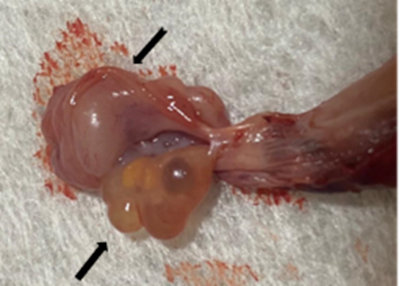
Fig. 1: Bitch ̓s ovary (black arrow) after bursa (notched arrow) removal (taken in the clinic of pet animals, Fac. Vet. Med., Benha University).
During proestrus, follicles appear by ultrasound as multiple anechoic structures that enlarge with time (up to > 1 cm in diameter). During diestrus, the ovaries may be lobular, and the corpora lutea are obvious hypoechoic structures of variable size.
2. Uterine tubes
the uterine tubes are quite narrow tubular structures, hanged by the mesosalpinx close to and above the ovary, caudal to the kidney. The infundibulum is the ovarian funnel-shaped extremity of the oviducts that is bounded by finger-like projections (known as fimbriae) to pick up the ova and ensure its entrance into the uterine tubes following ovulation.
3. Uterus
The uterus is located in the midline of the caudal abdominal cavity. It is a Y-shaped structure composed of two long uterine horns that are five times the length of the uterine body (Fig. 2).
The uterine wall consists of 3 layers;
1. The endometrium: consists of columnar epithelial cells, glandular tissue, and blood vessels.
2. The myometrium: consists of smooth muscle fibers that are responsible for uterine contractions at parturition.
3. Perimetrium: consists of connective tissue fibers.
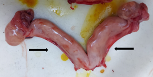
Fig. 2: Uterus of cat, Y-shape structure with two long uterine horns (black arrows) (taken in the clinic of pet animals, Fac. Vet. Med., Benha University).
The uterine size differs according to breed, animal size, age, parity, and stage of the estrous cycle. During proestrus and estrus, the uterine size increases, and the uterus becomes edematous, while in diestrus it reduces in size and becomes corkscrew in shape. Progesterone hormone stimulates endometrial thickening so able to receive and implant embryos during pregnancy.
4. Cervix
The cervix is a thick-walled tube that connects the uterus with the vagina so allowing the passage of sperm from the vagina to the uterus during mating, and the fetus from the uterus to the vagina during parturition. For fetal protection form infection during pregnancy, a mucoid plug closes the cervix.
5. Vagina
The vagina is a musculo-memberanous tube, extends from the cervix to the external urethral orifice. It is lined with squamous epithelial that is arranged in longitudinal folds to allow its dilatation during parturition. During the estrous cycle, in response to the circulating hormones, the epithelial lining changes. These changes can be detected by vaginal smears so help selection of correct mating time in bitches (Fig. 4).
6. Vestibule
The vestibule is located between the external urethral orifice and the vulva. It is a common pathway to the urinary tract and the reproductive tracts. Its structure is similar to that of the vagina except owing smooth walls rather than folded.
7. Vulva
The vulva is the external reproductive tract opening that consists of two lips fused ventrally and dorsally. It is normally tightly closed to prevent entering of infection. Enlargement of the vulva occurs during proestrus and estrus in the dog, but not in the cat. In its ventral angle, the clitoris is present which is an equivalent of the penis of male. The clitoris becomes erected during sexual excitement due to filling of the cavernous spaces with blood.