UPPER AND LOWER EYELIDS AFFECTION
- Structure of the eyelids
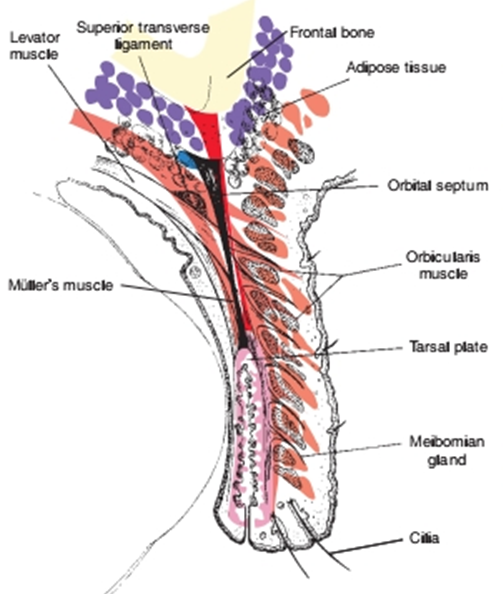
Fig.1:. Structure of the normal eyelid. (Remington, 2005).
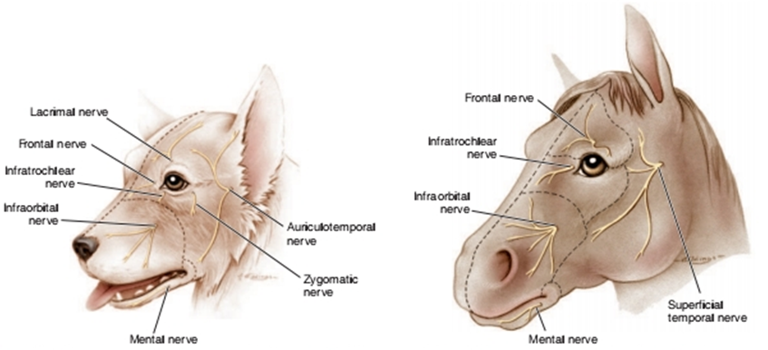
Fig.2: Sensory innervation of the canine periocular area. Fig.3: Sensory innervation of the equine
(Westhues and Fritsch, 1964 periocular area. (Westhues and Fritsch, 1964)
The eyelids protect the eye in the following ways
1. Sensory and protective effects of the cilia and sensory vibrissae surrounding the eye.
2. Secretions of the meibomian glands and conjunctival goblet cells, which contribute to the outer lipid and inner mucopolysaccharide layers of the precorneal tear film, respectively.
3. Physical protection against trauma.
4. Reduction of evaporation of tears.
5. Distribution of the precorneal tear film by eyelid movements.
6. “Pumping” of tears down the nasolacrimal duct, preventing epiphora and promoting a precorneal tear film of uniform thickness and optical properties.