Pigeon Diseases
| Site: | EHC | Egyptian Health Council |
| Course: | Avian and Rabbit Medicine Guidelines |
| Book: | Pigeon Diseases |
| Printed by: | Guest user |
| Date: | Tuesday, 2 June 2026, 7:47 PM |
Description
"last update: 7 April 2025" Download Guideline
Table of contents
- - Acknowledgement
- - Scope
- - Aim
- - Definitions
- - System affections and clinical problems
- - Viral diseases
- - Bacterial diseases
- - Protozoal diseases
- - Parasitic diseases
- - Fungal diseases
- - Other diseases
- - Vaccination program of pigeon
- - Types of pigeon breeds in Egypt
- - Viral diseases Paramyxovirus infection
- - Pigeon Pox Virus infection
- - Adenovirus infection
- - Circo Virus
- - Bacterial diseases
- - Chlamydophila's(ornithosis)
- - Streptococcus & Staphylococcal infection
- - E. coli or Collibacillosis
- - Mycoplasmosis
- - Protozoal diseases in pigeon
- - Trichomoniasis (canker)
- - Spironucleus columbae (hexamitiasis)
- - Coccidiosis
- - Parasitic diseases in pigeon
- - Ectoparasites in pigeon
- - Fungal diseases
- - Candida Infectionm sour, muguet, sour crop
- - Other diseases
- - References
- Acknowledgement
We would like to acknowledge the committee of National Egyptian Guidelines for Veterinary Medical Interventions, Egyptian Health Council for adapting this guideline.
Executive Chief of the Egyptian Health Council: Prof. Mohamed Mustafa Lotief.
Head of the Committee: Prof. Ahmed M Byomi
The rapporteur of the Committee: Prof. Mohamed Mohamedy Ghanem.
Scientific Group Members: Prof. Nabil Yassin, Prof. Ashraf Aldesoky Shamaa, Prof. Amany Abbass, Prof. Dalia Mansour, Dr. Essam Elmarakby, Dr. Mohamed Elsharkawy, Prof. Gamal A. Sosa., Dr. Naglaa Radwan, Dr. Hend El Sheikh
Authors: Prof. Dalia Mansour Hamed, Dr. Essam Elmarakby
- Scope
The guidelines concerned diagnosis, treatment, and prevention of pigeon diseases. The guidelines also provided landmarks for the evaluation of the severity and the most suitable antibiotics for therapeutic intervention.
- The target audience
The guideline is intended for all veterinarians who are intended to diagnose, treat, and control pigeon diseases.
- Aim
Pigeon production is essential to their economy and food security. When proper husbandry and management practices are used, pigeons are relatively resistant to disease.
- Definitions
Loft: nest of pigeon, a place where pigeons live
Squabs: newly hatched pigeon
- System affections and clinical problems
System/ problem | Symptom | Etiology |
Respiratory system | Respiratory infections
make it hard for a bird to breathe and fly so they are less active and
competing pigeons will perform poorly. pigeon sneezing and/or coughing will be detected. | Viral: Herpesvirus, Influenza A (acute), Adenovirus (some),
Coronavirus |
Digestive system | Skin form: scabs and
proliferations of wattle, legs, feet, and commissures of the beak. The first
signs are usually conjunctivitis with excess lacrimation and swelling of the
eye. diphtheroid form can occur,
causing lesions on the mucosal surface inside the mouth. Secondary bacterial
invasion can cause proliferative lesions, obstructing respiration and making
eating difficult. | Viral: POX, Circovirus |
Nervous system | polyuria (not diarrhea) and central nervous signs, ranging from incoordination, difficulty picking up grains, and mild head tilt, to severe ataxia and torticollis. | Bacterial: , P multocida,Salmonella |
Locomotor system | Joint abscess: Swelling of the
elbow, tibiotarsal tarsometatarsal joint and wing, causing lameness and
inability to fly (wing droop). | Bacterial: |
Skin and feather | Skin form: scabs and proliferations of wattle, legs, feet, and
commissures of the beak. | Viral: POX |
| A mild to necrotizing pharyngitis and esophagitis are the primary symptoms. Diphtheritic membranes and general signs of illness, including neurologic abnormalities, green droppings, and anorexia, can suggest PHV infection. Vomiting with no other symptoms and inclusion body hepatitis can occur.( hepatic necrosis of any cause. Basophilic intranuclear inclusion bodies are strongly suggestive of adenovirus) | PMV1 and pigeon pox virus, herpes virus and adenovirus are the most common. however, circovirus, rotavirus, parvovirus, and influenza virus cause |
Reproductive problems | embryonic or early squab death | Paratyphoid (Salmonellosis) |
Young bird sickness | slow crop emptying, regurgitation, diarrhea, weight loss, poor performance, and occasionally death. Lesions occur in the lymphoreticular system, alimentary tract, and respiratory system | circovirus infection, birds exhibit protozoal, fungal, and mixed bacterial infections associated with enteritis. Concurrent C. psittaci pneumonitis. |
Infectious diseases | Decreased appetite · Clear, watery droppings with little solid matter · Vomiting · Rapid weight loss · Death can occur rapidly, within days. | Salmonella ,
E coli , |
Diseases associated with poor management | Infected pigeons can display a variety of disease signs include weight loss, diarrhea, problems breathing, and difficulty flying. | Bacterial: Salmonella |
- Viral diseases
|
Viral diseases |
General Treatment |
|
No specific treatment, symptomatic treatment with vit. supply and antibiotic to prevent secondary bacterial infection |
|
|
Pox |
|
|
Adenovirus infection |
|
|
Adenovirus type infection (necrotizing hepatitis) |
|
|
Circovirus infection |
- Bacterial diseases
|
Bacterial diseases |
Specific Treatment |
|
Salmonellosis |
Oxytetracycline, enrofloxacin |
|
Chlamydophillosis(ornithosis) |
Chlortetracycline and doxycycline |
|
Streptococcus & Staphylococcal infection |
doxycycline, enrofloxacin trimethoprim |
|
E. coli |
Sulphonamides |
|
Pasteurella multocida |
Sulphonamides, Sulfamethazine. Norfloxacin |
|
Mycoplasma |
Tylosin |
- Protozoal diseases
|
Protozoal diseases |
Specific Treatment |
|
Trichomoniasis (canker) |
Ridzol ,Dimetridazole, Metronidazole, Copper Sulfate, Quaternary Ammonium, Carnidazole |
|
Spironucleus columbae (hexamitiasis) |
Ronidazole or metronidazole Dimetridazole |
|
Coccidiosis |
Amprolium, Sulphonamides, Clazuril, and Toltrazuril. |
- Parasitic diseases
|
|
Parasitic diseases
|
Treatment |
|
Worm infection |
Helminths |
Moxidectin , piperazine levamisole, ivermectin, moxidectin, selamectin |
|
Roundworms |
||
|
Hairworms |
||
|
Stomach worms |
||
|
Tapeworms |
||
|
Strongylids |
||
|
Ectoparasites |
Fleas |
spray with an insecticide, such as permethrin, Moxidectin orally |
|
Mosquitoes |
||
|
Lice |
||
|
Mites |
- Fungal diseases
Aspergillosis, Candidiasis
- Other diseases
One-eyed cold, Sour crop. Gout
- Vaccination program of pigeon
|
Route of vaccination |
Age of bird |
Time of vaccine |
Type of vaccine |
|
-wing web sticking or stabbing -brushing of feather follicles in the upper part of thigh or breast muscles |
6 weeks |
Annually between July and August |
Pox vaccine |
|
0.5 cm S/C of neck |
5 weeks |
Every 6 months |
Paramyxovirus vaccine |
|
0.5 cm S/C of neck |
4 weeks |
Every 4 months |
Salmonella vaccine |
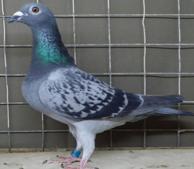
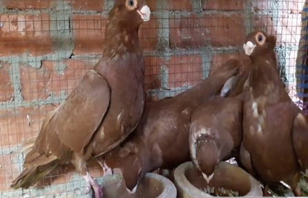
- Types of pigeon breeds in Egypt
1- Local pigeon
|
Types of pigeon in Egypt |
Characteristic features |
|
Baladi pigeons
|
This type of pigeon is characterized by its diverse colors, including red, gray and white, and weighs up to 600 grams. |
|
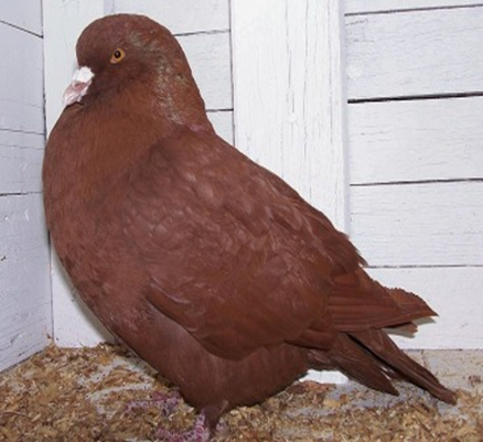
Kattawy pigeons
|
Similar in their form to Roman pigeons, but their weight reaches 400-500 grams and is characterized by a dark red color and there is a large amount of feathers accumulated around its beak. |
|
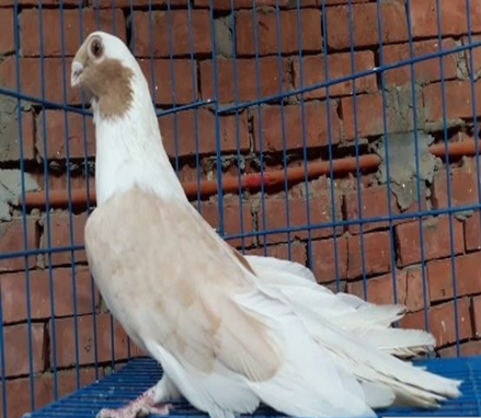
Maltese pigeons
|
This breed closely resembles chicken. Charchtrized by high standing on uniquely strong and straight legs. weight about 500 grams. its head is smooth and not encircled with feathers. |
|
Prolific black pigeon
|
having a round head, a thick, short and thick beak, a thick nose, and round and wide eyes. The eye color is dark ruby and his eyelids are white and thick |
|
Prolific red pigeon
|
Red color and has a white tail. It has a thick and short beak. His eyes are white tinged with red. He has a white obelisk on top of his beak, his nails are white. |
|
Spotted pigeons
|
Having a thin head and is one of the most beautiful types of Egyptian pigeons, its eyes are very wide and its color is bright white, it has a white obelisk in the middle of the head and has a short white beak, its legs are short and white, and the wings have a light fat color |
|
Al Safi
|
Characterized by its beauty and predominantly white coloration, except for its neck, which features continuous feathers in various colors such as yellow, red, or dark green. It has a rounded head with small eyes, a medium breadth, and a rounded, glossy black proximity. Its thin, white eyelids complement a short, thick, and wide beak. The neck is of medium length, and the overall body size is small to medium. The bird displays multiple color variations, including sugar, dark red, and sky blue. |
|
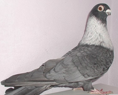
kotkaty pigeons
|
One of the most famous Egyptian pigeons and loved by people for the purpose of breeding because of its shape and size distinct and has three types: Mahlawi: The body and head are dark gray or very black and have a silver neck. Soapy: Light gray to silver, its head is darker than the rest of the body, and the neck is silver. Light and dark fish peel: the body color is silver or gray, the head is dark gray and the neck color is silver. |
- Viral diseases Paramyxovirus infection
Epidemiology:
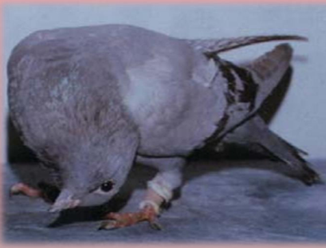
The viral disease affecting pigeons is characterized by neurological manifestations and respiratory symptoms, caused by the paramyxovirus (PMV) 1, specifically a mutant mesogenic strain of Newcastle Disease Virus (NDV). All age groups of pigeons are susceptible, but the condition poses greater risks to young birds. Infections can occur year-round, with increased incidence noted in unvaccinated flocks during the racing season. Transmission occurs via both direct and indirect contact with infected birds, primarily through ingestion of contaminated food and water following exposure to droppings or nasal secretions, as well as through inhalation of infectious respiratory secretions.
Clinical signs:
-Nervous manifestations, tremors in the head and neck, torticollis, opsthtinous, wing and neck paralysis,dropping of the head and neck.
- Greenish watery diarrhea with haemorrhages in severe cases.
- Respiratory signs are rare, morbidity and mortality rate ranged from (20-80%).
|
Torticollis |
Paralysis of wing, balance disorder |
Post-mortem:
- Hyperemia and congestion of the brain with petechial haemorrhages.
- Septicemic picture of all carcasses, catarrhal enteritis, enlarged kidneys.
- Haemorrhages on pancreas.
Diagnosis:
- The sample from the feces or respiratory secretions during acute phase of an infection.
- Isolation, identification or detection of antibodies titers are similar to other PMV infection (as NDV).
- Pigeon Pox Virus infection
Epidemiology:
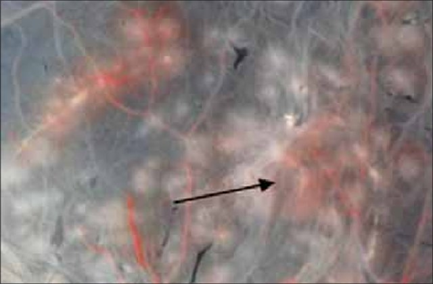
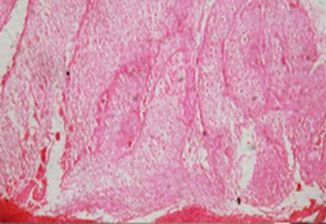
The disease is a highly contagious viral infection affecting poultry, distinguished by the presence of lesions on unfeathered skin areas and/or diphtheritic lesions on the mucous membranes of the upper digestive and respiratory tracts. The causative agent belongs to the Poxvirus family (Poxviridae) DNA virus.
The virus replicates on the chorioallantoic membrane (CAM) of embryonated chicken eggs (ECE), resulting in the formation of grayish focal areas of cellular proliferation, known as pock lesions.
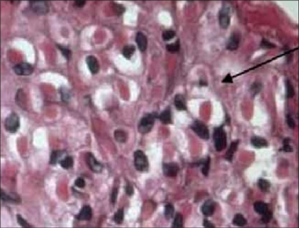
Histopathological examination reveals the presence of round to oval intracytoplasmic inclusion bodies, called ( Bollinger bodies).
Transmission: of the virus primarily occurs through wound infections due to skin abrasions, direct contact with infected birds, and mechanically via vectors such as mosquitoes. Additionally, airborne transmission may contribute to the spread of infection. Understanding these epidemiological aspects is crucial for controlling infection.
Clinical signs:
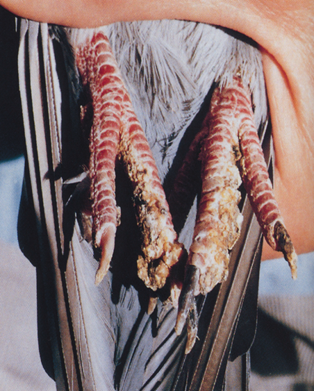
I.Skin (cutaneous or dry) form:
- Wart like nodules (grayish blister white spots) on the un-feathered parts of the bird’s body (around eyes, base of the peak, legs, under the wings and around the vent).
II. Mucous membrane (wet, diphtheritic) form:
- Diphtheritic membrane (pustule like nodules) on the upper digestive, upper respiratory tract and nasal cavity.
- Yellow pustules changed to caseous necrotic material forming what is called pseudo membrane (diphtheritic membrane).
III. Mixed form:
- Cutaneous lesion and diphtheritic lesion in the same time.
|
Pigeon pox (cutaneous form) |
Diphtheroid form of pigeon pox, yellowish membranes in the oral cavity |
|
Pigeon pox (cutaneous form) |
pocks lesions were produced by field strain of fowl pox virus on CAM |
|
Cutaneous pock lesion of pigeon pox showing vacuolated prickle cells and intracytoplasmic eosinophilic inclusions. |
Chorioallantoic membrane showing intracytoplasmic inclusion bodies (×100).( called Bollinger bodies |
Diagnosis:
- The lesions are suggestive.
- Histopathological sections from the lesions revealed presence of intracytoplasmic inclusion bodies (Bollingers bodies).
- Inoculation of the suspected material on CAM of ECE gives pock lesions after 5-7 days ten confirm by histopathology.
- Identification by IGPT, NT passive HA and ELISA test.
Prevention and Control:
1. Sanitation and Management: sanitation practices and sound flock management to inhibit disease spread.
2. Vaccination:
- Apply a live attenuated virus vaccine, specifically the pigeon pox vaccine (PPV), for chickens, turkeys, and pigeons, propagated in tissue culture (TC).
- Vaccination techniques include wing web sticking or stabbing for chickens and pigeons, and brushing of feather follicles in the upper thigh or breast muscles.
- Emergency Vaccination:
1. Initiate vaccination as soon as clinical signs appear during an outbreak.
2. Utilize the live virus (pigeon pox vaccine), as it typically induces milder post-vaccinal reactions compared to the fowl pox vaccine.
3. Administer the vaccine intradermally via the wing web or feather follicle methods.
- Ectoparasite and Insect Control: Eradicate ectoparasites and insects to reduce potential disease vectors.
- Avoidance of Trauma: Address and eliminate sources of abrasions and wounds, such as cannibalism and mechanical trauma.
- Mosquito Control: Eradicate standing water to eliminate primary mosquito vectors. Isolate or cull infected birds to remove sources of the virus.
- Decontaminate feeders, waterers, bird baths, and cages using a 10% bleach solution.
Treatment:
1. Remove lesions.
2. Cleanse or treat affected areas with a tincture of iodine mixed with glycerin (1:4).
3. Administer antibiotics to prevent or treat secondary bacterial infections.
4. Provide multi-vitamins, particularly vitamin A for epithelial repair and vitamin E to enhance resistance.
5. Stimulate appetite by offering wet mash.
- Adenovirus infection
Only birds whose immune systems are suppressed are vulnerable to this disease. There are two types of Adeno Virus that infect pigeons. Type 1 affects young pigeons primarily and causes vomiting and diarrhea, from which many birds recover.
Type 2 is contracted by older pigeons and strikes the liver, with most affected birds dying within 24 hours. Some birds display a fluid yellow diarrhea and vomiting before death. But the main sign is sudden death, occurring within 24 hours of the onset. None of the affected birds live longer than 48 hours. E. coli often accompanies Type 1 ( which is associated with young pigeons ) and complicates the infection, making the diarrhea and vomiting more severe and adding respiratory symptoms. This Type 1 Adeno Virus/E. coli sometimes successfully treated with antibiotics. Cases that are the best managed are those in which the loft environment is good and in which all secondary diseases like canker and coccidiosis are treated so that birds are best able to fight the virus.
Prevention
Adeno virus is always present in a young bird, and it erupts when the immune system or fails. It is important to wait until the 12-th week for any type of vaccination. There is currently no vaccine that is proven to be effective against Adeno Virus. It's also important to keep stress in the loft at a minimum with Antifungal and probiotics to boost pigeons' general health with vitamin supplements that are already included in Pigeon Vitality products, etc.
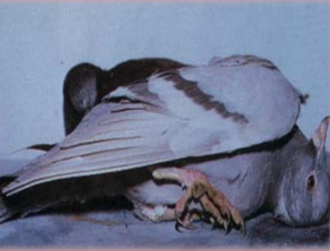
- Circo Virus
Circo Virus
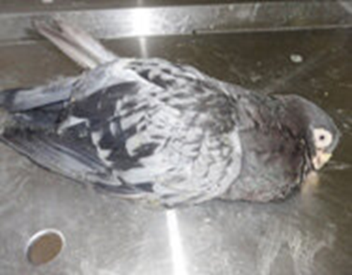
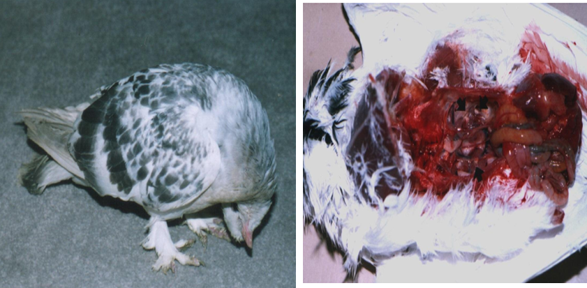
Circo Virus is sometimes called pigeon AIDS. Circo Virus damages the lymphocytes in the blood, which are closely associated with the immune system. With damaged lymphocytes, the pigeons become susceptible to secondary infections with other viruses, parasites and bacteria. Pigeons infected with Circo Virus can also have continuing problems with diseases like respiratory infections, chlamydia, or canker due to the fact that they cannot form natural immunities to them. Birds with Circo Virus have a yellowish discharge dried on the beak, and they are very reluctant to move, thin and dehydrated, and have no appetite and difficulty breathing.
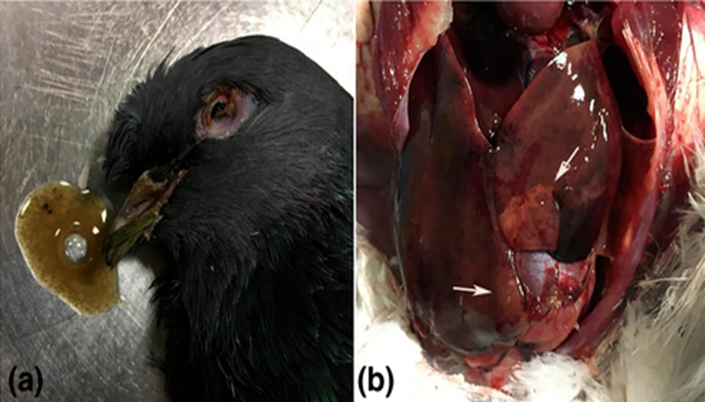
a) Picture of one of the infected birds that were necropsied. Vomit from the bird is shown beside the bird's head, characterised by mucous and watery content. The bird had shrunken eyes indicating dehydration and poor condition.
(b) Image of an example infected pigeon liver taken during necropsy. Widespread discoloration indicates severe necroses (white arrows) and haemorrhages on the liver
Prevention
- Use of probiotics and Antifungal while keeping the disease out of the loft by not introducing birds from lofts known to have health problems.
- Stray youngsters should be immediately removed if they do not look well, and try to identify carrier stock birds by re-pairing those whose offspring seem weak or die.
- If the virus comes into lofts, minimize its spread by taking sick birds out immediately and isolating them while giving them full doses of Improver, paying attention to ongoing hygiene.
- Good care, given day to day, places your pigeons in the best situation to resist infection, and gives those that become infected the best chances of recovery.
-The main defense against Circo Virus is to identify and treat secondary infections, allowing the birds to live long enough for immune system to repair itself.
- Probiotics help well birds resist the disease by maintaining a healthy bowel population of bacteria
Protocols for managing pigeon health and preventing most viral diseases transmission:
1. Implementing biosecurity to separate healthy from infected pigeons.
2. Avoiding the mixing of different age groups.
3. Restricting free-ranging pigeons from accessing captive areas.
4. Quarantining returning pigeons for six weeks post-competition.
5. Administering live attenuated vaccines via eye drops.
6. Recognizing the lack of specific treatments for adults, focusing on supportive care.
7. Providing specialized feeding and antibiotics to prevent secondary infections.
8. Conducting emergency vaccinations during outbreaks.
9. Isolating and removing clinically infected birds from the flock.
- Bacterial diseases
Paratyphoid- Salmonellosis
This very common and quite widespread is caused by a gram-negative bacterium which is flagellated, therefore mobile. It can be brought into a loft either through introduction of infected pigeons, by rodents, through inhalation of infected dust, on the soles of fancier’s shoes, by roaches, or through contact with wild pigeons. Often an adult bird that has overcome the disease remains a carrier and continues to produce infected droppings.
Clinical signs:
- Most adult birds will show rapid weight loss, along with somewhat loose, greenish droppings.
- Some birds may develop swelling in the leg joints or feet, or may develop wing boils.
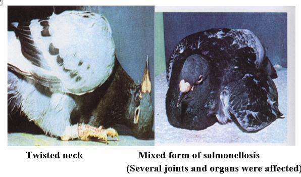
- Other birds may have the "twisted neck" syndrome commonly associated with PMV.
- Baby birds will often show labored breathing or die in the nest before the second week after hatching.
Prevention
- Loft hygiene is critical, because salmonella flagellates can live in the droppings for some time. But once Antibacterial is given in the drinking water of the pigeons, the droppings will stop being infected with salmonella.
- Regular cleaning and disinfecting of lofts, feeders and drinkers is imperative.
- Minimizing contact with rodents, roaches and wild birds, quarantining newly acquired birds, and maintaining an acid pH level below 4.0 in lofts are all helpful steps in keeping this disease under control.
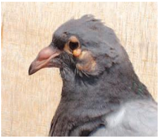
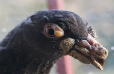
- Chlamydophila's(ornithosis)
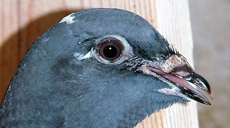
Ornithosis is an infectious disease that affects many bird species worldwide. It can also be transmitted to humans and other mammals. Ornithosis is a notifiable disease in pigeons caused by Chlamydia psittaci. Chlamydia are small, non-motile micro-organisms that invade cells parasitically. Infection occurs via inhalation of stirred-up dust containing the pathogen, uptake of fecal contaminated feed or water, or else billing or feeding of squabs.
Symptoms
Ornithosis occurs in 2 forms:
The acute form can be recognized in young pigeons from: wheezing noises, uni or bilateral conjunctivitis and muco-aqueous enteritis with diarrhea.
The chronic form is more often found in adult birds, which, however, show few or no signs of the disease. Pigeons that have recovered are a dangerous source of infection for young pigeons and for humans due to their latent shedding of the pathogen.
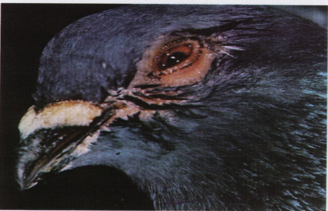
Severe conjunctivitis, weeping and sticking together of feathers
Diagnosis
The disease can be demonstrated in dead pigeons by microscopic examination of a smear or impression ("klatsch") preparation of spleen, liver, conjunctiva or air sac that has first been stained using the method according to Stamp. In live birds, the pathogen is demonstrated in feces, via a sink dab from the cloaca, or alternatively by serological identification of specific antibodies.
Treatment
Flocks are treated with chlortetracycline, which has been successfully used for many years to control ornithosis. In order to maintain effective blood levels, administration of chlortetracycline+ must not be interrupted during the 30-day treatment period.
- Streptococcus & Staphylococcal infection
Bacterial infections :Streptococcus faecalis, Streptococcus gallinarum, and Staphylococcus aureus. These bacteria are transmitted through contaminated feed or water and from infected birds. They can enter the host through body openings or skin lesions. While these bacteria are present globally and typically reside harmlessly on the skin and mucous membranes, infections may occur depending on the host's immune resistance.
Clincal signs:
Affected birds may exhibit signs similar to those of salmonellosis, including:
- Diarrhea, Listlessness, Paralysis, Emaciation, particularly in young pigeons
Abscess-like nodules may be observed in various organs, especially the intestines.
Diagnosis of bacterial infections is achieved through laboratory examination of droppings and tissue samples.
Prevention: To mitigate the risk of bacterial infections, it is crucial to maintain hygienic loft conditions. Implementing general preventive measures, such as regular disinfection with appropriate disinfectants, is essential for controlling the spread of these pathogens.
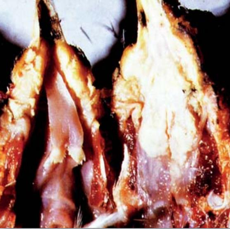
- E. coli or Collibacillosis
This disease, which is now thought to be more prevalent in pigeons than once suspected, is caused by gram negative bacteria's which can invade our lofts through infected dust particles, rodent droppings, and trough infected pigeon droppings coming into contact with eggs in the nest. Infected adult pigeons will emit the bacteria throughout a pigeon loft.
Clinical signs:
Since the E. coli bacteria can manifest themselves in any part of the pigeon's body, symptoms can be diverse. Most often young will die in the nest, adult birds will become listless and lose weight, and their droppings will become loose, mucous, and greenish-yellow in appearance. Sometimes the droppings will have a foul odor. Occasionally some birds may have nasal discharges and respiratory problems associated with this disease.
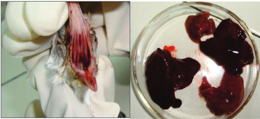
Bleeding from oral cavity (Left) and Post mortem lesions –necrosis of liver (Right).
Prevention
Maintaining good loft hygiene and keeping rodents away from feed and water are very important. Also keeping dust and ammonia levels down will help to control any outbreaks.
General Antibiotics
Any fancier would be well advised to have a good general antibiotic. They can be useful as "first choice" drugs if and when problems occur. General antibiotics are effective against a broad range of both gram positive and negative bacteria. But excessive use of them can really damage the microflora of the bird, therefore most veterinarian suggest a use of probiotic and will have the same effect that any antibiotic, but will leave the good bacteria and keep the balance of the pigeon intact.
- Mycoplasmosis
The chronical form of catarrh in pigeons often is called "Mycoplasmosis". It is caused by a multiple infection with pathogens: bacteria (e.g. Cocci), viruses (e.g. Herpes) and pathogens which belong to the group of mycoplasma organisms. It is assumed that mycoplasma causes severe conditions only in the presence of other infections.
Etiology: Mycoplasma organisms are viable only for a short period (approx. 17 days at 20°C, but only 20 minutes at 50°C). Low temperatures favor their survival. Mycoplasma organisms are killed by almost all commonly used disinfectants, e.g. disinfectants. Transmission takes place through the feces, the drinking-water, feed, equipment and by droplet infection from pigeon to pigeon.
Clinical signs: Mucopurulent discharge from the nose, reduced flying performance, unwillingness to fly, flying awkwardly, throat inflammation, rattling and wheezing sound of respiration particularly noticeable by night. Air sac inflammation.
Acute mycoplasmosis,lachrymation, panting
P.M. examination: air sac inflammation
Prevention
Elimination of possible factors that reduce the bird's resistance to infection. Such factors may be: overcrowding in the loft, lack of cleanliness, latent infections (e.g. ectoparasites, worm infestations, coccidial infection),
- Protozoal diseases in pigeon
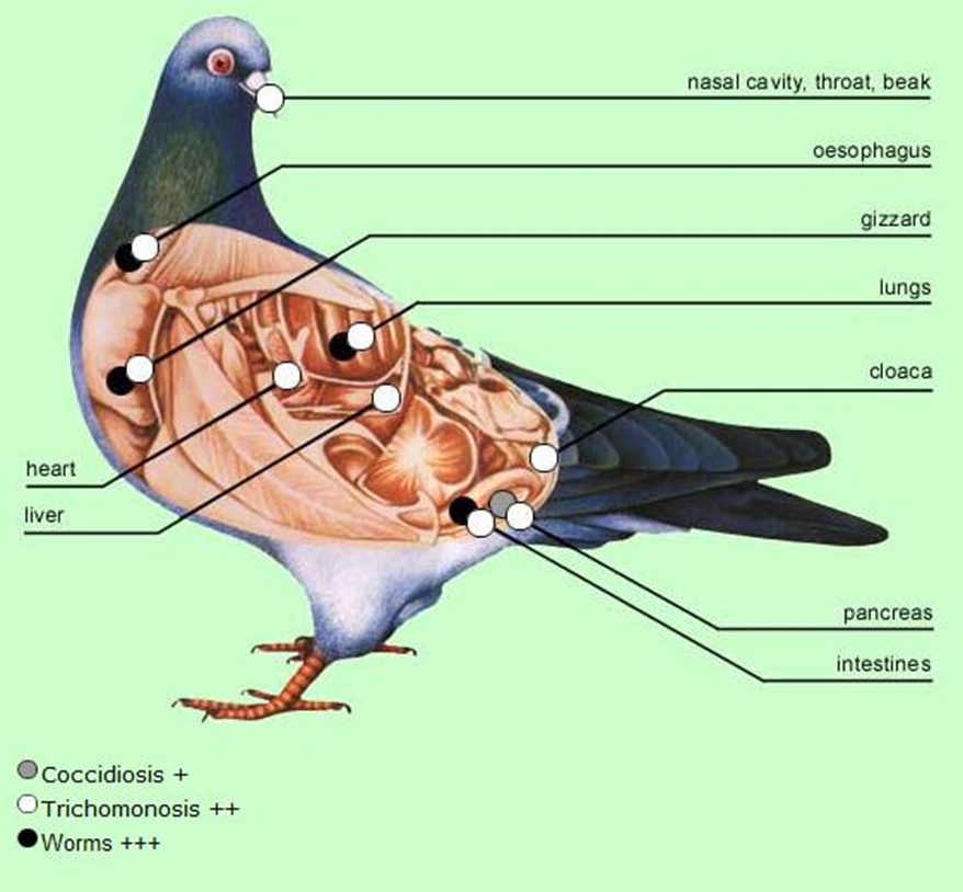
https://149357833.v2.pressablecdn.com/wp-content/uploads/2018/03/coccidiosis-.jpg
- Trichomoniasis (canker)
Canker is a protozoan disease that is the greatest killer of the pigeon diseases. It is found in domestic and wild pigeons and doves caused by a microscopic protozoan which is flagellated and, therefore, mobile. It can be transmitted from one bird to another usually through the drinking water, and parent birds can infect their young through milk feeding.
Clinical signs
A swelling in throat and cheesy growth in the mouth are certain signs of canker.
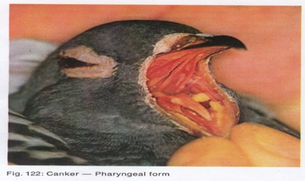
Canker (pharyngeal form)
Treatment :
All forms of canker can be treated if found early. Ridzol Soluble Powder For flock treatment in drinking water. or Ronidazole Tablets for individual treatment.
- Spironucleus columbae (hexamitiasis)
Hexamitiasis: is an intestinal disease of pigeons that is associated with mucoid, or even bloody feces caused by the flagellate, Hexamita columbae occurs in pigeon flocks mainly in the summer and autumn months. It primarily colonies the rectum. Especially susceptible are newly weaned squabs, whose resistance is still low. Infected adult pigeons do not normally show visible signs of the disease, but can excrete the pathogen in large quantities in their droppings (chronic carriers). The incubation period is 4-5 days.
Clinical signs
Acute catarrhal (or even bloody) enteritis with liquid, rice water-like or mucoid, malodorous diarrhea. Affected pigeons refuse feed and drink more water, resulting in emaciation and debility.
Recognition
Hexamita are demonstrated via microscopic examination smears from the intestinal mucosa of a recently killed, acutely affected pigeon. With extremely severe infestation, it is also possible to demonstrate the parasites in a cloacal swab from a live bird. They can be recognized from their characteristically rapid movements in a straight line - in contrast to trichomonads, which exhibit slow, circular movements around their own axis.
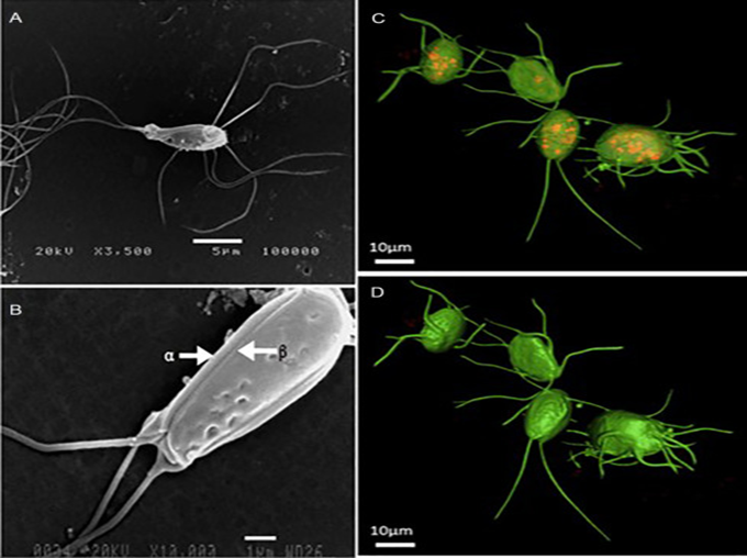
Spironucleus Hexamita columbae. Note shape of body and arrangement of the 8 flagella, each of which taper terminally culminating in a small bulb.
- Coccidiosis
- This highly infection and very common disease is caused by a protozoan that infects the intensities of our birds. It usually presents to some degree in all pigeons, but most adult birds have developed enough immunity to the disease to remain healthy. Most often infected are
young pigeons or birds that have been subjected to severe stress (i.e., racing, showing, lack of feed/water, or relocation). Adult birds may become infected from drinking unclean water or from being in contact with moist droppings.
Clinical signs
- Infected birds have little or no desire to eat or drink, will remain puffed up on perches, lack any desire to move and often close their eyes. Droppings are usually very loose, greenish in color and may become very watery. Loss of weight occurs, and death can occur in young birds.
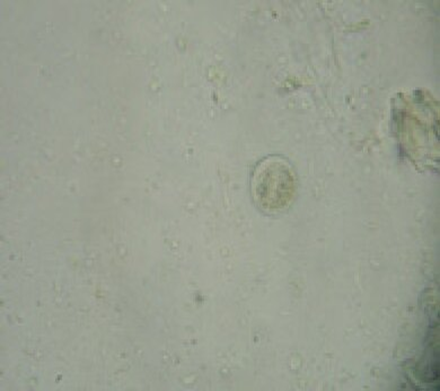
Pigeon with severe coccidiosis (Left) and
unsporulated Oocysts (Right; 400X).
Prevention
- Do not allow feed to come into contact with droppings, and regularly disinfect drinkers. Do not allow birds to drink from gutters or mud puddles, and keep feed and water free from contact with rodents. Always isolate new birds, as they are a primary target for the spread of coccidiosis. Returning race birds should be given a preventive treatment shortly after their return,. Baskets should be disinfected weekly.
- Parasitic diseases in pigeon
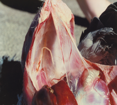
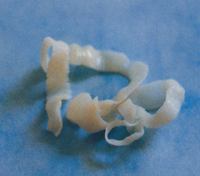
Worms in pigeon
Common Types Roundworms, Tapeworms, Hairworms, Gapeworms.
Etiology: Worm eggs are passed in droppings and by birds eating Slugs, Earthworms and pill bugs can get worms.
Roundworm: Most common found in pigeons. Worm lives in the intestine and feeds on digested food. Pigeons may become lethargic.
Tapeworm : Lives in the Small intestine. Pigeon may become sluggish. Insects are a common carrier.
Hairworm (threadworms) in pigeon
anther common pigeon parasite. Heavily infested pigeons have diarrhea and sluggish, loose interest in feeding and drinking.
Gapeworms in pigeon
Pigeons eating earthworms is one way a pigeon can get gapeworms. Gape worms can cause breathing problems.
keeping a clean loft and a preventive program of deworming at least every six months.
|
Round worn |
Tap worm |
|
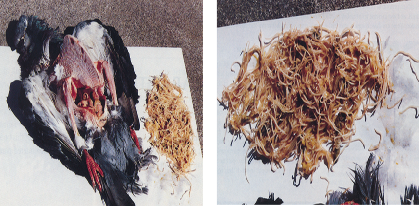
Autopsy of a pigeon with a heavy roundworm burden. More than 300 roundworms were removed from this pigeon’s bowel at autopsy and were the obvious cause of its emaciation. |
|
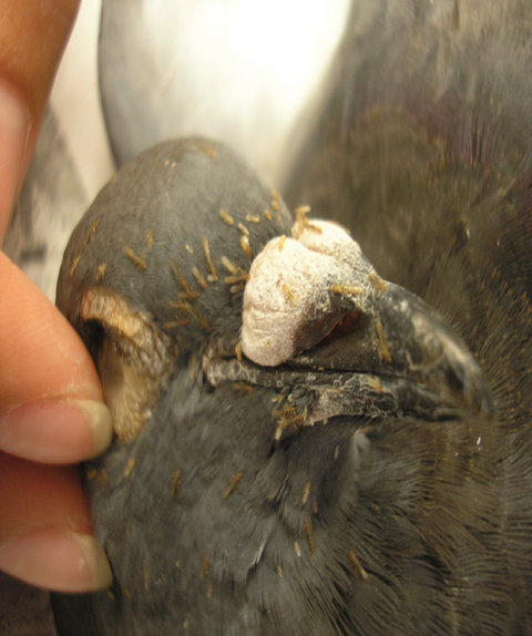
- Ectoparasites in pigeon
Ectoparasites are widespread in pigeon flocks.
Etiology:
Feather lice, scaly-leg mites and body mange mites live permanently on infested pigeons, leaving them only to seek new hosts. Pigeon ticks, bird ticks and red bird mites attack pigeons only periodically at night to suck blood. Otherwise, they conceal themselves in cracks in the loft. They can transmit pathogens.
Recognition of the disease:
Feather lice are visible in the pigeon's feathers with the naked eye. Infestation with body mange mites and scaly-leg mites can be confirmed by microscopic examination of a scraping from inflamed skin. Pigeon and bird ticks and red bird mites can be detected with the naked eye in cracks in the loft - ideally in the early hours of the morning, when the parasites leave the birds in search of a hiding place. They are also found under feeding troughs and nest bowls.
|
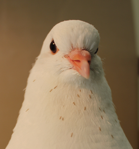
Pigeon lice |
Lice on the neck |
|
lice move to the surface of the feather. |
scaly mite infestation. |
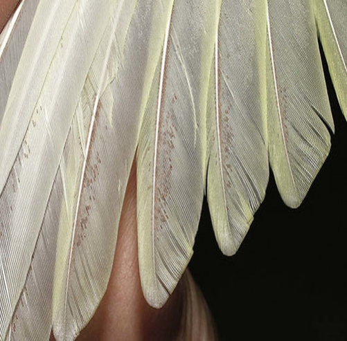
Ectoparasites on pigeon's feathers
Prevention
Clean feed and water vessels with hot water. Regular bathing in clean water at least once a week protects pigeons against parasite infestation
A- Long Louse
Found on feathers of the whole body. They feed on feather scuff and cause little damage.
Treatment: Dipping and dusting the loft and the birds with pesticides.
B- Small Louse
Do not like light and are found on the underside of the body feathers. They eat feather scurf and cause prickling and burning irritation.
Treatment: Dipping and dusting the loft and the birds with pesticides.
C- Feather Mite
Found on the feathers shaft of the flight feathers. Causes much irritation.
They appear as small black specks on the sides of the feather’s shafts.
Dipping and dusting the loft and the birds with pesticides.
D- Itch Mite
These mites cause feathers to fall out. They burrow through the feathers
shaft into the follicle. If feathers have a swollen root, it is probably Itch
Mite. The feather shaft swells and then the feather is shed. Small pale spots appear on the underside of the feathers on the breast, wings, back and neck.
Dipping and dusting the loft and the birds with pesticides.
E- Red Mite
These mites will note be found on the pigeon when examine it. The Red
Mite hides in small cracks and crannies during the day. They come out at
night to feed on the blood of the pigeon. The mite causes irritation and
damage through blood sucking.
Dipping and dusting the loft and the birds with pesticides.
- Fungal diseases
Aspergillosis
Aspergillosis is a fungal disease of birds, animals and humans. It is usually characterized in the pigeon as a chronic infection of the lungs and air sacs. Another name for this disease is pneumomycosis.
Etiology:
Aspergillus fungi, they grow as multicellular, fluffy mold colonies, free
living in the soil, on vegetation or parasitic living in or on birds, animals,
and humans.
Clinical signs
Respiratory form: difficulty in breathing, greenish deposits on tongue and palate.
Skin form: skin scaling off with breaking of feathers.
Postmortem examination: Fungal colony in lungs.
Microscopic examination of deposits and skin scrapings.
Treatment:
Elimination of source of infection (e.g. moldy feed), separation of affected
birds. No treatment is recommended for Aspergillus respiratory infections in pigeons. Skin infections can be successfully treated e.g. with copper sulfate (1:2000 dilution) or a solution of mercuric chloride (1:500 dilution).
A pigeon with neurological symptoms suffer from Aspergillosis (Left) and numerous granulomatous lesions on air sacks (Right).
Prevention:
Dry, well-ventilated loft, good quality feed, administration of vitamins.
- Candida Infectionm sour, muguet, sour crop
Thrush is a common acute or chronic fungus infection especially of the digestive tract. Other names for this disease are mycosis, muguet, sour crop.
Etiology
- Candida albicans, a yeast-like organism.
- Bird’s eating food that is wet, moldy, sour or contaminated. Dirty drinking water is another source of trouble.
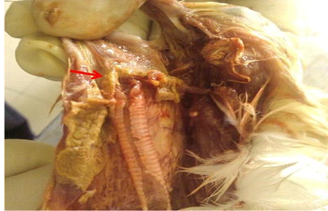
Pseudomembrane on esophagus, pharynx, and larynx (red arrow)
Symptoms
Poor growth of young pigeons, accumulation in the crop; whitish fungal growths in the throat.
Recognition
Microscopic examination of the fungal growths in the throat.
Treatment: Separation of affected birds. Administration of antifungal e.g. Nystatin and high levels of Vitamin A.
Prevention: Improving the environment.
- Other diseases
One-eyed cold
Etiology: Often confused with the onset of mycoplasmosis, one- eye colds are usually associated with a peck in the eye or some other type of physical injury affecting the eye. One-eye colds can also be caused by improper ventilation, dampness in the loft.
Clinical signs: A watery or mucous discharge in only one eye is usually the symptom most commonly noticed, but occasionally both eyes will have watery appearances. Sometimes one eye can become completely shut, depending upon the degree of infection.
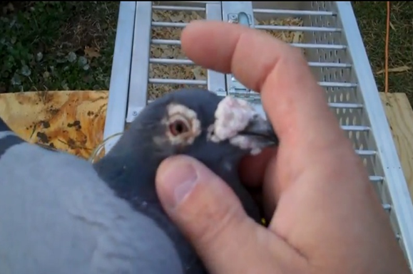
One-eyed cold in pigeon.
Prevention : Maintaining proper ventilation and not allowing overcrowded conditions, will go a long way in preventing one eye colds. It is also considered good loft hygiene to keep dust levels to a minimum, as many types of infectious bacteria are carried by dust particles.
Gout
Etiology: Water shortage, kidney damage, nutritional deficiency
Clinical signs: Nodular painful swelling of the joints. Liver and peritoneum, pericardium, air sacs appear as if dusted with lime (uric acid crystals). Kidneys swollen, interspersed with uric acid deposits.
Recognition : Microscopic examination of the deposits (crystals).
Prevention: Ensure adequate vitamin intake and exercise. Feed birds as required by performance.
Disinfecting Pigeon Lofts and Safe Disinfectants
Pigeon lofts, used for keeping domestic pigeons, require regular cleaning and disinfection to prevent disease and ensure a healthy environment.
Pigeons are susceptible to diseases, making effective disinfection is essential. The disinfection process includes several steps:
Preparation:
Remove pigeons, ensure ventilation, and wear personal protective equipment (PPE).
Cleaning: Remove organic matter using scrapers and brushes, then wash surfaces with soapy water.
Disinfecting:
Apply safe disinfectants, such as:
Vinegar: Natural and non-toxic.
Hydrogen Peroxide: Broad-spectrum antimicrobial.
Quaternary Ammonium Compounds: Effective but necessitates proper ventilation and dilution.
Rinsing and Drying: Rinse surfaces if required and allow drying to eliminate pathogens.
Routine Maintenance: Clean and disinfect every 2-4 weeks or as needed.
The study evaluates safe disinfectants, emphasizing their efficacy and safety profiles, and concludes that thorough cleaning and the use of safe agents are crucial for maintaining pigeon health and loft hygiene. Regular maintenance and monitoring are vital for the welfare of the birds.
- References
1- Abadie, 1., f Nguyen, C. Groizeleau, C. Amenna, B. Fernandez., C. GOtTIud, L. Guigand, P. Robart, B. lefebvre, and M. Wyen. 2001. Pigeon circovirus infection: Pathoklgy observations and suggested pathogenesis. Avian Pothqlogy 30: 149-1 58.
2- Alexander, D. J. and G. Parsons. 1986. Pathogenicity for chickens
3- Baele, M., L. A. Devriese, P. Butaye, and F. Haesebrouck. 2002. Composition of enterococcal and streptococcal flora from pigeon intestines. I of Appl. Microbiol. 92:348-351.
4- Biancifiori, F. and A. Fioroni. 1983. An occurrence of Newcastle disease in pigeons: Virological and serological studies on the isolates. Comp lmmuno, Microbiol lnfect. Dis 6:247-252. Great Britain during 1983-1985. Avian Patho/15:487-493. Of avian paramyxovirus type I isolates obtained from pigeons in
5- Garg, S. K., M. S. Sethi, and S. K. Negi. 1967. Hemagglutinating property of pigeon pox virus strains. Ind J Microbio /7:101-102.
6- Glover, 1. S. 1951. Ulcerative enteritis in pigeons. CanJComp Med J4t Sci 15:295-297.
7- McLoughlin, D. K. 1966. Observations on the treatment of Trichomonas gallinae in pigeons. Avian Dis 10:288-290.
8- Pearson, J. E., D. A. Senne, D. J. Alexander, W. D. Taylor, L. A. Peterson, and P, H, Russell. 1987. Characterization of Newcastle disease virus (avian paramyxovirus-I) isolated from pigeons. Avian Dis3I:I05-111.
9- Pock forming ability of fowl pox virus isolated from layer chicken and its adaptation in chicken embryo fibroblast cell cultureVeterinary World 8(3):245-250, DOI:10.14202/vetworld.2015.245-250
10- Pedersen, K., L. Clark, W. F. Andelt, and M. D. Salman. 2006. Prevalence of Shiga toxin-producing Escherichia coli and Salmonella enterica in rock pigeons captured in Fort Collins, Colorado. J Wildlife Dis 42:46--55.
11- Pigeons and Quails Diseases Prepared by: Prof. Dr. Wafaa Abd El-Ghany
12- Reece, R. L., L. Ireland, and P. C. Scott. 1986. Mycoplasmosis in racing pigeons. Aust ~tJ 63: 166-167.
13- Takase, K., N. Yoshinaga, T. Egashira, T. Uchimura, and M. Yamamoto. 1990. Avian adenovirus isolated from pigeons affected with inclusion body hepatitis. Jpn J Vet Sci 52:207-215.
14- Wehr, E. E., M. I. Colglazier, R. H. Burtner, and L. M. Wiest, Jr. 1967. Methyridine, an effective anthelmintic for intestinal thread wonn, capillaria obsignala in pigeons. Avian Dis 11:322-326.
15- Woods, L. W, K. S. Latimer, B. C. Barr, F. D. Niagro, R. P. Carnpagnoli, R. W. Nordhausen, and A. E. Castro. 1993. Circovirus like infection in a pigeon. Journal of Veterinary Diagnostic Investigation 5:609--612.
16- https://149357833.v2.pressablecdn.com/wp-content/uploads/2018/03/coccidiosis-.jpg.
17- https://www.auspigeonco.com.au/parasitic-diseases.html
18- American Veterinary Medical Association. (2021). Guidelines for Disinfection in Animal Care Facilities.
19- Centers for Disease Control and Prevention. (2020). Health Risks from Birds.
20- World Organization for Animal Health. (2019). Manual of Diagnostic Tests and Vaccines for Terrestrial Animals.