Lumpy Skin Disease (LSD)
| Site: | EHC | Egyptian Health Council |
| Course: | Large ruminant Medicine and surgery Guidelines |
| Book: | Lumpy Skin Disease (LSD) |
| Printed by: | Guest user |
| Date: | Saturday, 20 June 2026, 10:39 PM |
Description
"last update:
16 July 2025" Download Guideline
- Acknowledgement
We would like to acknowledge the committee of National Egyptian Guidelines for Veterinary Medical Interventions, Egyptian Health Council for adapting this guideline. Executive Chief of the Egyptian Health Council: Prof. Mohamed Mustafa Lotief.
Head of the Committee: Prof. Ahmed M Byomi
The rapporteur of the Committee: Prof. Mohamed Mohamedy Ghanem.
Scientific Group Members: Prof. Nabil Yassien, Prof. Ashraf Aldesoky Shamaa, Prof. Amany Abbass, Prof. Dalia Mansour, Dr. Essam Elmarakby, Dr. Mohamed Elsharkawy, Prof. Gamal A. Sosa., Dr. Naglaa Radwan, Dr. Hend El Sheikh
Author Prof. Mohamed Ghanem
- Scope
This guideline provides the veterinarian in large ruminant farms with a comprehensive understanding of LSD, including its etiology, epidemiology, pathogenesis, clinical features, diagnosis, treatment, control strategies, and economic impact.
- Abstract
Lumpy Skin Disease (LSD) is an acute, infectious viral disease of cattle caused by the Lumpy Skin Disease Virus (LSDV), belonging to the genus Capripoxvirus. Lumpy skin disease is an eruptive, occasionally fatal disease of cattle characterized by nodules on the skin and other parts of the body. Secondary bacterial infection often aggravates the condition. The disease was first identified in Africa, it has spread rapidly across the Middle East, Europe, and Asia. LSD causes significant economic losses due to reduced productivity, hide damage, reproductive losses, and trade restrictions. LSD is emerging viral transboundary disease which can cause acute or sub-acute disease in cattle and rarely in water buffalo. All ages and breeds of cattle are affected, but especially the young and cattle in the peak of lactation.
- Introduction
Lumpy Skin Disease (LSD) has emerged as a serious transboundary animal disease, particularly affecting countries with intensive cattle farming. It is characterized by the appearance of skin nodules, fever, enlarged lymph nodes, and general systemic illness. The disease is included in the list of notifiable diseases by the World Organization for Animal Health (WOAH). Due to its rapid spread and economic implications, understanding LSD is critical for veterinarians, farmers, and policymakers.
- Etiology
· LSD is caused by the Lumpy Skin Disease Virus (LSDV) (Neethling virus), a double-stranded DNA virus in the genus Capripoxvirus, family Poxviridae.
· LSDV is closely related antigenically to sheep pox virus (SPPV) and goat pox virus (GTPV), although it is host-specific to cattle and water buffaloes.
· The virus is highly stable in the environment and can remain infectious for long periods under favourable conditions.
- Epidemiology
➡️Historical Background
LSD was first reported in Zambia in 1929 and spread throughout Africa in subsequent decades. In recent years, outbreaks have been reported in the Middle East, Southeast Europe, the Indian subcontinent, and Southeast Asia. LSD was first reported in Egypt in 1988.
➡️Transmission
LSD is primarily transmitted through mechanical vectors (arthropod-borne) such as:
· Biting insects (e.g., Stomoxys calcitrans, mosquitoes)
· Ticks (e.g., Rhipicephalus, Amblyomma)
· Other possible routes include direct contact, contaminated feed and water, and iatrogenic transmission via contaminated instruments.
➡️Risk Factors
· High stocking density of cattle
· Warm, humid climates favouring vector proliferation
· Cross-border or smuggling movement of animals
· Lack of vaccination or weak veterinary infrastructure
· The introduction of new animals to a herd,
· Movement of infected animals into disease-free areas.
· Common pasture and water sources
➡️Pathogenesis
After inoculation via an insect bite or skin lesion, the virus replicates locally and spreads via lymphatic and blood circulation to multiple organs. Viremia develops within 3–5 days post-infection. LSDV has tropism for the skin, lymph nodes, lungs, and reproductive organs. Lesions are characterized by severe vasculitis, necrosis, and granulomatous inflammation.
➡️Clinical symptoms
➡️Symptoms
· Incubation Period: 4 to 14 days.
· High fever (up to 41°C)
· Anorexia and depression
· Firm skin nodules (1–7 cm in diameter), particularly on the neck, limbs, perineum, and udder.
· Enlargement of superficial lymph nodes: Regional lymph nodes are swollen,
· Edema develops in the udder, brisket, and legs.
· Nasal and ocular discharge
· Infertility and abortion in breeding animals
· Decreased milk production
· Secondary bacterial infections of skin lesions and other organs
➡️The development of the characteristic nodules in LSD
· The nodules are well circumscribed, round, slightly raised, firm, and painful and involve the entire cutis and the mucosa of the GI, respiratory, and genital tracts.
· Nodules may develop on the muzzle and within the nasal and buccal mucous membranes. The skin nodules contain a firm, creamy-gray or yellow mass of tissue.
· Secondary infection sometimes occurs and causes extensive suppuration and sloughing; as a result, the animal may become extremely emaciated, and euthanasia may be warranted.
· The nodules either regress, or necrosis of the skin results in hard, raised areas (“sit-fasts”) clearly separated from the surrounding skin. These areas slough to leave ulcers, which heal and scar.
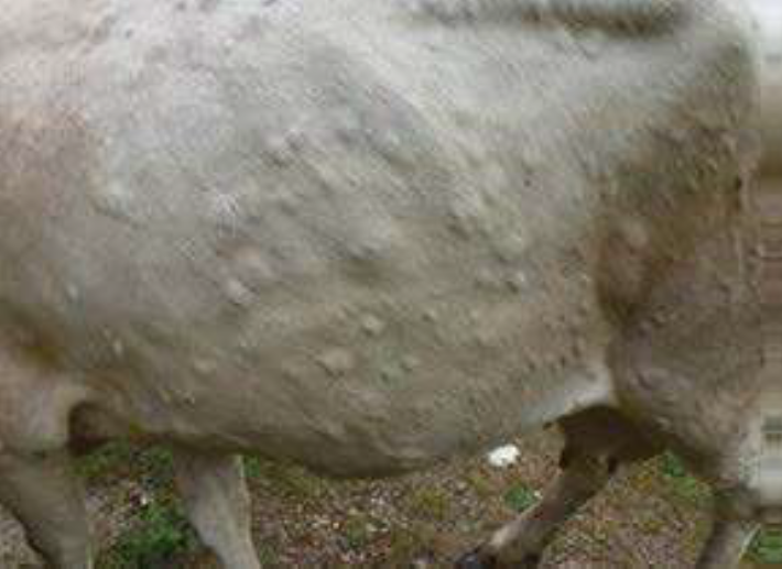
Severely case of LSD in a cow with nodular skin lesions covering the entire body, and enlarged lymph node (Al-Salihi, K. A., & Hassan, I. Q. (2015).
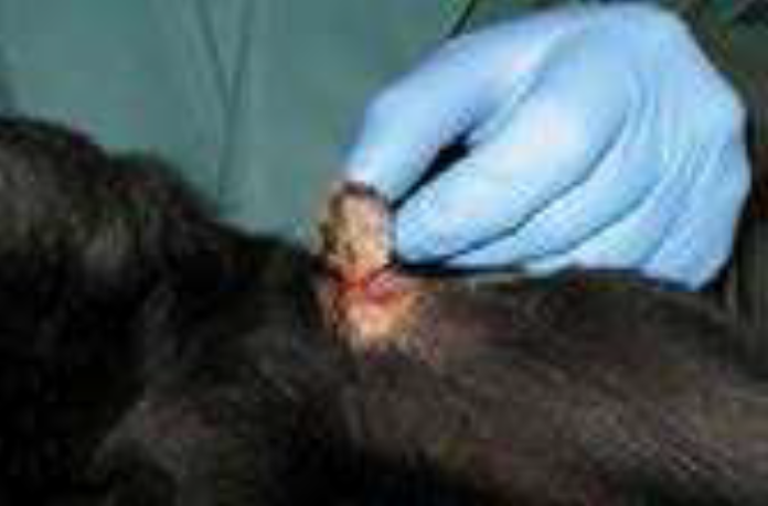
Core of necrotic tissue forms a plug (sit-fast) (Tuppurainen et al., 2017)
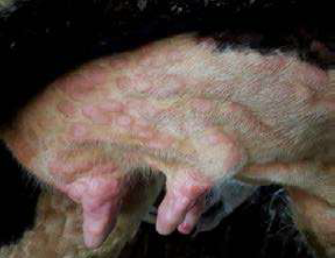
Severe case of LSD with skin nodules covering the udder and teats (Tuppurainen et al., 2017)
➡️Morbidity and Mortality
· Morbidity: 10% to 85%
· Mortality: usually 1% to 5%, higher in susceptible populations
➡️Economic Impact
LSD causes severe economic impact due to the following adverse effects:
· Loss of milk production
· Reduced weight gain and fertility
· Cost of vaccination and treatment
· Trade restrictions due to notifiable
status
In developing countries.
➡️Zoonotic Potential
· LSD is not a zoonotic disease and does not pose a direct risk to human health
- Diagnosis
➡️Clinical Diagnosis
The characteristic skin nodules and systemic signs are often sufficient during outbreaks.
➡️Laboratory Diagnosis
· Polymerase Chain Reaction (PCR): highly specific and sensitive
· Virus Isolation: in cell cultures or embryonated chicken eggs
· Serological Tests: ELISA, virus neutralization test
· Histopathology: necrotizing dermatitis, vasculitis, and inclusion bodies
➡️Differential Diagnosis
· Pseudolumpy skin disease (Bovine Herpesvirus 2): The disease may be confused with the less clinically important pseudo-lumpy skin disease, which is caused by a herpesvirus (bovine herpesvirus 2). These diseases can be similar clinically, although in some parts of the world the herpesvirus lesions seem confined to the teats and udder of cows
· Dermatophilosis
· Bovine papular stomatitis
· Photosensitization
· Insect bite hypersensitivity
- Complications of LSD in cattle
The complications of severe disease were reported as keratitis, dysentery, lameness, pneumonia, mastitis and myiasis
➡️Treatment
There is no specific antiviral treatment for LSD.
Management is supportive and symptomatic and includes:
· Non-steroidal anti-inflammatory drugs (NSAIDs)
· Broad-spectrum antibiotics for secondary infections
· Antiseptic dressing of lesions
· Nutritional supplementation to elevate animal immunity
- Prevention and Control
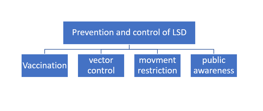
- Vaccination
Vaccination is the most effective control strategy. Types of vaccines include:
· Live attenuated LSDV vaccines
· Heterologous vaccines (e.g., sheep/goat pox vaccines)
➡️Control of vectors
· Use of insecticides and repellents
· Eliminating vector breeding sites
➡️Quarantine and Movement Restrictions
· Quarantine and culling of infected animals
· Restricting animal transport during outbreaks
· Hygienic disposal of dead animals and their waste
➡️Public Awareness
· Training farmers and veterinarians
· Surveillance and reporting systems
- References
1. World Organisation for Animal Health (WOAH). Lumpy Skin Disease Fact Sheet.
2. FAO (2021). Manual on LSD Surveillance and Control.
3. Tuppurainen, E.S.M., et al. (2017). “Lumpy Skin Disease.” Transboundary and Emerging Diseases.
4. European Food Safety Authority (EFSA). (2020). “Scientific Opinion on LSD.”
5. Tuppurainen, E. S. M. , Alexandrov, T. , & Beltran‐Alcrudo, D. (2017). Lumpy skin disease field manual ‐ A manual for veterinarians. FAO Animal Production and Health Manual, 20, 1–60.
6. Givens, M. D. ( 2018 ). Review: Risks of disease transmission through semen in cattle. Animal, 12 (S 1 ), s 165 s 171
7. SALIB, F.A., and OSMAN, A.H., 2011 . Incidence of lumpy skin disease among Egyptian cattle in Giza Governorate, Egypt. Veterinary world, 4 4 162 167
8. Tuppurainen, E. S., Stoltsz, W. H., Troskie, M., Wallace, D. B., Oura, C. A., Mellor, P. S., Coetzer, J. A., & Venter, E. H. (2011). A potential role for ixodid(Hard) tick vectors in the transmission of lumpy skin disease virus in cattle. Transboundaryand Emerging Diseases, 58, 93–104.
9. Al-Salihi, K. A., & Hassan, I. Q. (2015). Lumpy skin disease in Iraq: Study of the disease emergence. Transboundaryand Emerging Diseases, 62, 457–462. https://doi.org/10.1111/tbed.12386
10. Tuppurainen, E., Alexandrov, T. & Beltrán-Alcrudo, D. 2017. Lumpy skin disease field manual –A manual for veterinarians. FAO Animal Production and Health Manual No. 20. Rome. Food and Agriculture Organization of the United Nations (FAO).