Affections Of Specific Parts and Organs of Food Animals
| الموقع: | EHC | Egyptian Health Council |
| المقرر الدراسي: | Food hygiene Guidelines |
| كتاب: | Affections Of Specific Parts and Organs of Food Animals |
| طبع بواسطة: | Guest user |
| التاريخ: | السبت، 20 يونيو 2026، 9:35 PM |
الوصف
"last update:
2 Sep 2025" Download Guideline
جدول المحتويات
- - Acknowledgement
- - Glossary
- - Scope
- - I. Affections of the Skin
- - II. Affections of the Head and Tongue
- - III. Affections of the Liver
- - IV. Affections of the Lungs
- - V-Affections of the Pleura
- - VI. Affections of the Heart
- - VII Affections of the Kidney
- - VIII. Affections of the Spleen
- - IX. Affections of the Udder
- - X. Affections of the Blood
- - XI Affections of the Lymphatic System
- - XII. Affections of the Muscular System
- - References
- Acknowledgement
We would like to acknowledge the committee of
National Egyptian Guidelines for Veterinary Medical Interventions, Egyptian
Health Council for adapting this guideline. Executive Chief of the Egyptian
Health Council: Prof. Mohamed Mustafa Lotief.
Head of the Committee: Prof. Ahmed M Byomi
The rapporteur of the Committee: Prof. Mohamed
Mohamedy Ghanem.
Scientific Group Members: Prof. Nabil Yassien,
Prof. Ashraf Aldesoky Shamaa, Prof. Amany Abbass, Prof. Dalia Mansour, Dr.
Essam Elmarakby, Dr. Mohamed Elsharkawy, Prof. Gamal A. Sosa., Dr. Naglaa
Radwan, Dr. Hend El Sheikh
Author Prof. Nabil Yassien, Dr.
Hamdy M.B.A. Zaki
- Glossary
|
| |
|
Causative agent |
The organism or factor responsible for causing the disease. |
|
Judgment |
The decision taken by an inspector, based on ante and post-mortem inspection. |
|
Emaciation |
Extreme weight loss and muscle wasting. |
|
Localized |
Disease limited to a specific region without systemic signs. |
|
Generalized |
Disease spread throughout the body with systemic signs. |
|
Approved for human consumption |
Meat that has been inspected and passed without any restrictions |
|
Carcass |
The body of any slaughtered animals after bleeding and dressing |
|
Meat |
The edible part of any mammal slaughtered in an abattoir. |
|
Condemned |
Meat that has been inspected and judged as unfit for human consumption |
|
Total condemnation |
If the entire carcass and offal are condemned. |
|
Partial condemnation |
If only parts of the slaughtered animal are condemned, while others are judged otherwise |
|
Approved for distribution in restricted areas |
Meat that has been inspected and approved for human consumption with the requirement that the distribution shall be limited to restricted area. |
|
Conditionally approved for human consumption |
Meat that has been inspected and approved for human consumption after certain treatments ( freezing , boiling , steaming , pickling ) |
|
Offal |
Any edible or non-edible part of the animal other than carcass |
- Scope
This document provides a practical reference for veterinary meat inspectors. It lists common diseases and affections found in food-producing animals during post-mortem inspection, organized by anatomical region or organ. For each affection, the causative agent and the recommended judgment are summarized in accordance with standard meat hygiene practices.
- I. Affections of the Skin
1. Dermatitis
Causative agent: Infectious, allergic, nutritional, or parasitic.
Judgment: Case-dependent.
2. Sheep and Swine pox
Causative agent: Poxvirus
Judgment: With fever: Total condemnation; Without: Condemn affected parts.
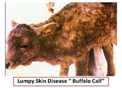
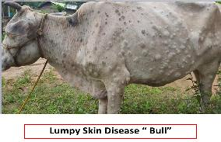
3. Lumpy Skin Disease
Lumpy skin disease is highly infectious viral disease of cattle characterized by the presence of firm nodules in the skin, lung, digestive and reproductive tracts accompanied by fever and lymphangitis. The nodules may be ruptured leaving ulcers or become indurated..
Causative agent: Mild form of herbs virus.
Appearance: Thickening of the skin with red raised spots become scabs.
Judgment: As in pox, or Removal of affected parts with heat treatment of carcass and viscera.
|
Fig. 1 |
Fig. 2 |
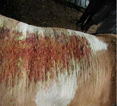
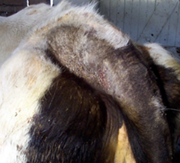
4. Mange
Causative agent: Parasites.
Appearance: Thickening of skin with red raised spots become scabs.
Judgment: In cattle and sheep, condemn the carcass if there is emaciation, while if there is no emaciation, condemn the skin only / In swine stripping of affected skin.
|
Fig. 3. Severe crusting lesions caused by psoroptic mange in a bull |
Fig. 4. Mange at the trailhead of a dairy cow causing scaling, itching and hair loss |
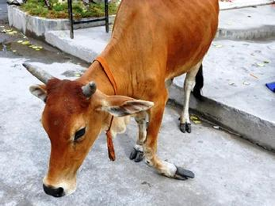
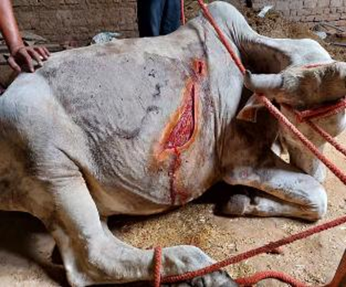
5. Wounds and Injuries
Causative agent: Trauma, diseases.
Appearance: May be local lesions or multiple.
Judgment: Total condemnation in the presence of systemic disturbances. / Condemnation of affected parts in absence of systemic disturbances.
|
Fig. 5 Fracture |
Fig. 6 Wound |
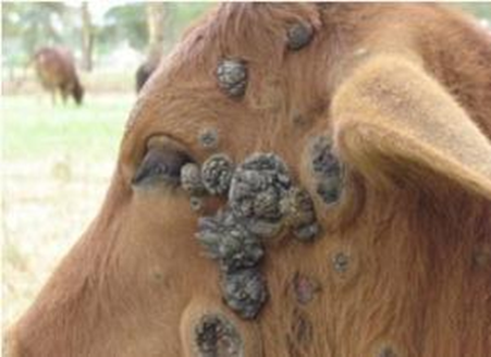
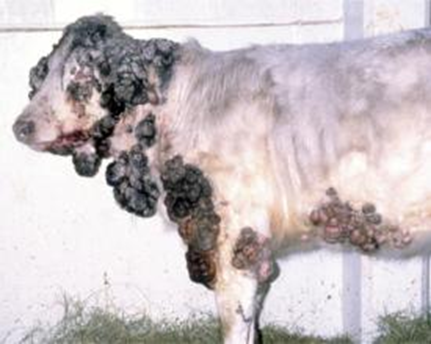
6. Tumors
As papilloma, melanoma, squamous cell carcinoma.
Appearance: May be benign or malignant
Judgment:
- Total condemnation in malignant tumors.
- Condemnation of affected parts in benign tumors.
|
Fig. 7 Squamous cell carcinoma |
Fig. 8 Squamous cell carcinoma |
7. Skin tuberculosis
Causative agent: Mycobacterium tuberculosis.
Appearance:
· Hard painless nodules up to hen's egg. Subcutaneous or intra-dermal of to lymphatic vessels.
· Lesions are found in limbs, fore arm, rarely on chest and shoulders. On section fibrous wall enclosing a caseo-calcareous center or thick yellow gelatinous pus or dried material like powder maize.
Judgment:
· Total condemnation in case of generalization, or localization and emaciation.
· Condemnation of skin in case of localized affection.
8. Acute skin congestion
Acute skin congestion in the pig may be widespread or localized. As in; swine fever, swine erysipelas, urticaria, pig paratyphoid.
9. Transit erythema
Affects pig on long rail journeys in the form of red patches on the skin in contact with floor due to irritant effect of disinfectant and urine on the floor. The condition is localized and the affected areas should be condemned. In severe cases extravasations of blood into the subcutaneous fat occur which only required removal of all colored fatty tissues. While in very severe cases there is congestion of the lymph nodes and fevered appearance of the carcass, which required total condemnation.
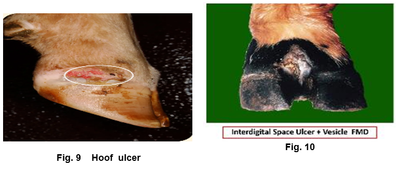
10. Foot and Mouth Disease
The lesions appear on the lips, muzzle or on the skin of the hoofs in the form of vesicles between the clefts of the feet, on the heel near the digits and / or around the cornet.
11. Ring worm
Ringworm is a common skin lesion affection but of no significance in meat inspection
12. Contagious foot rot ‘Foul of the foot’, Interdigital necrobacillosis “
Foot rot is an infectious disease of cattle associated with Sphaerophorus necrophorus. One or more of the feet may be involved with inflammation and necrosis at the top of the interdigital clefts. In acute stage there is edema and hyperemia of the affected part. In early stages there is sever lameness often with fever and loss of condition. In sheep the condition may be seen around the coronet and may cause emaciation. In local condition the carcass approved for food after condemnation of the affected part.
13. Contagious pustule dermatitis “ORF”
ORF is a highly contagious viral disease of sheep and appears in the form of papule, vesicle, pustule and crust chiefly involving lips and feet. It is a local condition necessitating condemnation of the affected parts, but care should be taken in handling of the infected material, as the disease is transmissible to man.
14. Warble fly
The ox hide may be damaged by the warble fly larvae or ticks bites. These bites may be left a white spots on the skin after tanning.
- II. Affections of the Head and Tongue
1. Cattle plague
Acute febrile infectious viral disease characterized by the presence of diphtheritic inflammation with cheese like deposits in the mouth and pharynx.
Free area: Total condemnation. (Diseased and contact animals).
Enzootic area: Condemnation of the viscera with heat treatment of the carcass and distribution in limited area.
2. Calf diphtheria:
The lesions in the form of diphtheritic patches and ulceration on tongue, mouth, pharynx or gums. These lesions appear as grey necrotic areas, which can only be scraped or peeled off with difficulty.
Localized: Heat treatment of the carcass after condemnation of the head.
Generalized: Total condemnation due to fever and emaciation.
3. Haemorrghic septicemia
The disease occur in two forms:
· Skin form which showed fever, marked swelling of the head, neck, throat and dewlap with the adjacent lymph nodes are enlarged and may be petechial hemorrhages.
· Pectoral form, which is characterized by high fever, petechial, hemorrhages in the serous and mucous membranes as well as all the organs. It show marked pneumonia. In both forms hemorrhagic enteritis is evident.
Judgment: Total condemnation
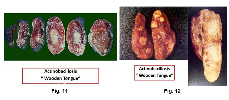
4. Actinobacillosis “Wooden tongue”
Actinobacillosis is a chronic granulomatous disease of the soft tissues (tongue, skin, and parenchymatous organs) of cattle and pig. Actinobacillus lignireesi is the main cause of the disease. The lesions in the tongue and muscles are associated with the lesions in the associated lymph nodes. The lymph node lesions are convex with small yellowish glistening nodules embedded in fibrous connective tissue and arranged characteristically in the peripheral portion of the cortex in irregular clusters toward one end of the node. The tongue showed enlargement and hardening.
Localized: Condemnation of head, Condemnation of liver, lung, stomach, peritoneum and shoulder if affected.
Generalized: Total condemnation due to fever and ill bleeding.
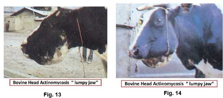
5. Actinomycosis “Lumpy jaw”
Actinomycosis is a chronic suppurative disease of bone tissue of cattle and udder tissue of pig. The lesions are usually confirmed to the head and involved the jaw bones to produce ‘”Lumpy Jaw”. The lower jaw is more affected than the upper one. The bone being thickened and presenting a bony combed appearance with small abscesses and suppuration tracts and fistulae on section.
In cattle: Condemnation of head and the carcass approved.
In Saw: Condemnation of udder and the carcass approved.
6. Tuberculosis
The common lesions of tuberculosis in the bovine head are mainly in the retropharengeal lymph node while in pig the sub maxillary nodes are commonly affected.
Localized: Without emaciation, condemnation of the head / with emaciation total Condemnation
Generalized: Total condemnation.
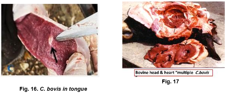
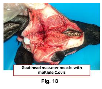
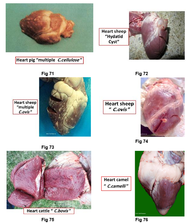
7. Parasitic
|
Pig |
Total condemnation even if one cyst present |
|
|
Sheep |
C. ovis |
Condemnation of the cyst |
|
Camel |
C. camelii |
Condemnation of the cyst |
|
Cattle |
C. bovis |
|
|
|
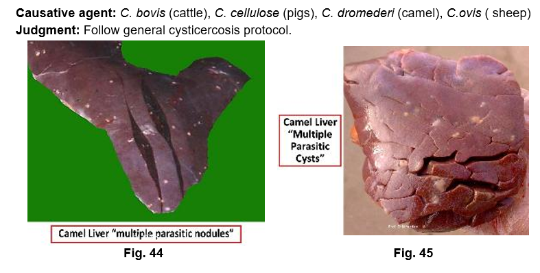
Cattle: C. bovis may be found in masseter muscle, tongue Oestrus bovis. Pig: C. cellulose. Sheep: C. ovis, Coenuris cerebralis, Oestrus ovis. Camel: C. dromedarii, C. bovis, C. ovis. |
|
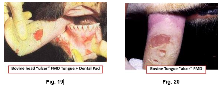
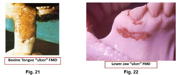
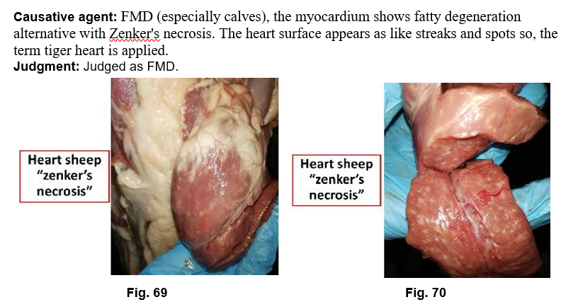
8. Foot and Mouth Disease (FMD)
The vesicular lesions are found on the tongue, lips and dental pad. The vesicles eventually ruptured leaving red painful erosions. In sheep the vesicles are much smaller than in the cattle.
Causative agent: Aphthovirus
Judgment: Free countries: Total condemnation. Enzootic countries: When slaughter of contact is practicable, diseased animal showed fever (T), and those which show no fever and contacts (Kh+ D) When immediate slaughter is not economic, diseased animal (T), contact and recovered 3 months after last case or vaccination (L + D).
NB: D = partial condemnation of the head, feet, pharynx, esophagus, testicles, udder and bones
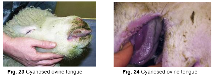
9. Blue tongue
Blue tongue is an infectious viral disease of sheep and pigs characterized by stomatitis and severe loss of condition. The mouth lesions vary from superficial inflammation to ulcers, erosions and necrosis of the buccal mucosa. The tongue is swollen and cyanosed. Acute cases with widespread lesions necessitate total condemnation of the carcass.
Causative agent: Orbivirus
Judgment: Acute with widespread lesions: Total condemnation.
10. Ulcers
Ulcers may be of varying size and may be seen in actinobacillosis and FMD.
Causative agent: Various (e.g., FMD, actinobacillosis etc.)
Judgment: Based on primary cause
11. Pharyngeal abscesses
Pharyngeal abscesses in pig and sheep are generally traumatic in origin caused by swallowing of sharp objects.
Causative agent: Traumatic origin
Judgment: Condemn head.
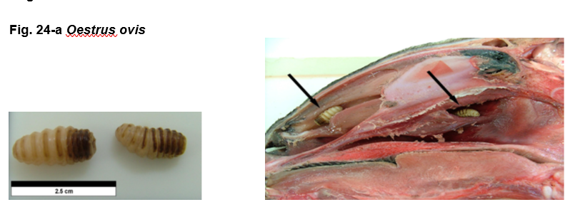
12. Oestrus ovis
Causative agent: also known as the sheep nasal bot, is a fly approximately 10mm in length whose larval stage is parasitic in the nasal passages of sheep. The female adult fly is viviparus, the young hatching within the adult who then squirts fluid containing approximately 15-20 first stage larvae (L1) at the muzzle of the animal. The L1 larvae, approximately 1mm in length then migrates through the nostrils to the frontal sinuses where they feed on the mucoid secretion stimulated by their presence. After the first moult the L2 larvae crawl into the frontal passages where the second moult to the L3 larvae occurs.
Judgment: Condemn of head.
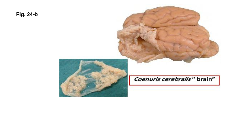
13. Coenuriosis (Coenurus cerebralis)
Causative agent: Also known as ‘gid’ and ‘sturdy’, coenuriosis is a condition of sheep caused by the presence of the migrating larvae and metacestode stage of the canine tapeworm Taenia coenurus (T. multiceps) of the central nervous system. In canines the adult tapeworm is 40-100cm in length .
Judgment: Condemn of head.
- III. Affections of the Liver
A- Pathological affections
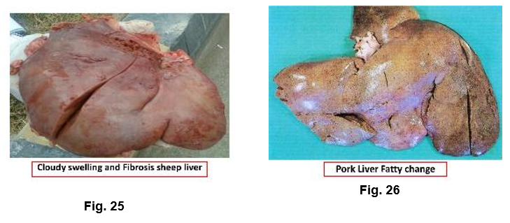
1. Degeneration:
Animal suffers from acute infectious disease, the liver is the first organ undergoes macroscopic changes. These changes are manifested by cloudy swelling and or pathological fatty change.
Organ: Condemn the affected organ.
Carcass: Total condemnation of carcass showed systemic reactions.
(Fever, septicemia, pyaemia). Normal carcass approved.
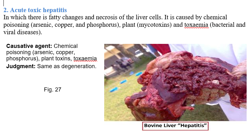
2. Acute toxic hepatitis
In which there is fatty changes and necrosis of the liver cells. It is caused by chemical poisoning (arsenic, copper, and phosphorus), plant (mycotoxins) and toxaemia (bacterial and viral diseases).
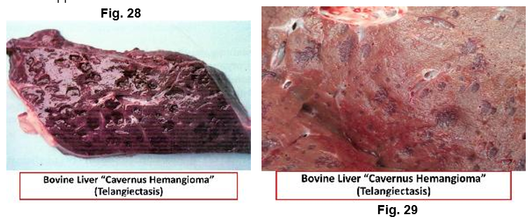
3. Cavernous-haemangioma “Telengiectasis’ ‘Plum pudding liver’
It is a common affection in imported animals. The cause is not well known but feeding factor and Sphaerophorus necrophorus may be concerned. On the other hand the lesion may have neurogenic origin caused by stretching of the spinal nerves, which may cause vasodilatation of the blood vessels of the liver. It is more common in intensive fed animals. Bluish black areas beneath the capsule and throughout the parenchyma characterize it. Lesions consist of dilated blood capillaries filled with blood.
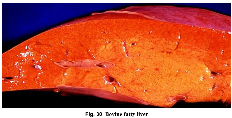
4. Fatty liver
Fatty liver is due to the mobilization of lipids from exterahepatic sources and from increased synthesis of lipids in the liver itself. The condition commonly seen in well-nourished lambs and very fatty aged cows. Affected liver is larger and heavier than normal. The liver edges are rounded, yellow or yellowish brown in color & soft in consistency.
Affected liver is fit for food, but the changes in color and consistency may make it unmarketable.
5. Liver Enlargement
Causative agent: Fatty infiltration, cirrhosis, abscess, tumor, plant poisoning.
Judgment: Case-dependent.
6. Rupture of Liver
Causative agent: Trauma, tumors, cysts, fascioliasis
Judgment: Condemn affected organ.
7. Adenoma
Causative agent: Benign tumor.
Judgment: Condemn organ and its lymph nodes.
8. Leukemia
The liver enlarged with proliferation of its interlobular connective tissue.
. Causative agent: Neoplastic proliferation.
Judgment: Total condemnation.
9. White liver disease
The condition occurs in sheep where the liver is enlarged and shows fatty changes. It is usually associated with cobalt /vitamin B12 deficiency, pregnancy toxemia in ewes and poor nutrition of cows in late pregnancy.
Causative agent: Cobalt/vitamin B12 deficiency.
Judgment: Condemn affected liver.
10. Black Liver
Causative agent: It is due to presence of the pigment lipofuscin from feeding on leaves of the mulga tree.
Judgment: Condemn affected liver.
11. Saw dust liver
The liver showed minute yellowish necrotic foci scattered throughout the substance and surface of the liver of young fatty cattle. It believed to be either early abscesses or the result of vit. E deficiency. Affected liver must be condemned.
B- Bacterial affections of the liver:
1.Tuberculosis
Tuberculosis of cattle and pig is common, while that of sheep and camel is rare. Congenital TB occurs in calves less than 2 weeks old.
Causative agent: Mycobacterium spp.
Judgment:
Localized: Condemnation of liver and its lymph nodes
Localized and emaciation: Total condemnation
Generalized: Total condemnation
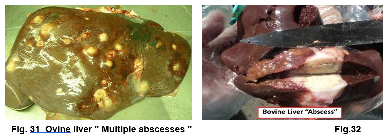
2.Liver Abscess
Liver abscess may occurs in young cattle due to umbilical infection while in older one, liver abscess may occur due to pyaemia, penetration of foreign body from reticulum or fasciola encysted metacercaria with pyaemic microorganisms from gastrointestinal tract.
Judgment: Small: Remove abscess / Multiple:
Condemn the liver.
Carcass: Total condemnation if there is systemic disturbance; otherwise,
bacteriological examination of the affected carcass.
3. Focal necrosis ‘Bacterial necrosis’
Occur in sheep and cattle but rare in pig and camel. It is caused by Seropherous necropherous which inhabitant intestinal tract of herbivores. The microorganisms penetrate the intestinal wall to reach the liver through portal vein. In acute cases the liver enlarged with formation of numerous necrotic foci and fever, while in chronic form necrotic foci with coagulated material.
Judgment: Localized: Condemn affected part /
Heavy: Condemn the liver.
Carcass: Approved if normal, total condemnation if there is fever or jaundice.
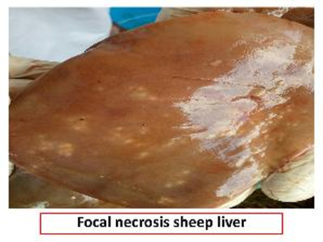
Fig.33
C-Parasitic affections of the liver:
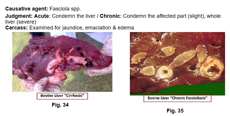
1. Fascioliasis
It is a common affection in the liver of cattle and sheep.
In acute stage, the liver is congested and swollen with petechial hemorrhages under the capsule. In chronic stage: cirrhosis and formation of connective tissue in the wall of the bile ducts and the surrounding liver tissues. In cattle the bile ducts become calcareous or pipes due to deposition of calcium salts. The flukes fail to reach the bile ducts & become encapsulated in the liver parenchyma. In sheep, there is no calcium precipitation but the liver irregularly lobulated and distorted due to connective tissue proliferation. The bile ducts thickened, dilated and have bluish color & does not undergo calcareous infiltration.
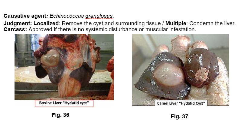
2.Hydatid Cyst
It is the larval stage of Echinococcus granulosus, which inhabit small intestine of carnivore chiefly dog. It is a common affection in the liver of camel.
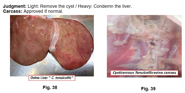
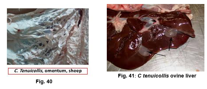
3.Cysticercus tenuicollis
It is the larval stage of Taenia hydatigena of dog. Usually present under the capsule of liver, mostly in sheep & pig, rarely in cattle and camel.
4.Linguatula larvae
Yellowish or greenish nodule under the liver capsule. It could be identified by microscopic examination to identify the typical hooks of the parasite.
Causative agent: Linguatula serrate.
Judgment: Condemn the affected part.
5.Milk Spots
It affects liver of pig and caused by the migration of Ascaris summ larvae. Milk spots are focal interstitial hepatitis and found on both surface of the liver. It is consists of white opaque center from which radiate interlobular septa. This affection may be confused with lesions caused by avian TB. In such condition grey flat like areas are found on the liver surface. Malchoire's palpation test is used to differentiate between lesion of avian TB and milk spots. The test includes, palpation of the upper part of the small intestine to manipulate Ascaris or not. Smear stained with Zeil Nelson to detect acid-fast bacilli of TB.
Judgment: Affected parts should be condemned in light cases, while the whole liver to be condemned in heavy affected infestation.

6.Coccidiosis
It is caused by Eimeria stiedea and affect rabbit liver. Appear in the form of irregular white spots on liver parenchyma.
Judgment: Condemn the liver& carcass condemned if there is emaciation.
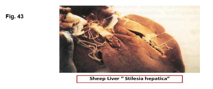
7. Stilesia hepatica
It is a cestode resembles Monzia. It affects the bile duct of sheep, goats, cattle and wild ruminants. The intermediate host is Oribatid mites. The bile duct may be occluded or form a sac-like dilatation filled with worms. Affected liver may show slight cirrhosis and the wall of the bile duct is thickened.
Judgment: Condemn the affected liver.
7. Cysticercosis
- IV. Affections of the Lungs
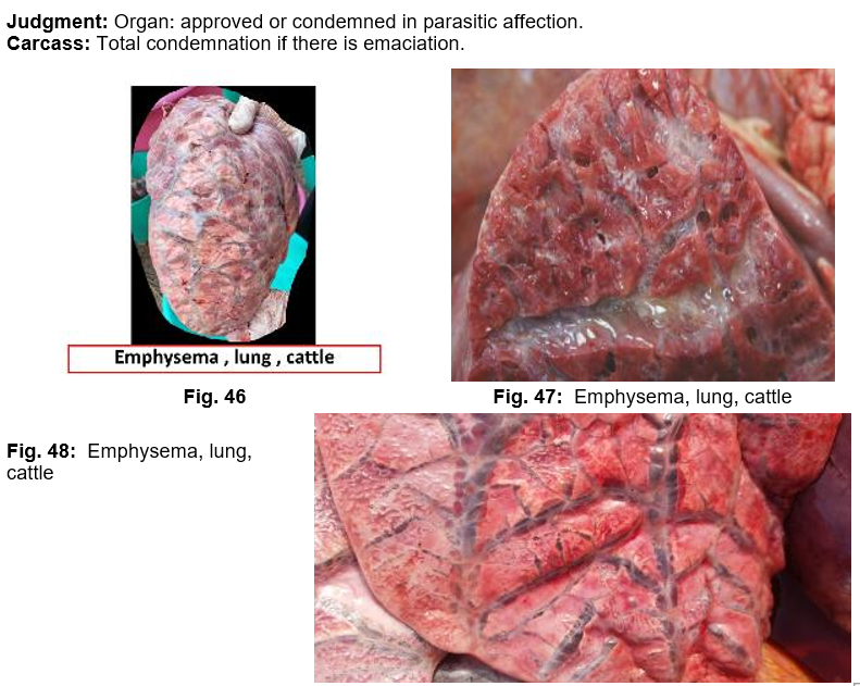
1. Interstitial or interlobular emphysema
It is a common affection in the lung of aged animals, accompanied parasitic pneumonia, and also in young animals severely affected with Dictycolus viviparous. Rupture of air alveoli results in escape of air bubbles into interstitial tissue. Emphysema may be extending to mediastinal tissue and subcutaneous tissue of thorax and abdomen.
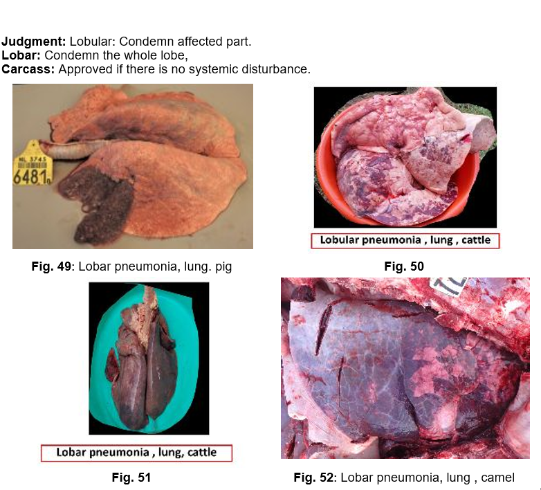
2. Pneumonia
The cause may be virus, bacteria, parasite, fungi, bad weather, inhalation of drugs or penetration of foreign bodies from reticulum. Pneumonia commonly seen in connection with specific diseases such as: bovine TB, shipping fever, enzootic pneumonia of calves and pigs, swine fever, extensive parasitic infestation of respiratory tract e.g., Ascaris. Fungi as Candida albicans and Mucor rarely give rise to pneumonia. In broncho-pneumonia (Lobular pneumonia), small pneumonic foci are intermixed with healthy tissue, while in lobar pneumonia the whole lobe or its greater part is in one stage of pneumonia.
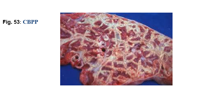
3. Contagious bovine pleuropneumonia CBPP
Contagious bovine pleuropneumonia is stages of hepatization, which surrounded by connective tissue. On cut section give marble appearance. Pneumonic foci undergo necrosis and become firm, dry and encapsulated to form sequesteraum.
Causative agent: Mycoplasma mycoides.
Judgment: Condemn the affected lung.
Carcass totally condemned if there is fever.
4. Pneumomycosis
Pneumomycosis is a fungal affection of lungs due to Aspergillus or Mucor, most common in young fowl, ducks and geese (brooder pneumonia) and occasionally seen in lambs, pig and cattle. It is characterized by diffuse hepatization of the lungs with numerous grey or greenish spots scattered throughout the lungs with a thin deposits of mould in the bronchi.
Judgment: The affected lung must be condemned, and also the carcass in case of septic
or gangrenous reactions.
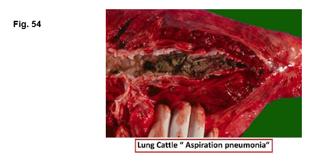
5. Contamination of trachea and lungs
Trachea and lungs may be contaminated with ingesta or blood.
Judgment: The affected trachea and lungs are condemned.
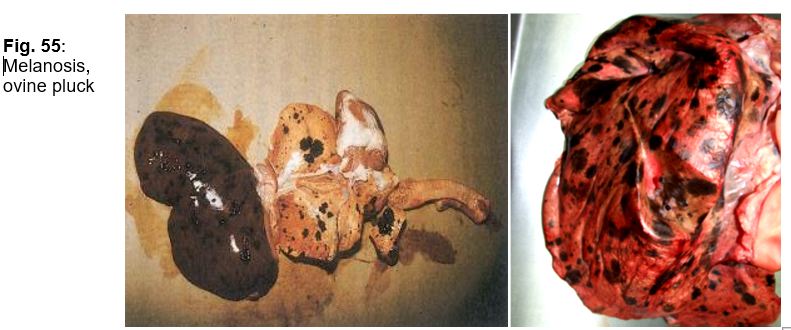
6. Melanosis
Black pigment on the lung tissue and bronchial lymph node.
Causative agent: Pigmentation disorder.
Judgment: Condemn lungs and its bronchial lymph nodes.
7. Parasitic
A.Parasitic Nematodes
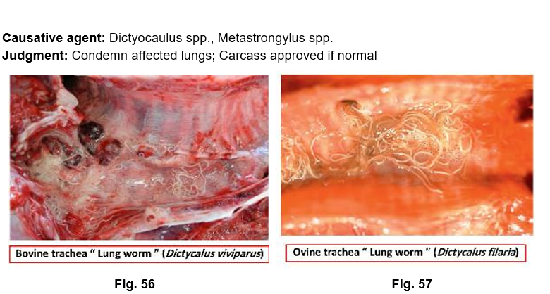
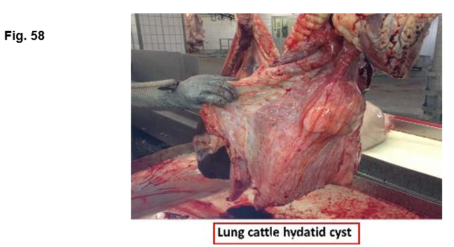
B. Hydatid Cyst
Causative agent: Larval stage of Echinococcus granulosus,
Judgment: Condemn affected part (light), condemn whole lung (heavy); Carcass approved if there is no emaciation, edema and or, muscular infestation.
C-Immature form of Fasciola.
Judgment: Condemnation of affected part of the organ (small number, or
Condemnation of lungs (large number).The carcass approved if there is no emaciation.
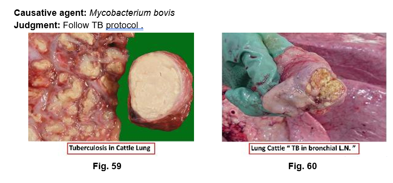
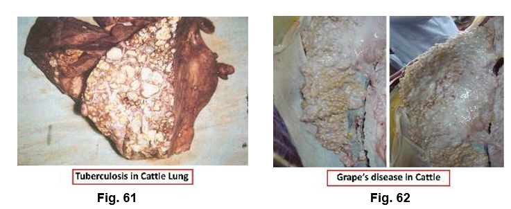
8. Tuberculosis
· Miliary TB.
· Acute early generalization.
· Acute late generalization.
· Caseous pneumonia.
· Acute acino-nodular TB.
· Chronic acino-nodular TB.
· Lung cavitation.
· Pearls or grapes disease.
7. Anthracosis
Causative agent: Bluish-black pigment of lung and its lymph nodes due to inhalation of coal dust.
Judgment: Condemn affected lungs and its lymph nodes.
8. Pulmonary Edema
Causative agent: Occurs in congestive heart failure, atypical interstitial pneumonia
(Fusarium) and organophosphate poisoning.
Judgment: Case-dependent.
9. Pulmonary Abscess
Causative agent: Single or small multiple abscesses may originate from emboli from other organs as in cases of: septic meteritis, septic mastitis, omphalophlebitis and /or penetration of foreign body. It may also occur as a part of primary diseases as: TB. actinobacillosis and aspergillosis.
Judgment: Condemn affected lungs; total condemnation if there is systemic disturbance.
10. Neoplasms
Causative agent: Lymphosarcoma, adenoma, melanoma
Judgment: Condemn affected lung or total condemnation in malignant tumors
- V-Affections of the Pleura
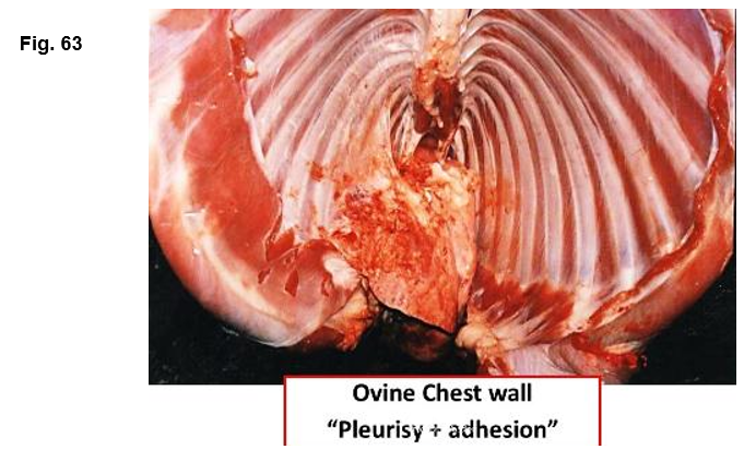
1. Pleurisy
It is the inflammation of the pleura. Usually associated with pneumonia and in acute stage is characterized by fibrinous exudates, which in cattle has red and velvety appearance. Acute pleurisy may tend to assume a chronic form with the production of fibrinous adhesions between the partial pleura and lung surface. This condition required stripping of the pleura.
2. Sub-pleural petechial hemorrhage
The condition may be seen in many septicemic diseases including swine fever, swine erysipelas, & requires total condemnation.
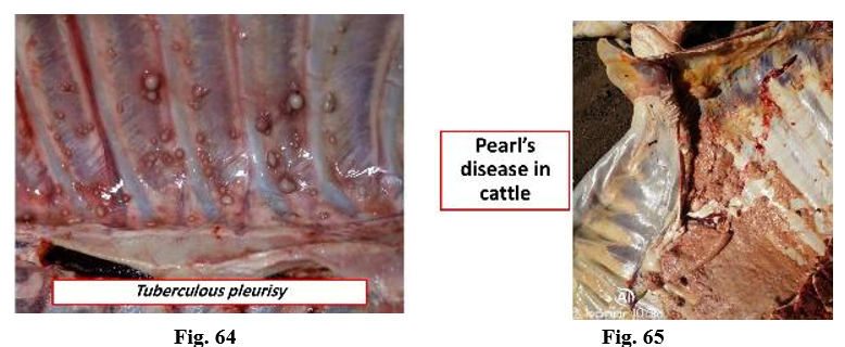
3. Tuberculous pleurisy
|
Localized |
Stripping of the pleura and flaming of the affected part. |
|
Diffuse Acute Chronic
|
Condemnation of full forequarter at 11th intercostal space. Axillary’s negative: Ribbing, oystering, racking. Axillary’s positive: Condemnation of full forequarter at 9th intercostal space. |
Oystering means removal of the dorsal vertebrae, sternum and ribs from the outer muscular mass which is passed for food if the axillary lymph node not affected.
4. Hydrothorax
It is an extensive accumulation of fluids in the pleural cavity. Occurs in congestive heart failure, bovine viral leucosis and generalized edema due to malnutrition.
5. Empyemia
Empyemia refers to the collection of pus in the pleural cavity.
6. Abscesses
Small abscesses containing pale green pus may be seen on the pleura.
7. Back bleeding
Occurs when the pleural membrane at the entrance of the chest is punctured during slaughtering with the aspiration of blood into the thoracic cavity.
- VI. Affections of the Heart
Pericardium
1.Traumatic Pericarditis
Causative agent: Foreign body (wire).
Judgment: Total
condemnation if there is fever, emaciation and or serous infiltration.
2.Tuberculous
Pericarditis
Causative agent: Mycobacterium spp.
Judgment: Follow TB protocol.
3. Hydropericardium
Causative agent: Generalized edema.
Judgment: Condemn affected heart.
4.Pericarditis
Causative agent: Swine fever, salmonellosis, pasteurellosis
Judgment: Condemn affected heart; total condemnation if there is systemic disturbance.
Epicardium
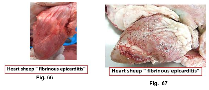
1.Fibrinous Epicarditis
|
Causative agent: In pig , swine erysipelas ,However, in cattle the condition occurs as a result of traumatic pericarditis. Judgment: Condemn affected heart. |
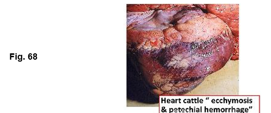
2. Subepicardial petechial Hemorrhage
Causative agent: Septicemia.
Judgment: Total condemnation.
Myocardium
1.Myocardial Degeneration
In the form of cloudy swelling and pathological fatty changes. It is associated with toxic affections of the carcass and infectious febrile conditions.
Judgment: Condemn affected heart.
2.Myocardial Abscesses
It may be seen in the myocardium due to penetration of foreign bodies from the reticulum and /or pyemia.
3.Myocarditis
Causative agent: It occurs in all infectious febrile conditions as septic meteritis, septic mastitis, listeriosis and /or toxoplasmosis.
Judgment: Condemn affected heart; total condemnation if there is systemic disturbance.
4.Tiger Heart
5.Brown Atrophy
Causative agent: The heart reduced in size, flabby, wrinkled, and brown in color due to accumulation of numerous pigment granules. The affection accompanied wasting diseases as TB.
Judgment: Condemn affected heart.
6.Parasitic Affections
|
Causative agent: Cysticercus spp., Hydatid cysts, Linguatula, Sarcocystis Judgment: Condemn affected heart / carcass: Depend on causative agent. |
Endocardium
1.Vegetative Endocarditis
Causative agent: Chronic swine erysipelas.
Judgment: Total condemnation.
2.Ulcerative Endocarditis
Causative agent: Streptococci, C. pyogenes
Judgment: Total condemnation if there is systemic disturbance.
3.Subendocardial Hemorrhage
Causative agent: Septicemia, enterotoxaemia
Judgment: Total condemnation.
- VII Affections of the Kidney
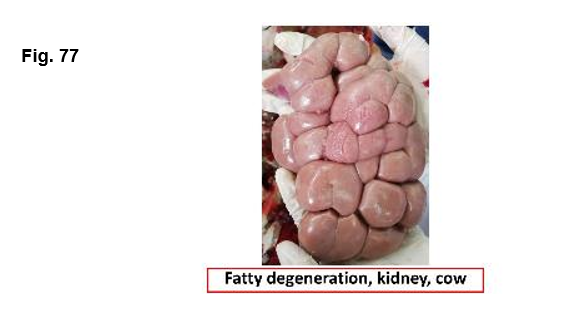
1.Degeneration
Causative agent: Cloudy swelling and pathological fatty change occur due to toxic affections .
Judgment: Condemn affected kidney.
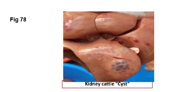
2.Congenital Cysts
Causative agent: Cyst free from urinary constituents. They may be single or multiple cysts. The cysts contained clear fluid within their thin transparent walls, which usually protrude from the surface. However, cysts may be found in the kidney parenchyma.
Judgment: 1-2 cysts:
Approved.
Extensive: Condemn the whole kidney.
Carcass: Approved if normal.
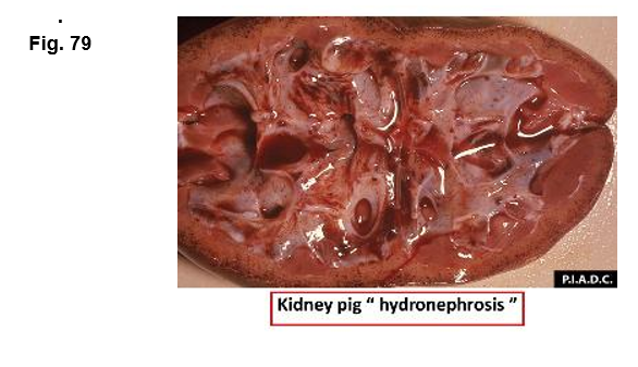
3.Hydronephrosis
Causative agent: Urinary tract obstruction.
Judgment: Unilateral: Condemn affected
kidney / Bilateral: Condemn both kidneys.
Carcass: Total condemnation if there is uriniferous odor.
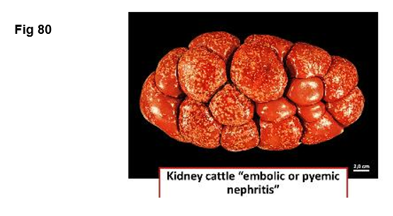
4.Embolic or pyaemic nephritis
Causative agent: Characterized by multiple abscesses, small in size in the renal cortex due to circulation of pyogenic microorganisms in the blood. Kidney is flappy, pale color and multiple abscesses in the renal cortex.
Judgment: Condemn affected kidney.
Carcass: Total condemnation if there is pyaemia; or bacteriological examination
if there is no pyaemia.
5.Pyelonephritis
Causative agent: Pyelonephritis occurs as a result of ascending infection with Corynebacterium renal or C. pyogen. Renal pelvis is dilated and contain slimy glassing fluid intermixed with pus and has strong smell of ammonia. Infection may be extended and give rise to large abscess in the kidney substance which may be uni or bilateral.
Judgment: Unilateral: Condemn affected
kidney / Bilateral: Condemn both kidneys.
Carcass: Total condemnation if there is emaciation ,edema , pyaemia or uremia.
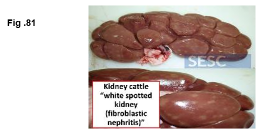
6. Fibroblastic nephritis “White spotted kidney”
This condition is common in young calf suffering from brucellosis or salmonellosis. It is due to arrested in the embryonic kidney development. Affected kidney shows numerous white nodules that are circular in cross section.
Judgment: Condemn affected
kidney.
Carcass: approved unless
brucellosis.
7.Black Spotted Kidney
Causative agent: In the cortex of calf kidney assumes olive-green or black spots due to accumulation of the bile pigment (Biliverdin).
Judgment: Condemn affected kidney.
8.Pulpy Kidney Disease
Causative agent: Common affection in sheep suffering from enterotoxaemia, which caused by Cl. welchii type D. Kidney is soft and has hemorrhagic streaks.
Judgment: Total condemnation.
9.Tuberculous nephritis
Causative agent: Mycobacterium spp.
Judgment: (Generalized) Total condemnation
- If the kidney substance and its lymph node is affected with T.B
- If the kidney substance is free and the lymph node is affected with evidence of generalized TB.
- If the kidney substance is free and the lymph node is affected & the carcass free from emaciation. Condemn affected kidney& its lymph node only.
10.Parasitic Affections
Causative agent: Cysticerci, Hydatid cyst (In heavy infested carcass only)
Judgment: Condemn affected kidney.
11.Tumours
Judgment: Benign: Condemn affected kidney Malignant: Total condemnation
12.. Pigmentation changes
· Brown pigmentation: The condition mainly occurs in cattle due to accumulation of hemosiderin and lipofuscin pigments.
· Black discoloration: The condition occurs in sheep as a result of haemolysis caused by chronic copper poisoning.
Judgment: Condemn affected kidney.
13.Lymphomatosis
Causative agent: Lymphosarcoma.
Judgment: Condemn affected kidney.
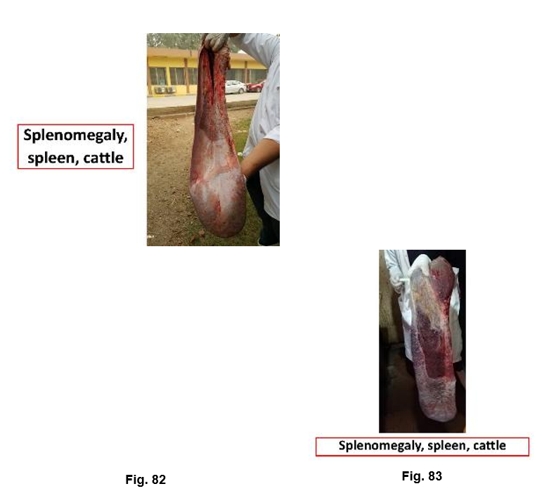
- VIII. Affections of the Spleen
1.Enlargement
|
Causative agent: Various diseases (e.g. anthrax, swine erysipelas, leukemia, Bl. parasites) Judgment: Condemn affected spleen ,if associated with systemic disease total condemnation |
2.Tuberculosis
Causative agent: Mycobacterium spp.
Judgment: Total condemnation if there is generalized TB.
3.Parasitic Affections
Causative agent: Hydatid cyst, Linguatula, liver flukes, cysticerci.
Judgment: Condemn spleen in heavy infestation.
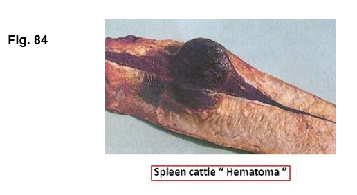
s4.Haematoma
Causative agent: Trauma or foreign body.
Judgment: Condemn affected spleen.
5.Slaughter Spleen
Causative agent: The condition occurs in cattle due to long pitting cane (more than 60 cm), which cause destruction of vasoconstrictor nerve (splenic nerve) followed by dilatation and congestion of spleen. The condition encountered in pig as a result of post mortem finding of live scalding.
Judgment: Condemn affected spleen.
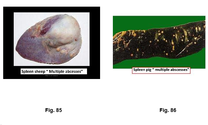
6.Abscess
Causative agent: Abscesses of spleen may be occurs due to penetration of foreign bodies from the reticulum and lodgments of septic emboli.
Judgment: Condemn affected spleen.
7. Infarctions
Causative agent: End artery occlusion.
Judgment: Condemn affected spleen.
8. Neoplasms
Causative agent: Leukaemia, lymphosarcoma, melanoma
Judgment: Condemn affected spleen.
- IX. Affections of the Udder
1.Simple Catarrhal Mastitis
Causative agent: Streptococcus agalactiae
Judgment: Condemn affected quarter or udder.
2.Septic Mastitis
Causative agent: Staphylococcus aureus, C. pyogenes
Judgment: Condemn affected udder; total condemnation if there is systemic reaction.
3.Tuberculous Mastitis –
a-Chronic Nodular
Causative agent: Mycobacterium spp.
Judgment: Condemn affected udder.
b-Tuberculous Mastitis ( Caseated Form )
Causative agent: Mycobacterium spp.
Judgment: Total condemnation
c-Tuberculous Mastitis ( Miliary Form )
Causative agent: Mycobacterium spp.
Judgment: Total condemnation
- X. Affections of the Blood
1.Anaemia
Causative agent: TB., Johne’s disease, red water fever, parasites.
Judgment: Total condemnation if there is systemic disturbance.
2.Leukaemia and Leucosis
Causative agent: Lymphoid malignancy.
Judgment: Total condemnation.
3.Hemoglobinemia
Causative agent: Babesiasis, leptospirosis, Cl. haemolyticum
Judgment: Total condemnation if there is icterus or ill bleeding.
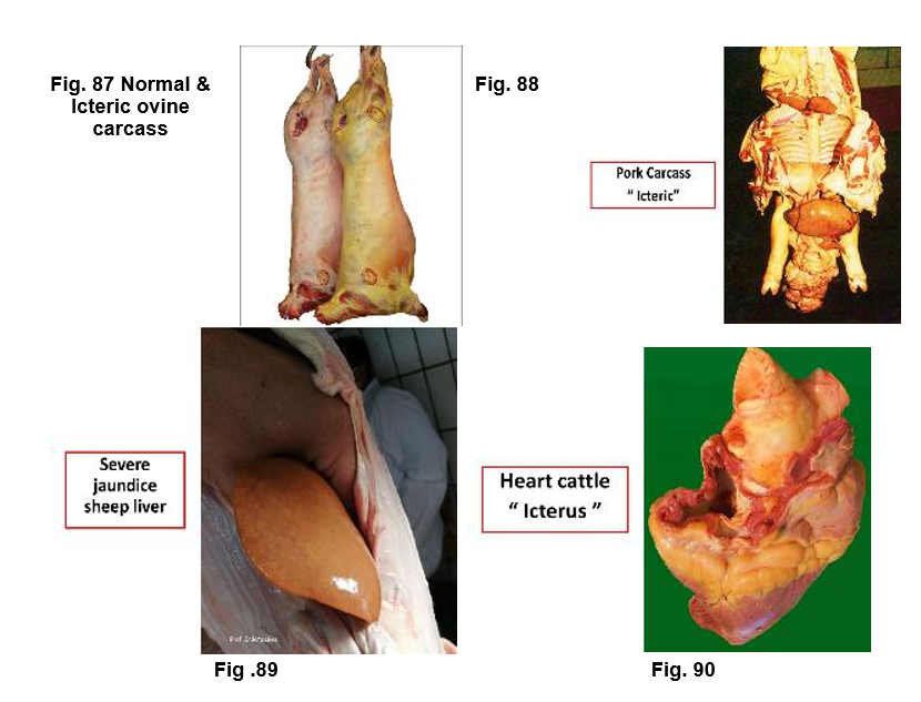
4.Icterus
Causative agent: Obstruction, hemolysis, liver damage.
Judgment: Total condemnation if severe or persistent after detention in detained meat room for 24 hrs.
5.Uraemia
Causative agent: Urinary tract obstruction.
Judgment: Total condemnation if there is strong uriniferous odor or positive boiling test.
- XI Affections of the Lymphatic System
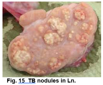
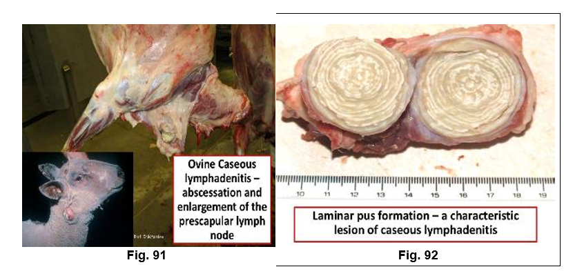
1.Lymphadenitis
Causative agent:
a) Acute lymphadenitis is manifested by swelling, congestion and edema of the node, which is painful and softer than normal e.g. Anthrax, and acute swine fever.
b) Chronic lymphadenitis is manifested by the development of fibrous connective tissue in the affected node, which becomes enlarged and indurated. These changes may be seen in caseous lymphadenitis of the sheep, TB and actinobacillosis of the cattle.
Judgment: Condemn affected lymph nodes; total condemnation if generalized.
2.Pigmentary Changes
Causative agent: Anthracosis, xanthosis, melanosis
Judgment: Condemn affected nodes.
3.Parasitic Infestation
Causative agent: Linguatula, Muellerius, F. hepatica
Judgment: Condemn affected nodes.
4.Lymphangitis
Causative agent: Johne’s disease, mycotic lymphangitis
Judgment: Condemn affected vessels and associated tissues.
- XII. Affections of the Muscular System
1.Xanthosis
Causative agent: Unknown (common in cattle)
Judgment: Condemn affected muscles.
2.Degenerative Changes
Causative agent: Toxins, milk fever.
Judgment: Condemn affected muscles if there is systemic disturbance.
3.Muscular Fibrosis (Myositis Interstitialis)
Causative agent: Trauma.
Judgment: Condemn affected muscles.
4.Myositis
Causative agent: FMD, toxoplasmosis.
Judgment: Condemn affected muscles.
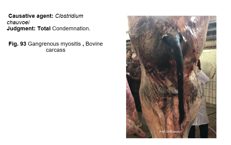
5.Gangrenous myositis
6.Muscular Dystrophy (White Muscle Disease)
Causative agent: Vitamin E / Selenium deficiency
Judgment: Condemn affected muscles.
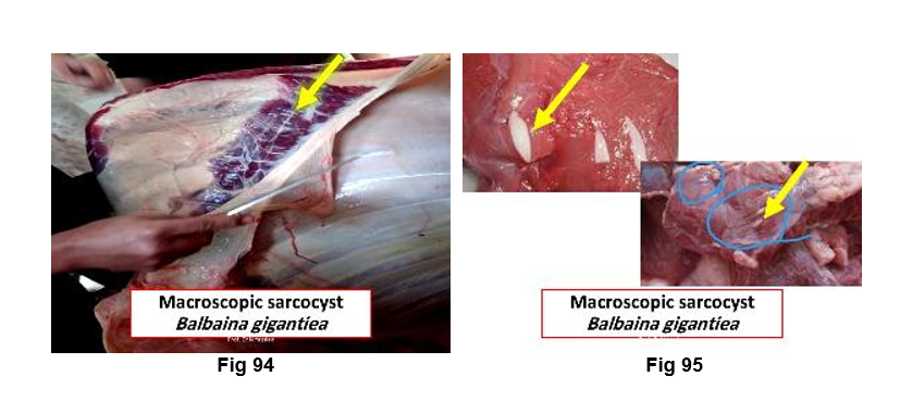
7.Parasitic Infestations
Causative agent: C. bovis, C. cellulosae, Trichinella, Sarcosporidia, Hydatid cysts
Judgment: Condemn affected muscles.
8.Tumors
Causative agent: Lymphosarcoma, rhabdomyoma, rhabdomyosarcoma
Judgment: Condemn affected muscles.
7. Actinobacillosis
Causative agent: Actinobacillus spp.
Judgment: Condemn affected muscle (head).
8. Muscular Atrophy
Causative agent: Nerve injury (e.g., radial nerve)
Judgment: Condemn affected muscle.
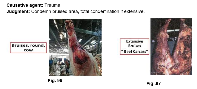
9. Bruising
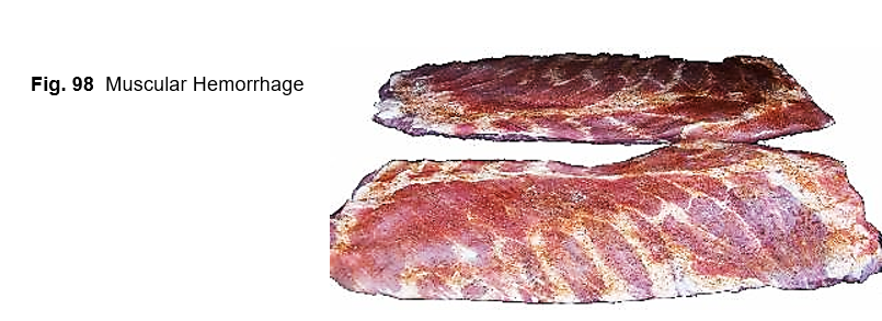
12.Muscular Hemorrhage (Shot Meat)
Causative agent: Pre-slaughter stress or poor stunning.
Judgment: Condemn affected parts if extensive.
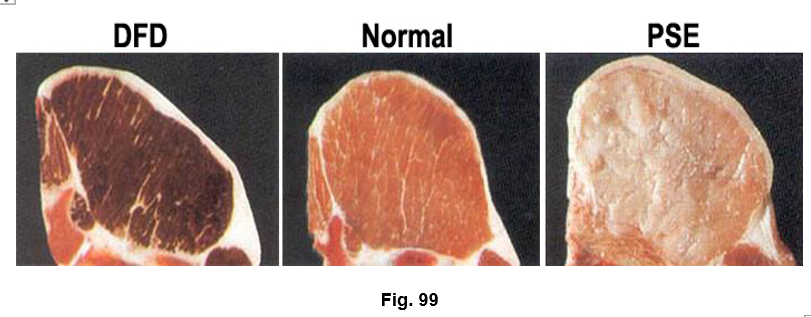
7. Dark Cutting Beef (DFD)
Causative agent: Pre-slaughter stress.
Judgment: Approved as inferior quality & used for meat processing.
8. Pale Soft Exudative (PSE)
Causative agent: Rapid pH drop post-slaughter.
Judgment: Approved as inferior quality meat & used for meat processing.
- References
1. Gracey, J.F., Collins, D.S., & Huey, R.J. (2015). Gracey’s Meat Hygiene, 11th Edition London: W.B. Saunders.
2. Jones, T.C., Hunt, R.D., & King, N.W. (2006). Veterinary Pathology (6th ed.). Blackwell Publishing.
3. Meat Inspection Manual for Developing Countries. Food and Agriculture Organization of the United Nations (FAO), Rome. Available at: https://www.fao.org/4/t0756e/t0756e00.htm
4. Radostits,
O.M., Gay, C.C., Hinchcliff, K.W., & Constable, P.D. (2007).
Veterinary Medicine: A Textbook of the Diseases of Cattle, Horses, Sheep,
Pigs and Goats (10th ed.). Saunders Elsevier.
5. Meat
Inspection and Control in the Slaughterhouse
by Michael D. O'Grady & Ian J. Blanchfield (2012). Wiley-Blackwell.
6. Figures Credit:
|
Figure |
Reference |
|
Fig.1 |
Manual on meat inspection for developing countries (FAO), 1994, CHAPTER 3 SPECIFIC DISEASES OF CATTLE, Available at: https://www.fao.org/4/t0756e/T0756E00.htm#TOC |
|
Fig.2 |
Fenemore, C., Swinson, V. and Foster, A. (2024), Nodular skin disease in cattle. Veterinary Record, 195: 288-289. https://doi.org/10.1002/vetr.4768 |
|
Fig 3 |
National Animal Disease Information Service available at: https://clients.nadis.org.uk/planner-articles/mites-in-cattle/ |
|
.Fig. 4 |
National Animal Disease Information Service available at: https://clients.nadis.org.uk/planner-articles/mites-in-cattle/ |
|
Fig. 5 |
Image details: Available at: https://www.shutterstock.com/image-photo/one-cow-injured-wat-hua-krabue-1136512415 Asset id: 1136512415, One cow was injured at Wat Hua Krabue, Thailand, Upload date: July 17, 2018, Categories: Animals/Wildlife |
|
Fig.6 |
Swati Goel Sharma (2024) Multiple Cows With Grievous Wounds Found in Haryana Village After Lok Sabha Results, Activists Demand Deeper Probe, Available at: https://swarajyamag.com/society/was-it-done-to-disturb-peace-activists-demand-deeper-probe-after-multiple-cows-with-grievous-wounds-found-in-haryana-village-after-election-results |
|
Fig.7 |
Available at: https://infonet-biovision.org/animal-health-and-disease/skin-problems-new/skin-tumours-and-warts |
|
Fig. 8 |
Danny W. Scott, Skin Diseases, Chapter 4 Available at: https://veteriankey.com/skin-diseases-2/ |
|
Fig. 9 |
Carrie Hammer, Taw Scaff, Miranda Meehan, Mary Keena, (2025)Foot-and-Mouth Disease (FMD), North Dakota State University publication, Available at: https://www.ndsu.edu/agriculture/extension/publications/foot-and-mouth-disease-fmd |
|
Fig. 10 |
Available at: https://www.ndda.nd.gov/divisions/animal-health/diseases/foot-and-mouth-disease |
|
Fig. 11 |
Available at: https://vetmed.uodiyala.edu.iq/wp-content/uploads/2023/01/ACTINOBACILLOSIS.pdf |
|
Fig. 12 |
Manual on meat inspection for developing countries (FAO), 1994, CHAPTER 3 SPECIFIC DISEASES OF CATTLE, Available at: https://www.fao.org/4/t0756e/T0756E00.htm#TOC |
|
Fig. 13 |
Available at: https://vetmed.uodiyala.edu.iq/wp-content/uploads/2023/01/ACTINOMYCOSIS.pdf |
|
Fig. 14 |
Available at: https://vetmed.uodiyala.edu.iq/wp-content/uploads/2023/01/ACTINOMYCOSIS.pdf |
|
Fig. 15 |
Crawshaw, T., de la Rua‐Domenech, R., & Brown, E. (2013). Recognising the gross pathology of tuberculosis in South American camelids, deer, goats, pigs and sheep. In Practice, 35(9), 490-502. https://doi.org/10.1136/inp.f5683 |
|
Fig. 16 |
Elbarbary, N. K., Gareh, A., Abdelhaseib, M., Fotouh, A., Abdelmotilib, N. M., Ragab, M. F., & Dandrawy, M. K. (2025). Cysticercus bovis in slaughtered cattle in upper Egypt: implications for food safety. BMC Veterinary Research, 21(1), 344. Available at: https://bmcvetres.biomedcentral.com/articles/10.1186/s12917-025-04768-y |
|
Fig. 17 |
---------------------------------- |
|
Fig. 18 |
The authors (Prof. Nabil Yassien, Dr. Hamdy M.B.A. Zaki) own photo from Basatin Abattoir Affections. |
|
Fig. 19 |
Available at: https://libyaherald.com/2012/02/fmd-hits-north-west-libya/ |
|
Fig. 20 |
Credit: Dr. D. Gregg, Noah's Arkive, PIADC, Photo ID: FMD_010, Available at: https://www.cfsph.iastate.edu/diseaseinfo/disease-images/?disease=foot-and-mouth-disease |
|
Fig. 21 |
Manual on meat inspection for developing countries (FAO), 1994, CHAPTER 3 SPECIFIC DISEASES OF CATTLE, Available at: https://www.fao.org/4/t0756e/T0756E00.htm#TOC |
|
Fig. 22 |
Credit: PIADC, Photo ID: FMD_001, Available at: https://www.cfsph.iastate.edu/diseaseinfo/disease-images/?disease=foot-and-mouth-disease |
|
Fig. 23 |
Enserink, M. (2008). Exotic disease of farm animals tests Europe's responses. Science, Vol. 319, No. 5864, available at: https://www.science.org/doi/10.1126/science.319.5864.710 |
|
Fig. 24 |
The Pirbright Institute, Available at: https://www.hooknortonvets.co.uk/bluetongue-virus-update/ |
|
Fig. 24-a |
Ovine Meat Inspection - 2nd Edition: Anatomy, Physiology and Disease Conditions, Nottingham University Press, Second edition, 2011, by Andy Grist |
|
Fig. 24-b |
Ovine Meat Inspection - 2nd Edition: Anatomy, Physiology and Disease Conditions, Nottingham University Press, Second edition, 2011, by Andy Grist |
|
Fig. 25 |
The authors (Prof. Nabil Yassien, Dr. Hamdy M.B.A. Zaki) own photo from Basatin Abattoir Affections. |
|
Fig. 26 |
Manual on meat inspection for developing countries (FAO), 1994, CHAPTER 3 SPECIFIC DISEASES OF CATTLE, Available at: https://www.fao.org/4/t0756e/T0756E00.htm#TOC |
|
Fig. 27 |
The authors (Prof. Nabil Yassien, Dr. Hamdy M.B.A. Zaki) own photo from Basatin Abattoir Affections. |
|
Fig. 28 |
Manual on meat inspection for developing countries (FAO), 1994, CHAPTER 2 SPECIFIC DISEASES OF CATTLE, Available at: https://www.fao.org/4/t0756e/T0756E00.htm#TOC |
|
Fig. 29 |
The authors (Prof. Nabil Yassien, Dr. Hamdy M.B.A. Zaki) own photo from Basatin Abattoir Affections. |
|
Fig. 30 |
Available at: Fatty liver in fat cow syndrome. Blowey RW, Weaver AD, Diseases and Disorders of Cattle, Mosby, 1997 |
|
Fig. 31 |
In the beef industry, liver abscesses result in significant financial loss (Credit: T. Lawrence) Available at: https://www.sciencedirect.com/journal/applied-animal-science/about/news/understanding-losses-from-liver-abscesses-in-the-beef-industry |
|
Fig. 32 |
The authors (Prof. Nabil Yassien, Dr. Hamdy M.B.A. Zaki) own photo from Basatin Abattoir Affections. |
|
Fig. 33 |
The authors (Prof. Nabil Yassien, Dr. Hamdy M.B.A. Zaki) own photo from Basatin Abattoir Affections. |
|
Fig. 34 |
The authors (Prof. Nabil Yassien, Dr. Hamdy M.B.A. Zaki) own photo from Basatin Abattoir Affections. |
|
Fig. 35 |
Available at: https://vetjournal.it/it/parassitologia/impatto-della-fasciola-epatica-sul-valore-della-carcassa.html |
|
Fig. 36 |
Available at: https://sesc.cat/en/what-is-your-diagnosis-2/ |
|
Fig. 37 |
The authors (Prof. Nabil Yassien, Dr. Hamdy M.B.A. Zaki) own photo from Basatin Abattoir Affections. |
|
Fig. 38 |
----------- |
|
Fig. 39 |
The authors (Prof. Nabil Yassien, Dr. Hamdy M.B.A. Zaki) own photo from Basatin Abattoir Affections. |
|
Fig. 40 |
The authors (Prof. Nabil Yassien, Dr. Hamdy M.B.A. Zaki) own photo from Basatin Abattoir Affections. |
|
Fig. 41 |
Borai, M. G., Nagi, A. A., Gab-Allah, M. S., El-Mashad, I. A., & Moustafa, S. A. (2013). Comparative pathological studies on parasitic affections of liver in farm animals. Benha Vet. Med. J, 25(2), 284-295. |
|
Fig. 42 |
Mark White, 2007, Available at: https://www.nadis.org.uk/disease-a-z/pigs/ascariasis/ |
|
Fig. 43 |
Manual on meat inspection for developing countries (FAO), 1994, CHAPTER 5 SPECIFIC DISEASES OF CATTLE, Available at: https://www.fao.org/4/t0756e/T0756E00.htm#TOC |
|
Fig. 44 |
----------------- |
|
Fig. 45 |
The authors (Prof. Nabil Yassien, Dr. Hamdy M.B.A. Zaki) own photo from Basatin Abattoir Affections. |
|
Fig. 46 |
The authors (Prof. Nabil Yassien, Dr. Hamdy M.B.A. Zaki) own photo from Basatin Abattoir Affections. |
|
Fig. 47 |
Otter, A., & Brzozowska, A. (2022). Pneumonia in adult cattle. The Veterinary Record, 190(5), 191-193. https://doi.org/10.1002/vetr.1551 |
|
Fig. 48 |
Available at: https://www.awanuivets.co.nz/case-of-the-month-5/ |
|
Fig. 49 |
Available at: https://www.msd-animal-health.ie/species/cattle/bovine-respiratory-disease/ |
|
Fig. 50 |
The authors (Prof. Nabil Yassien, Dr. Hamdy M.B.A. Zaki) own photo from Basatin Abattoir Affections. |
|
Fig. 51 |
The authors (Prof. Nabil Yassien, Dr. Hamdy M.B.A. Zaki) own photo from Basatin Abattoir Affections. |
|
Fig. 52 |
Available at: https://ecampusontario.pressbooks.pub /pathologyoftherespiratorysystem/chapter/respiratory-diseases-of-cattle/ |
|
Fig. 53 |
Subacute contagious bovine pleuropneumonia (CBPP) : marbling of the lungs and distended interstitial septa, http://hdl.handle.net/2263/32927 |
|
Fig. 54 |
--------------- |
|
Fig. 55 |
Manual on meat inspection for developing countries (FAO), 1994, CHAPTER 2 GENERAL PATHOLOGICAL CONDITIONS, Available at: https://www.fao.org/4/t0756e/T0756E00.htm#TOC & Ovine Meat Inspection - 2nd Edition: Anatomy, Physiology and Disease Conditions, Nottingham University Press, Second edition, 2011, by Andy Grist |
|
Fig. 56 |
Available at: http://old.okologi.dk/media/236102/samgraesning-2014-randi-worm-powerpoint-oktober-2014.pdf |
|
Fig. 57 |
Available at: https://www.metropolevet.cz/plicnivky-u-kocek/ |
|
Fig. 58 |
Available at: https://sesc.cat/en/what-is-your-diagnosis-2/ |
|
Fig. 59 |
Available at: https://www.revistaveterinaria.com.br/testes-de-diagnostico-de-doencas-em-touro-a-necessidade-do-controle-sanitario/ |
|
Fig. 60 |
Available at: https://www.cdfa.ca.gov/ahfss/Animal_Health/images/gross_lesion.jpg |
|
Fig. 61 |
Manual on meat inspection for developing countries (FAO), 1994, CHAPTER 3 SPECIFIC DISEASES OF CATTLE, Available at: https://www.fao.org/4/t0756e/T0756E00.htm#TOC |
|
Fig. 62 |
------------------------ |
|
Fig. 63 |
The authors (Prof. Nabil Yassien, Dr. Hamdy M.B.A. Zaki) own photo |
|
Fig. 64 |
by MADELINE CIAK, WSMH StaffTue, October 9th 2018 at 9:07 AM Updated Wed, October 10th 2018 at 7:55 AM Available at: https://komonews.com/news/nation-world/michigan-government-warns-hunters-to-lookout-for-bovine-tuberculosis-in-deer |
|
Fig. 65 |
---------------------------- |
|
Fig. 66 |
The authors (Prof. Nabil Yassien, Dr. Hamdy M.B.A. Zaki) own photo from Basatin Abattoir Affections. |
|
Fig. 67 |
Ovine Meat Inspection - 2nd Edition: Anatomy, Physiology and Disease Conditions, Nottingham University Press, Second edition, 2011, by Andy Grist |
|
Fig. 68 |
Credit: PIADC, Photo ID: Available at: https://diseaeseshows.com/hs_disease_pictures-2/ |
|
Fig. 69 |
The authors (Prof. Nabil Yassien, Dr. Hamdy M.B.A. Zaki) own photo from Basatin Abattoir Affections. |
|
Fig. 70 |
The authors (Prof. Nabil Yassien, Dr. Hamdy M.B.A. Zaki) own photo from Basatin Abattoir Affections. |
|
Fig. 71 |
Manual on meat inspection for developing countries (FAO), 1994, CHAPTER 4 SPECIFIC DISEASES OF Pig, Available at: https://www.fao.org/4/t0756e/T0756E00.htm#TOC |
|
Fig. 72 |
------------------------------------ |
|
Fig. 73 |
The authors (Prof. Nabil Yassien, Dr. Hamdy M.B.A. Zaki) own photo from Basatin Abattoir Affections. |
|
Fig. 74 |
The authors (Prof. Nabil Yassien, Dr. Hamdy M.B.A. Zaki) own photo from Basatin Abattoir Affections. |
|
Fig. 75 |
The authors (Prof. Nabil Yassien, Dr. Hamdy M.B.A. Zaki) own photo from Basatin Abattoir Affections. |
|
Fig. 76 |
------------------- |
|
Fig. 77 |
The authors (Prof. Nabil Yassien, Dr. Hamdy M.B.A. Zaki) own photo from Basatin Abattoir Affections. |
|
Fig. 78 |
The authors (Prof. Nabil Yassien, Dr. Hamdy M.B.A. Zaki) own photo from Basatin Abattoir Affections. |
|
Fig. 79 |
Credit: PIADC, Photo ID: CSF_004, Available at: https://www.cfsph.iastate.edu/diseaseinfo/disease-images/?disease=classical-swine-fever https://www.cfsph.iastate.edu/DiseaseInfo/ImageDB/CSF/CSF_004.jpg |
|
Fig. 80 |
Available at: https://www.academia.edu/figures/25501276/figure-23-descriptive-human-health-risk-assessment-of |
|
Fig. 81 |
Available at: https://sesc.cat/en/white-spotted-kidney-in-a-bovine/ |
|
Fig. 82 |
The authors (Prof. Nabil Yassien, Dr. Hamdy M.B.A. Zaki) own photo from Basatin Abattoir Affections. |
|
Fig. 83 |
The authors (Prof. Nabil Yassien, Dr. Hamdy M.B.A. Zaki) own photo from Basatin Abattoir Affections. |
|
Fig. 84 |
Manual on meat inspection for developing countries (FAO), 1994, CHAPTER 2 GENERAL PATHOLOGICAL CONDITIONS, Available at: https://www.fao.org/4/t0756e/T0756E00.htm#TOC |
|
Fig. 85 |
Ovine Meat Inspection - 2nd Edition: Anatomy, Physiology and Disease Conditions, Nottingham University Press, Second edition, 2011, by Andy Grist |
|
Fig. 86 |
Manual on meat inspection for developing countries (FAO), 1994, CHAPTER 2 SPECIFIC DISEASES OF PIGS, Available at: https://www.fao.org/4/t0756e/T0756E00.htm#TOC |
|
Fig. 87 |
Våge, D. I., & Boman, I. A. (2010). A nonsense mutation in the beta-carotene oxygenase 2 (BCO2) gene is tightly associated with accumulation of carotenoids in adipose tissue in sheep (Ovis aries). BMC genetics, 11(1), 10. |
|
Fig. 88 |
Manual on meat inspection for developing countries (FAO), 1994, CHAPTER 2 GENERAL PATHOLOGICAL CONDITIONS, Available at: https://www.fao.org/4/t0756e/T0756E00.htm#TOC |
|
Fig. 89 |
The authors (Prof. Nabil Yassien, Dr. Hamdy M.B.A. Zaki) own photo from Basatin Abattoir Affections. |
|
Fig. 90 |
-------------------------------------- |
|
Fig. 91 |
Ovine Meat Inspection - 2nd Edition: Anatomy, Physiology and Disease Conditions, Nottingham University Press, Second edition, 2011, by Andy Grist |
|
Fig. 92 |
Ovine Meat Inspection - 2nd Edition: Anatomy, Physiology and Disease Conditions, Nottingham University Press, Second edition, 2011, by Andy Grist |
|
Fig. 93 |
The authors (Prof. Nabil Yassien, Dr. Hamdy M.B.A. Zaki) own photo from Basatin Abattoir Affections. |
|
Fig. 94 |
----------------------------- |
|
Fig. 95 |
----------------- |
|
Fig. 96 |
The authors (Prof. Nabil Yassien, Dr. Hamdy M.B.A. Zaki) own photo from Basatin Abattoir Affections. |
|
Fig. 97 |
Manual on meat inspection for developing countries (FAO), 1994, CHAPTER 2 GENERAL PATHOLOGICAL CONDITIONS, Available at: https://www.fao.org/4/t0756e/T0756E00.htm#TOC |
|
Fig. 98 |
-------------------------- |
|
Fig. 99 |
FAO. (2019). Effects of stress and injury on meat and by-product quality. Retrieved from: https://dx.doi.org/10.1002%2Fcphy.c150015 |