Predict, Prevent and Manage Severe Hyperbilirubinemia in Term and Late Preterm Newborns
| Site: | EHC | Egyptian Health Council |
| Course: | Pediatrics Guidelines |
| Book: | Predict, Prevent and Manage Severe Hyperbilirubinemia in Term and Late Preterm Newborns |
| Printed by: | Guest user |
| Date: | Sunday, 20 September 2026, 5:52 PM |
Description
"last update: 22 June 2026" Download Guideline
Table of contents
- - Executive summary
- - Recommendations
- - Acknowledgments
- - ABBREVIATIONS
- - INTRODUCTION
- - Definitions
- - SCOPE AND PURPOSE
- - Evidence
- - Implementation Considerations
- - Implementation strategies
- - IMPLEMENTATION TOOLS
- - Parent Information leaflet
- - Appendix
- - Glossary
- - Limitation of the guidelines
- - Further research needs
- - Monitoring and evaluating the impact of the guideline
- - REFERENCES
- - Annexes
- Executive summary
A practical guideline for Prediction, Prevention and Management of Severe Hyperbilirubinemia in Term and Late Preterm Newborns has been adapted to fit the Egyptian healthcare system. This process of customizing existing evidence-based clinical practice guidelines for local contexts offers a practical alternative to creating new ones from scratch, potentially enhancing their usefulness while conserving resources. This guideline aims to provide practical guidance for Prediction, Prevention and Management of Severe Hyperbilirubinemia in Term and Late Preterm Newborns in Egypt, as well as the adaptation methods employed to create Egyptian National Guideline for Prediction, Prevention and Management of Severe Hyperbilirubinemia in Term and Late Preterm Newborns using the Adapted ADAPTE method. The entire adaptation process, encompassing the setup, adaptation, and finalization phases, is thoroughly described. This involved a guideline adaptation group (GAG) and an external review group by experts in clinical content.
The finalized adapted CPG provides pediatricians and healthcare workers in the field of neonatology in Egypt with practical, evidence-based guidance for Prediction, Prevention and Management of Severe Hyperbilirubinemia in Term and Late Preterm Newborns. This initiative underscores the effectiveness of the Adapted ADAPTE method and emphasizes the significance of collaboration between clinical and methodological experts in adapting national guidelines.
Guideline’s development and methods:
· After reviewing all the inclusion and exclusion criteria and quality appraisal results, the GDG/ GAG recommended using the following source original clinical practice guidelines (CPGs):
Clinical practice guideline revision: Management of hyperbilirubinemia in the newborn infant 35 or more weeks of gestation. Pediatrics. AAP 2022.
· We conducted Adolopment for these guidelines: (Adoption and Development):
Ø Adoption for most of the guideline recommendations.
Ø Development of Good Practice Statements.
Guideline registration:
Will be registered in: http://www.guidelines-registry.org.
Recommendations and Good Practice Statements (GPS):
· All pregnant women should be tested to determine their ABO blood group and Rh (D) type and receive an antibody screen to determine the need for Rh (D) immunoglobulin (RhIG) and to assess the potential for isoimmune hemolytic disease of the fetus or newborn. SoR: GPS
· If the maternal antibody screen is positive or unknown because the mother did not have prenatal antibody screening, the infant should have a direct antiglobulin test (DAT) and the infant’s blood type should be determined as soon as possible using either cord or peripheral blood. SoR: Strong
· If the maternal blood group is unknown, testing for maternal and infant blood group as well as DAT is essential in any case of early jaundice. SoR: GPS
· Clinicians should promote frequent breastfeeding on demand and recommend starting within the first hour after birth and should provide breastfeeding support for all mothers. SoR: GPS
· Oral supplementation with water or dextrose water should NOT be provided to prevent hyperbilirubinemia or decrease bilirubin concentrations. SoR: Strong
· All infants should be visually assessed for jaundice at least every 12 hours following delivery until discharge. TSB or TcB should be measured as soon as possible for infants noted to be jaundiced < 24 hours after birth. SoR: GPS
· TcB or TSB should be measured for ALL babies between 24 and 48 hours after birth or before discharge if that occurs earlier. SoR: Conditional
· If appropriate follow up cannot be arranged for an infant recommended to have an outpatient follow up bilirubin measure, discharge may be delayed. SoR: Conditional
· Before discharge, all families should receive WRITTEN and VERBAL EDUCATION about neonatal jaundice. SoR: GPS
· Beginning at least 12 hours after birth, if discharge is being considered, the difference between the bilirubin concentration measured closest to discharge and the phototherapy threshold at the time of the bilirubin measurement should be calculated and used to determine follow-up appointments. SoR: conditional
· Infants with risk factors for hyperbilirubinemia (Table-8) as determined by history of risk factors, family history, examination and laboratory results require closer monitoring than infants without risk factors. SoR: GPS
· Elevated TSB in a formula fed infant or late onset jaundice should raise the possibility of G6PD. SoR: GPS
· Use TSB as the definitive test to guide phototherapy and escalation-of-care decisions, including exchange transfusion. SoR: GPS
· Decisions to initiate phototherapy or escalate care are guided by the gestational age, the TSB, and the presence of risk factors for bilirubin neurotoxicity (Table-9). SoR: GPS
· A TSB measurement should be performed on every infant who is jaundiced in the first 24 hours after birth. SoR: Strong
· TSB should be measured if TCB exceeds or is within 3 mg /dl of phototherapy treatment threshold or if TcB is ≥ 15mg/dL. SoR: Conditional
· If more than 1 TcB or TSB measure is available, the rate of increase of bilirubin may be used to identify infants at higher risk of subsequent hyperbilirubinemia. A rapid rate of increase (≥ 0.3 mg/dL per hour in the first 24 hours or ≥ 0.2 mg/dL per hour thereafter) is exceptional and suggests hemolysis. In this case, a DAT should be performed if not previously done. SoR: Conditional
· The formal assessment of babies with gestational age ≥35 weeks presenting with indirect neonatal hyperbilirubinemia:
- History and family history (risk factors for hyperbilirubinemia and risk factors for neurotoxicity)
- Clinical examination (general / signs of neurotoxicity (BIND score).
- Laboratory assessment:
- Complete blood count and reticulocyte count.
- TSB
- Maternal and child blood group and Rh.
- DAT test.
- Further investigations as required. SoR: GPS
· - Assessment for acute bilirubin encephalopathy should be done in every jaundiced baby with risk factors for neurotoxicity (Table-9) or any severely jaundiced baby using modified BIND score. SoR: Conditional
· For breastfed infants who are still jaundiced at 3-4 weeks of age and for formula fed infants who are still jaundiced at 2 weeks of age, the total and direct reacting or (conjugated) bilirubin concentration should be measured to identify possible pathologic cholestasis. SoR: Conditional
· Infants who have an elevation of direct reacting or conjugated bilirubin should have a urine analysis and culture. Additional laboratory evaluation for sepsis should be performed if indicated by history and physical examination (Table-11). SoR: Stong
· All nurseries and NICUs treating infants should have the necessary equipment to provide intensive phototherapy (Table-12). SoR: Conditional
· Intensive phototherapy is recommended at the TSB threshold in (Figures-4 & 5) on the basis of gestational age, hyperbilirubinemia neurotoxicity risk factors and age of infant in hours. SoR: GPS
· The direct reacting or conjugated bilirubin value should not be subtracted from the total bilirubin value when determining management. SoR: GPS
· In breastfed infants on conventional phototherapy, it is recommended that, if possible, breastfeeding should be continued while withholding phototherapy for the duration of the feed (max 20 minutes). If TSB is approaching escalation of care level, the infant should not come out of phototherapy to feed, as this is a medical emergency. SoR: Strong
· In breastfed infants receiving phototherapy, supplementation with expressed breast milk or formula is appropriate if the infant’s intake seems inadequate, weight loss is excessive, or the infant is dehydrated. SoR: Strong
· Routine intravenous fluids are NOT necessary for term or near-term infants receiving phototherapy unless there is evidence of dehydration or if TSB is approaching escalation of care threshold. SoR: Strong
· For hospitalized infants, TSB should be measured within 12 hours after starting phototherapy. The timing of the initial TSB measurement after starting phototherapy and the frequency of monitoring during phototherapy should be guided by the age of the child, the presence of hyperbilirubinemia neurotoxicity risk factors, the TSB concentration and TSB trajectory. SoR: GPS
· For infant requiring phototherapy measure the hemoglobin concentration, hematocrit, or complete blood count to assess the presence of anemia and to provide a base line in case subsequent anemia develops. SoR: GPS
· Evaluate the underlying cause or causes of hyperbilirubinemia:
- In infants who require phototherapy by obtaining a DAT.
- In infants whose mother had a positive antibody screen.
- Or whose mother is blood group O, regardless of Rh status.
- Or whose mother is Rh (D) negative. SoR: GPS
· G6PD activity should be measured in:
· Any infant with jaundice of unknown cause whose TSB:
- increases despite intensive phototherapy.
- increases suddenly
- increases after an initial decline or who requires escalation of care.
· Any infant who requires escalation of care. SoR: GPS
· Monitoring TSB for infants receiving intensive phototherapy without signs of ABE:
· If TSB ≥ 25 mg/dL, repeat TSB within 2–3 h.
· If TSB 20–25 mg/dL, repeat within 3–4 h.
· If TSB <20 mg/dL repeat in 4–6 h.
· If TSB continues to fall, repeat in 8–12 h.
· Consider exchange transfusion if TSB is not decreasing or is moving closer to the level for exchange transfusion. SoR: Strong
· Discontinuing phototherapy is an option when TSB has decreased by at least 2 mg / dL below the hour specific threshold at the initiation of phototherapy. A longer period of phototherapy is an option if there are risk factors for rebound hyperbilirubinemia (e.g. Gestational age < 38 weeks, age < 48 hours at the start of phototherapy, hemolytic disease. SoR: Conditional
· Repeat bilirubin measurement after phototherapy is based on the risk of rebound hyperbilirubinemia. Risk factors are:
· Infants who received phototherapy before 48 hours.
· Infants who had a positive DAT.
· Infants who had known or suspected hemolytic disease.
· Infants with risk for hyperbilirubinemia and hyperbilirubinemia neurotoxicity risk factors (Table-8 and 9). SoR: GPS
· In the presence of one of the above risk factors: TSB should be measured for rebound at 6-12 hours after phototherapy discontinuation. All other infants should be followed up for TSB after 24 hours of discontinuation of phototherapy. SoR: GPS
· Care should be escalated when an infant’s TSB reaches or exceeds the escalation of care threshold defined as 2 mg /dl below the exchange transfusion threshold. The direct reacting or conjugated bilirubin value should not be subtracted from the total bilirubin value when determining management. SoR: GPS
· For infants requiring escalation of care, blood should be sent STAT for total and direct reacting serum bilirubin, a complete blood count, serum albumin, serum chemistries and blood type and cross match. SoR: GPS
· Infants requiring escalation of care should receive intravenous hydration and emergent intensive phototherapy. A neonatologist should be consulted about urgent transfer to a NICU that can perform an exchange transfusion. SoR: Conditional
· TSB should be measured at least every 2 hours during the escalation period. Once the TSB is lower than the escalation of care threshold, continue phototherapy according to the bilirubin level. SoR: GPS
· Intravenous immunoglobulin (IVIG) (0.5-1 g/kg over 2 hours) may be provided to infants with isoimmune hemolytic disease (i.e., positive DAT) whose TSB reaches or exceeds the escalation of care threshold. This dose can be repeated in 12 hours. SoR: Conditional
· Urgent exchange transfusion should be performed for infants with signs of intermediate or advanced stages of acute bilirubin encephalopathy (Table-10). SoR: Conditional
· An urgent exchange transfusion should be performed for infants if the TSB ≥ the exchange transfusion threshold (Figure-7 and 8). If while preparing for exchange transfusion but before starting the exchange transfusions, a TSB concentration is below the exchange transfusion threshold and the infant does NOT show signs or intermediate or advanced stages of ABE, then the exchange transfusion may be deferred while continuing intensive phototherapy and following TSB every 2 hours until the TSB is below the escalation of care threshold. SoR: Conditional
· Use the following medications with caution in a baby with hyper-bilirubinemia as they may cause bilirubin to be displaced from albumin binding sites and increase the risk of ABE:
· Konakion.
· Digoxin.
· Diazepam.
· Salicylates.
· Diuretics e.g., furosemide and hydrochlorothiazide.
· Ceftriaxone.
· Ibuprofen.
· Sulfamethoxazole such as in trimethoprim/sulfamethoxazole (cotrimoxazole). SoR: Conditional
· The use of any of the following medications for the treatment of hyperbilirubinemia is NOT RECOMMENDED (Phenobarbitone, Agar, Clofibrate, albumin, charcoal, cholestyramine, D penicillamine, glycerine, riboflavin, homeopathy, and metalloporphyrins). SoR: Conditional
· Any infant with severe neonatal hyperbilirubinemia should receive a hearing screen including brainstem auditory evoked potentials (ABR) for the early diagnosis of auditory dys-synchrony or sensory neural hearing loss and timely intervention. SoR: Conditional
· Infants who required exchange transfusion or those who exhibit neurological abnormalities with history of neonatal jaundice require regular neurological follow up. SoR: Conditional
· Infants with isoimmunization are at risk of severe anemia after several weeks (up to 8-12 weeks of age); Repeat hemoglobin measurement should be performed at two weeks if it was low at discharge and at four weeks if it was normal. SoR: Conditional
· Infants suspected to have G6PD deficiency (elevated TSB in formula infants or late onset jaundice) should be tested at 3 months of age. SoR: Conditional
- Recommendations
Table-3: Recommendation statements with level of evidence & strength of recommendations:
|
Source guideline |
Recommendation |
QoE |
SoR |
|
A. Prevention of severe hyperbilirubinemia: |
|||
|
Before Delivery: |
|||
|
Q1: How can SNH be prevented among newborns with gestational age ≥35 weeks with possible isoimmune hemolytic disease? |
|||
|
ACOG (30) Vats and Watchco (31) |
All pregnant women should be tested to determine their ABO blood group and Rh (D) type and receive an antibody screen to determine the need for Rh (D) immunoglobulin (RhIG) and to assess the potential for isoimmune hemolytic disease of the fetus or newborn. |
Very low |
GPS |
|
AAP 2022 |
If the maternal antibody screen is positive or unknown because the mother did not have prenatal antibody screening, the infant should have a direct antiglobulin test (DAT) and the infant’s blood type should be determined as soon as possible using either cord or peripheral blood (Figure-1). |
Moderate |
Strong |
|
Erdeve O (32) |
If the maternal blood group is unknown, testing for maternal and infant blood group as well as DAT is essential in any case of early jaundice. |
Very low |
GPS |
|
Following delivery: |
|||
|
Q2: What is the feeding counselling required for mothers to decrease risk of severe neonatal hyperbilirubinemia among newborns with gestational age ≥ 35 weeks? |
|||
|
Chen et al (33) |
Clinicians should promote frequent breastfeeding on demand and to start within the first hour after birth and should provide breastfeeding support for all mothers. |
Very low |
GPS |
|
AAP 2022 |
Oral supplementation with water or dextrose water should NOT be provided to prevent hyperbilirubinemia or decrease bilirubin concentrations. |
Moderate |
Strong |
|
Q3: How to screen for neonatal jaundice among healthy neonates with gestational age ≥ 35 weeks in the maternity unit? |
|||
|
AAP 2022 |
All infants should be visually assessed for jaundice at least every 12 hours following delivery until discharge. TSB or TcB should be measured as soon as possible for infants noted to be jaundiced < 24 hours after birth. |
Very low |
GPS |
|
AAP 2022 |
TcB or TSB should be measured for ALL babies between 24 and 48 hours after birth or before discharge if that occurs earlier. |
Low |
Conditional |
|
AAP 2022 |
If appropriate follow up cannot be arranged for an infant recommended to have an outpatient follow up bilirubin measure, discharge may be delayed.
|
Very low |
Conditional |
|
Monitoring for hyperbilirubinemia: |
|||
|
Q4: What message should parents of newborns with GA ≥ 35 weeks, receive before discharge from maternity hospital? What is the time of primary care follow up? |
|||
|
AAP 2022 |
Before discharge, all families should receive WRITTEN and VERBAL EDUCATION about neonatal jaundice. (implementation tool) |
Very low |
GPS |
|
AAP 2022 |
Beginning at least 12 hours after birth, if discharge is being considered, the difference between the bilirubin concentration measured closest to discharge and the phototherapy threshold at the time of the bilirubin measurement should be calculated and used to guide follow-up appointments. |
Low |
Conditional |
|
Q5: What are the risk factors for SNH and how to identify them in every newborn with gestational age ≥ 35 weeks? |
|||
|
AAP 2022 |
· Infants with risk factors for hyperbilirubinemia (Table-8) as determined by history of risk factors, family history, examination and laboratory results require closer monitoring than infants without risk factors do. · Elevated TSB in a formula fed infant or late onset jaundice should raise the possibility of G6PD. |
Very low |
GPS |
|
Q6: How to identify need for treatment for babies with gestational age ≥ 35 weeks presenting with indirect neonatal hyperbilirubinemia? |
|||
|
AAP 2022 |
· Use TSB as the definitive test to guide phototherapy and escalation-of-care decisions, including exchange transfusion. · Decisions to initiate phototherapy or escalate care are guided by the gestational age, the hour-specific TSB, and the presence of risk factors for bilirubin neurotoxicity (Table-9) |
Very low |
GPS |
|
AAP (2004,2009,2011) |
· A TSB measurement should be performed on every infant who is jaundiced in the first 24 hours after birth. |
Moderate |
Strong |
|
AAP 2022 |
TSB should be measured if TCB exceeds or is within 3 mg /dl of phototherapy treatment threshold or if TcB is ≥ 15mg/dL. |
Low |
Conditional |
|
AAP 2022 |
If more than one TcB or TSB measure is available, the rate of increase may be used to identify infants at higher risk of subsequent hyperbilirubinemia. A rapid rate of increase (≥ 0.3 mg/dL per hour in the first 24 hours or ≥ 0.2 mg/dL per hour thereafter) is exceptional and suggests hemolysis. In this case, perform a DAT if not previously done. |
Very low |
Conditional |
|
B- Assessment: |
|||
|
Q7: What should be included in the formal assessment of babies with gestational age ≥35 weeks presenting with indirect neonatal hyperbilirubinemia? |
|||
|
Porter and Dennis (34) |
· History and family history (risk factors for hyperbilirubinemia and risk factors for neurotoxicity). (Table-9) · Clinical examination (general / signs of neurotoxicity (BIND score). · Laboratory assessment: Ø Complete blood count and reticulocyte count. Ø Maternal and child blood group and Rh. Ø DAT test. Further investigations as required. |
Very low |
GPS |
|
Q8: When and how to assess for ABE among newborns with gestational age ≥ 35 weeks presenting with indirect neonatal hyperbilirubinemia? |
|||
|
AAP (2004,2009,2011) |
Assessment for acute bilirubin encephalopathy should be done in every jaundiced baby with risk factors for neurotoxicity (Table-9) or any severely jaundiced baby using modified BIND score. (Table-10) |
Low |
Conditional |
|
Q9: How to suspect and evaluate elevated conjugated bilirubin concentration, for newborns with gestational age ≥ 35 weeks presenting with neonatal hyperbilirubinemia? |
|||
|
AAP 2022 |
For breastfed infants who are still jaundiced at 3-4 weeks of age and for formula fed infants who are still jaundiced at 2 weeks of age, the total and direct reacting or (conjugated) bilirubin concentration should be measured to identify possible pathologic cholestasis. |
Very low |
Conditional |
|
AAP 2004-2009-20011 |
Infants who have an elevation of direct reacting or conjugated bilirubin should have a urine analysis and culture. Additional laboratory evaluation for sepsis should be performed if indicated by history and physical examination. (Table-11). |
Moderate |
Strong |
|
C- Treatment of neonatal hyperbilirubinemia: |
|||
|
Phototherapy: |
|||
|
Q10: In babies ≥ 35 weeks gestation with neonatal hyperbilirubinemia receiving phototherapy: |
|||
|
AAP 2004-2009-2011 |
All nurseries and NICUs treating infants should have the necessary equipment to provide intensive phototherapy. (Tables-12) |
Low |
Conditional |
|
Q10-A: When should phototherapy be prescribed? |
|||
|
AAP 2022 |
· Intensive phototherapy is recommended at the TSB threshold in (Figures-4 & 5) on the basis of gestational age, hyperbilirubinemia neurotoxicity risk factors and age of infant in hours. · The direct reacting or conjugated bilirubin value should not be subtracted from the total bilirubin value when determining management. |
Very low |
GPS |
|
Q10-B: What is the breast feeding recommendation for babies under phototherapy? |
|||
|
AAP 2004-2009-2011 |
In breastfed infants on conventional phototherapy, it is recommended that, if possible, breastfeeding should be continued while withholding phototherapy for the duration of the feed (max 20 minutes). If TSB is approaching escalation of care level, the infant should not come out of phototherapy to feed, as this is a medical emergency |
Moderate |
Strong |
|
AAP 2004-2009-2011 |
In breastfed infants receiving phototherapy, supplementation with expressed breast milk or formula is appropriate if the infant’s intake seems inadequate, weight loss is excessive, or the infant is dehydrated. |
Moderate |
Strong |
|
AAP 2004-2009-2011-2022 |
Routine intravenous fluids are NOT necessary for term or near term infants receiving phototherapy unless there is evidence of dehydration or if TSB is approaching escalation of care threshold. |
Moderate |
Strong |
|
Q10-C: How should babies under phototherapy be monitored? |
|||
|
AAP 2022 |
For hospitalized infants, TSB should be measured within 12 hours after starting phototherapy. The timing of the initial TSB measurement after starting phototherapy and the frequency of monitoring during phototherapy should be guided by the age of the child, the presence of hyperbilirubinemia neurotoxicity risk factors, the TSB concentration and TSB trajectory. |
Very low |
GPS |
|
AAP 2022 |
For infant requiring phototherapy measure the hemoglobin concentration, hematocrit, or complete blood count to assess the presence of anemia and to provide a base line in case subsequent anemia develops. · Evaluate the underlying cause or causes of hyperbilirubinemia: - In infants who require phototherapy by obtaining a DAT. - In infants whose mother had a positive antibody screen. - Or whose mother is blood group O, regardless of Rh status. - Or whose mother is Rh (D) negative. · G6PD activity should be measured in: - Any infant with jaundice of unknown cause whose TSB increases despite intensive phototherapy. - Whose TSB increases suddenly? - Or increases after an initial decline or who requires escalation of care. |
Very low |
GPS |
|
AAP 2004-2009-2011 |
Monitoring TSB for infants receiving intensive phototherapy without signs of ABE:
Consider exchange transfusion if TSB is not decreasing or is moving closer to the level for exchange transfusion. |
Moderate |
Strong |
|
Q10-D: When should phototherapy be discontinued? |
|||
|
AAP 2022 |
Discontinuing phototherapy is an option when TSB has decreased by at least 2 mg / dL below the hour specific threshold at the initiation of phototherapy. A longer period of phototherapy is an option if there are risk factors for rebound hyperbilirubinemia (e.g. Gestational age < 38 weeks, age < 48 hours at the start of phototherapy, hemolytic disease. |
Low |
Conditional |
|
Q 10-E: When to monitor for rebound hyperbilirubinemia following discontinuation of phototherapy? |
|||
|
AAP 2022 |
Repeat bilirubin measurement after phototherapy is based on the risk of rebound hyperbilirubinemia. Risk factors are: - Infants who received phototherapy before 48 hours - Infants who had a positive DAT. - Infants who had known or suspected hemolytic disease. Infants with risk for hyperbilirubinemia and hyperbilirubinemia neurotoxicity risk factors. (Table-8 and 9). |
Very low |
GPS |
|
Kaplan M et al (35) |
In the presence of one of the above risk factors: TSB should be measured for rebound at 6-12 hours after phototherapy discontinuation. All other infants should be followed up for TSB after 24 hours of discontinuation of phototherapy. |
Very low |
GPS |
|
Q11: Escalation of care: In babies ≥ 35 weeks gestation with neonatal hyperbilirubinemia requiring escalation of care, |
|||
|
Q11-A: What is the threshold for escalation of care? |
|||
|
AAP 2022 |
Care should be escalated when an infant’s TSB reaches or exceeds the escalation of care threshold defined as 2 mg /dl below the exchange transfusion threshold (figures-7 & 8). The direct reacting or conjugated bilirubin value should not be subtracted from the total bilirubin value when determining management. |
Very low |
GPS |
|
Q11-B: What is the approach for escalation of care? (Figure-6) (6) |
|||
|
APP 2022 |
For infants requiring escalation of care, blood should be sent STAT for total and direct reacting serum bilirubin, a complete blood count, serum albumin, serum chemistries and blood type and cross match. |
Very low |
GPS |
|
APP 2022 |
Infants requiring escalation of care should receive intravenous hydration and emergent intensive phototherapy. A neonatologist should be consulted about urgent transfer to a NICU that can perform an exchange transfusion. |
Low |
Conditional |
|
AAP 2022 |
TSB should be measured at least every 2 hours during the escalation period. Once the TSB is lower than the escalation of care threshold, continue phototherapy according to the bilirubin level. |
Very low |
GPS |
|
AAP 2022 |
IVIG: Intravenous immunoglobulin (IVIG) (0.5-1 g/kg over 2 hours) may be provided to infants with isoimmune hemolytic disease (i.e. positive DAT) whose TSB reaches or exceeds the escalation of care threshold. This dose can be repeated in 12 hours. |
Low |
Conditional |
|
Exchange Transfusion: |
|||
|
Q12: In babies ≥ 35 weeks’ gestation with neonatal hyperbilirubinemia, when is exchange transfusion indicated? |
|||
|
AAP 2022 |
Urgent exchange transfusion should be performed for infants with signs of intermediate or advanced stages of acute bilirubin encephalopathy (Table-10) |
Low |
Conditional |
|
AAP 2022 |
An urgent exchange transfusion should be performed for infants if the TSB ≥ the exchange transfusion threshold. (Figures-7 & 8) If while preparing for exchange transfusion but before starting the exchange transfusions, a TSB concentration is below the exchange transfusion threshold and the infant does NOT show signs or intermediate or advanced stages of ABE, then the exchange transfusion may be deferred while continuing intensive phototherapy and following TSB every 2 hours until the TSB is below the escalation of care threshold. |
Low |
Conditional |
|
Drugs (adjuvant therapy): |
|||
|
Q13: Which drugs can increase the risk of bilirubin neurotoxicity, in newborns with gestational age ≥ 35 weeks suffering from significant hyperbilirubinemia? |
|||
|
GPP |
Use the following medications with caution in a baby with hyper-bilirubinaemia as they may cause bilirubin to be displaced from albumin binding sites and increase the risk of ABE: · Konakion. · Digoxin. · Diazepam. · Salicylates. · Diuretics e.g. furosemide and hydrochlorothiazide. · Ceftriaxone. · Ibuprofen. Sulfamethoxazole such as in trimethoprim/sulfamethoxazole (cotrimoxazole). |
Low |
Conditional |
|
Q14: Which drugs are NOT recommended for the treatment of neonatal hyperbilirubinemia in newborns with gestational age ≥35 weeks? |
|||
|
GPP |
The use of any of the following medications for the treatment of hyperbilirubinemia is NOT recommended: (Phenobarbitone, Agar, Clofibrate, albumin, charcoal, cholestyramine, D penicillamine, glycerine, riboflavin, homeopathy, metalloporphyrins). |
Low |
Conditional |
|
D- Follow up after discharge: |
|||
|
Q15: How to follow up the baby≥ 35 weeks gestation with severe hyperbilirubinemia after discharge from the NICU? |
|||
|
Expert opinion (GPP) |
Any infant with severe neonatal hyperbilirubinemia should receive a hearing screen including brainstem auditory evoked potentials (ABR) for the early diagnosis of auditory dys-synchrony or sensory neural hearing loss and timely intervention. |
Low |
Conditional |
|
Expert opinion (GPP) |
Infants who required exchange transfusion or those who exhibit neurological abnormalities with history of neonatal jaundice require regular neurological follow up. |
Low |
Conditional |
|
Expert opinion (GPP) |
Infants with isoimmunization are at risk of severe anemia after several weeks (up to 8-12 weeks of age); Repeat hemoglobin measurement should be performed at two weeks if it was low at discharge and at four weeks if it was normal. Infants suspected to have G6PD deficiency (elevated TSB in formula infants or late onset jaundice) should be tested at 3 months of age. |
Low |
Conditional |
Table-4: Detailed Recommendations:
|
Detailed Recommendations |
|
A. Prevention of severe hyperbilirubinemia: |
|
Before Delivery: |
|
Q1: How can SNH be prevented among newborns with gestational age ≥35 weeks with possible isoimmune hemolytic disease? |
|
· All pregnant women should be tested for ABO and Rh (D) blood types. An antibody titer should be performed for mothers with suspected incompatibility. · If the mother was not screened for anti-erythrocyte antibodies during pregnancy, evaluation and treatment should occur shortly after delivery. · The American College of Obstetricians and Gynecologists recommends that pregnant women be tested to determine their ABO blood group and Rh (D) type and receive an antibody screen to determine the need for Rh (D) immunoglobulin (RhIG) and to assess the potential for isoimmune hemolytic disease of the fetus or newborn. (29) · If the maternal antibody screen is positive or unknown because the mother did not have prenatal antibody screening, the infant should have a direct antiglobulin test (DAT) and the infant’s blood type should be determined as soon as possible using either cord or peripheral blood. (Aggregate Evidence Quality Grade B, Recommendation). (29,30) · The DAT helps to identify infants at risk for hyperbilirubinemia attributable to hemolysis. DAT negative infants may be managed with usual care. · Mothers who received RhIG can have a positive antibody screen for anti-Rh (D), and RhIG can cause a positive DAT (anti Rh [D]) in the infant but generally no hemolysis. (36) · If the maternal blood type is Rh (D) negative, the Rh type of the infant should be determined to assess the need for administration of RhIG to the mother. · If the maternal blood is O positive and the maternal antibody screen is negative, it is an option to test the cord blood for the infant’s blood type and/or DAT. (30) |
|
Following delivery: |
|
Q2: What is the feeding counselling required for mothers to decrease risk of severe neonatal hyperbilirubinemia among newborns with gestational age ≥ 35 weeks? |
|
· Breast milk is the best means of nutrition for the newborn. · It is contraindicated to supplement infants with anything other than mother’s own milk in the absence of a specific clinical indication. · Jaundice in breastfed infants falls into 2 main categories, depending on its timing of onset: - Suboptimal intake can lead to hyperbilirubinemia, the so called “breastfeeding jaundice,” which typically peaks on days 3 to 5 after birth and is frequently associated with excess weight loss. it is more correctly described as “suboptimal intake hyperbilirubinemia”(37) Breastfeeding fewer than 8 times per day has been associated with higher TSB concentrations. (33) Low milk and low caloric intake contribute to decreased stool frequency and increased enterohepatic circulation of bilirubin.(37) - Hyperbilirubinemia that persists with adequate human milk intake and weight gain is referred to as “breast milk jaundice” or the “breast milk jaundice syndrome.” This cause of prolonged unconjugated hyperbilirubinemia, which can last up to 3 months, is almost always non-pathologic and not associated with direct or conjugated hyperbilirubinemia. which can last up to 3 months. (38) · Clinicians should promote breastfeeding support for all mothers and breast milk feeding within the first hour after birth with frequent feeding on demand (i.e., at least 8 times in 24 hours (33). Evidence of adequate breastfeeding includes the following: - Normal weight loss by hour of age and delivery method. Maximum weight loss by day 3 is on average 5% - 10% of the birth weight then they begin to gain 15 to 30 g daily, regaining birthweight by 10 to 14 days of age. - Appropriate urine output and transitional stooling (Four to six thoroughly wet diapers in 24 hours and the passage of 3 to 4 stools per day by the fourth day). - Absence of maternal discomfort, and audible swallowing as the mother’s milk volumes increase. (40) · Loss of >10% of the birth weight by day 3, suggests inadequacy of intake. (40) · Breastfed infants who are adequately hydrated should not routinely receive supplementation with commercially available infant formula. (39) |
|
Q3: How to screen for neonatal jaundice among healthy neonates with gestational age ≥ 35 weeks in the maternity unit? |
|
· All infants should be visually assessed for jaundice at least every 12 hours following delivery until discharge. TSB or TcB should be measured as soon as possible for infants noted to be jaundiced. · Although jaundice before 24 hours of age may not have an identifiable cause, (41) when a cause is identified, it is most likely to be a hemolytic process. The consequences of missing early jaundice attributable to significant hemolysis justify TSB or TcB measurement. This recommendation for visual assessment DOES NOT replace the need to obtain at least 1 screening TSB or TcB. |
|
· The TcB or TSB should be measured between 24 and 48 hours after birth or before discharge if that occurs earlier. (Aggregate Evidence Quality Grade C, Recommendation). · TcB measurements are valid and reliable when used as a screening test to identify infants who require a TSB measurement.(42) This may result in a reduction in blood draws and easier follow up of neonatal jaundice (43) There is a good correlation between TcB measures and TSB concentrations, with the TSB generally within 3 mg/dL of the TcB among newborn infants with TSB concentrations<15 mg /dl. (44) |
|
· TSB should be measured if the TcB exceeds or is within 3 mg/dL of the phototherapy treatment threshold or if the TcB is ≥15 mg/dL. (Aggregate Evidence Quality Grade C, Recommendation) |
|
· If more than 1 TcB or TSB measure is available, the rate of increase may be used to identify infants at higher risk of subsequent hyperbilirubinemia.(45) A rapid rate of increase (≥0.3 mg/dL per hour in the first 24 hours or ≥0.2 mg/dL per hour thereafter) is exceptional (46) and suggests hemolysis. In this case, perform a DAT if not previously done. (Aggregate Evidence Quality Grade D, Option) · If available, measurement of end tidal carbon monoxide production, corrected for ambient carbon monoxide (ETCOc), is a potentially useful method for quantifying hemolysis.(47) Carbon monoxide is produced in equimolar amounts with bilirubin when heme is catabolized to bilirubin. |
|
· If appropriate follow up cannot be arranged for an infant recommended to have an outpatient follow up bilirubin measure, discharge may be delayed. (Aggregate evidence Quality Grade D) · Once a spontaneous decline in TcB or TSB over at least 6 hours is noted, the risk of subsequent hyperbilirubinemia is low and it is not necessary to obtain additional bilirubin measurements unless there are worrying signs. |
|
Monitoring for hyperbilirubinemia: |
|
Q4: What message should parents of newborns with GA ≥ 35 weeks, receive before discharge from maternity hospital? What is the time of primary care follow up? |
|
· In the antenatal clinic, expecting mothers should be offered information about neonatal jaundice especially those with risk of incompatibility. Information should be provided through verbal discussion backed by a handout. Care should be taken to avoid causing unnecessary anxiety to parents or caregivers. (48) · Before discharge, all families should receive written and verbal education about neonatal jaundice. Parents should be provided written information to facilitate post-discharge care, including the date, time, and place of the follow-up appointment and, when necessary, a prescription and appointment for a follow-up TcB or TSB. Birth hospitalization information, including the last TcB or TSB and the age at which it was measured, and DAT results (if any) should be transmitted to the primary care provider who will see the infant at follow-up. (Aggregate Evidence Quality Grade X, Strong Recommendation) · The timing of pediatric primary care follow-up be based on the difference between the bilirubin concentration and the phototherapy threshold at the time of bilirubin measurement to determine the interval between discharge and follow-up and the need for additional TSB or TcB measurements. This approach incorporates both gestational age and other hyperbilirubinemia neurotoxicity risk factors. (45) · If appropriate follow-up cannot be arranged for an infant recommended to have an outpatient follow-up bilirubin measure, discharge may be delayed. (Aggregate Evidence Quality Grade D, Option). · For infants who required phototherapy before nursery discharge, the timing of follow-up bilirubin testing after discontinuing phototherapy should be based on the risk of rebound hyperbilirubinemia. In most instances, a follow-up bilirubin concentration should be obtained at least 12 hours, and preferably 24 hours, after phototherapy is discontinued to allow for sufficient time for rebound hyperbilirubinemia. (35) |
|
Q5: What are the risk factors for SNH and how to identify them in every newborn with gestational age ≥ 35 weeks? |
|
· Infants with risk factors for hyperbilirubinemia (Table-8) require closer monitoring than infants without risk factors. Determining the presence of these risk factors requires examining the infant, assessing laboratory data, and obtaining a family history of blood disorders or neonatal jaundice. Laboratory assessment: - Complete blood count and reticulocyte count. - Maternal and child blood group and Rh. - DAT test. - Further investigation as required Identification of predisposing factors. · Before discharge, every newborn should be assessed for the risk of developing significant hyperbilirubinemia (Table-8), and all nurseries should establish protocols for assessing this risk. This is particularly important in infants who are discharged before the age of 72 hours. · These risk factors should be explained to the mother before discharge so she can attend timely follow-up and understand the need for additional clinical and laboratory evaluation. |
|
Q6: How to identify need for treatment for babies with gestational age ≥ 35 weeks presenting with indirect neonatal hyperbilirubinemia? |
|
· Visual estimation of bilirubin levels: Ø Visual estimation is routinely used to guide decisions about obtaining TCB or TSB measures a term-born outpatients 3 or more days old, for whom treatment thresholds are high enough that distinguishing between milder degrees of jaundice is not important. However, all infants should have at least 1 TCB or TSB measured. (6) · Estimation of bilirubin level either by skin (TcB) or TSB measured in the laboratory: - TcB (Bili-Check) devices measure the yellowness of reflected light transmitted from the skin and use an algorithm to predict the TSB level from the objective measurement of skin color. Although TcB measurements do not directly assess bilirubin levels, they are valid and reliable when used as a screening test to identify infants who require a TSB measurement (42) Using TcB measures in this way may result in a reduction in blood draws. (43) Implementing universal TcB screening during the nursery stay and at subsequent public health nurse visits has been associated with a reduction in both blood draws and the likelihood of having a TSB level ≥20 mg/dL. (46) Ø There is a good correlation between TcB measures and TSB concentrations, with the TSB generally within 3 mg/dL of the TcB among newborn infants with TSB concentrations <15 mg/dL. (43-46) - The magnitude and direction of the average difference between TcB measures and TSB concentrations may depend on skin melanin concentration and the instrument used to measure TcB. For example, Bili-Chek instruments may underestimate TSB at higher levels (e.g., above about 15 mg/dL) in infants with greater skin melanin concentration by an average of about 1 to 2 mg/dL. In contrast, JM instruments may overestimate the TSB infants with greater skin melanin concentration by an average of about 0.7 to 2.5 mg/dL. (49) The recommendations for the use of TcB measures takes into account the degree of uncertainty related to skin melanin concentration. (46,49) - TcB is NOT recommended in case of: (50) -Making a treatment decision. -Prolonged jaundice. - Conjugated hyperbilirubinemia. - During or after phototherapy. -Following an exchange transfusion. · Plot total serum bilirubin on Hour-Specific Phototherapy Nomogram and Exchange Nomogram: (www.bilitool.org or www.peditools.org/bili2022) (6) - TcB is resorted to if TSB is not available. - The early onset of jaundice (detectable < 24 hours of age) is a risk factor for severe hyperbilirubinemia requiring early treatment. (6) - Babies who develop jaundice in the first 24 hours of life, usually due to hemolysis, are at risk of developing SNH and acute and chronic bilirubin encephalopathy. (41) - The risk of jaundice in the first 24 hours of life is higher in babies 35 to 36 weeks' gestation. (41) · In Egypt where babies are discharged early (before 12 hours from delivery), assessment of jaundice should be performed by nurses or health workers during postpartum home visits at Days 2,4,7 of life or in the Maternal Child health center (MCHC) during the administration of first dose of Hepatitis B vaccine on day 1 of life. |
|
B- Assessment: |
|
Q7: What should be included in the formal assessment of babies with gestational age ≥35 weeks presenting with indirect neonatal hyperbilirubinemia? |
|
· History and family history (risk factors for significant hyperbilirubinemia and neurotoxicity risk factors (Table-9). · Clinical examination (Modified BIND SCORE). (51) · Laboratory assessment: -Complete blood count and reticulocyte count. -Maternal and child blood group and Rh. -DAT test. · Further investigation as required. |
|
Q8: When and how to assess for ABE among newborns with gestational age ≥ 35 weeks presenting with indirect neonatal hyperbilirubinemia? |
|
· ABE may occur in the first days of life and causes a temporary alteration in neurologic state. (52-55) · The risk and severity of ABE increase with rising bilirubin level. · Neurotoxicity risk factors include: (6) (Table-9) - Isoimmune hemolytic disease, G6PD Deficiency, or other hemolytic conditions. - Sepsis. - Significant clinical instability in the past 24h. - Preterm < 38 wks. - Sepsis. - Clinical instability and Albumin Level <3.0 Mg/dl. · Clinical presentation of ABE is subtle and non-specific initially and manifests with poor feeding. This progresses to high pitched cry, high temperature and lethargy. Subsequently disturbance in neurobehavioral state, hypertonia, retrocollis and opisthotonus may occur. (53) · The modified bilirubin-induced neurologic dysfunction scale (M-BIND) (51) is a 12-point score, incorporating eye abnormalities with the original BIND score (53) to assess bilirubin induced neurological damage in an objective manner (Table-10). It facilitates diagnosis of ABE as well as monitors the neurological progress in neonates with severe hyperbilirubinemia. · Any infant with evidence of acute bilirubin encephalopathy should have an immediate exchange transfusion. (56) · Severe neonatal hyperbilirubinemia is a medical emergency that should be handled promptly to prevent ABE. Severely jaundiced babies should NOT be allowed to wait. |
|
Q9: How to suspect and evaluate elevated conjugated bilirubin concentration, for newborns with gestational age ≥ 35 weeks presenting with neonatal hyperbilirubinemia? |
|
· For breastfed infants who are still jaundiced at 3-4 weeks of age and 2 weeks for formula -fed infants, the total and conjugated bilirubin concentration should be measured to identify possible pathologic cholestasis. (57) · Infants who have an elevation of direct reacting or conjugated bilirubin should have a urine analysis and culture. Additional laboratory evaluation for sepsis and other possible causes should be performed if indicated by history and physical examination. (58) · Infection: -C-reactive protein (may be false negative near onset of infection). -Blood culture–unwell baby of any age. -Urine–microscopy and culture. -Investigate for congenital infections if there are other indications, e.g. clinical signs of suggestive history, severe jaundice, elevated conjugated bilirubin, thrombocytopenia (Toxoplasmosis, Rubella, Cytomegalovirus (CMV) Herpes simplex virus, Syphilis). · Inborn errors of metabolism: if baby looks unwell and the jaundice is severe, e.g. galactosemia, tyrosinemia. · Liver disease: -Albumin (decreased levels result in poor bilirubin binding capacity and risk of bilirubin toxicity). -Liver function tests (LFT) as liver enzymes may be increased, e.g. in congenital infections, inborn errors of metabolism. - If the direct reacting or conjugated bilirubin is elevated, additional evaluation for the causes of cholestasis is recommended. |
|
C- Treatment of neonatal hyperbilirubinemia: |
|
Phototherapy: |
|
Q10: In babies ≥ 35 weeks gestation with neonatal hyperbilirubinemia receiving phototherapy: |
|
Q10-A: When should phototherapy be prescribed? (4, 5, 59) |
|
· In using the guidelines for phototherapy and exchange transfusion (Figures-4, 5, 9 & 10), the direct reacting (or conjugated) bilirubin level should not be subtracted from the total bilirubin. · Measurement of direct bilirubin is not accurate and there is a large variability between labs. There is no harm to treat according to total bilirubin if the direct portion is < 50% of total. In unusual situations in which the direct bilirubin level is 50% or more of the total bilirubin, there are no good data to provide guidance for therapy, and consultation with an expert in the field is recommended. (6) · All nurseries and services treating infants should have the necessary equipment to provide intensive phototherapy (appendix). · Intensive phototherapy means an irradiance >30 µW/cm2 per nm at a wavelength around 475 nm. · Recommendations for phototherapy treatment are given in Table-14 & Figures-4 & 5. · Do not use phototherapy in babies unless indicated. · The amount of irradiance received by infants is higher directly below the light source than at the periphery. · If the TSB does not fall or continues to rise despite intensive phototherapy, it is likely that hemolysis is occurring. Examine baby daily for signs of ABE. |
|
Q10-B: What is the breast-feeding recommendation for babies under phototherapy? |
|
· Factors affecting phototherapy, side effects, precautions, dose and equipment (phototherapy appendix-). · In breastfed infants who require phototherapy, it is recommended that, breastfeeding should be continued unless there is a clear contraindication. · Feeding should be maintained during phototherapy to promote bilirubin clearance and avoid dehydration. Interrupting phototherapy for breastfeeding does not impact the overall effectiveness of phototherapy if it is otherwise appropriately used. (60) These interruptions should be minimized if the bilirubin concentration is approaching the need to escalate care. · If a breastfed infant’s intake is inadequate as proven by weight loss > 10% of the birth weight or the infant is dehydrated while on phototherapy, supplementation with expressed breast milk or formula is appropriate. · Poor feeding leads to reduced caloric intake and dehydration resulting in elevated TSB. · Intravenous fluids are NOT necessary for term or near-term infants receiving phototherapy unless there is evidence of dehydration not corrected by adequate oral intake. (6) |
|
Q10-C: How should babies under phototherapy be monitored ? |
|
· For infants receiving intensive phototherapy: (4) - If TSB ≥ 25 mg/dL, repeat TSB within 2–3 h. - If TSB 20–25 mg/dL, repeat within 3–4 h. - If TSB <20 mg/dL repeat in 4–6 h. If TSB continues to fall, repeat in 8–12 h. · For infant requiring phototherapy measure the hemoglobin concentration, hematocrit, or complete blood count to assess the presence of anemia and to provide a baseline in case subsequent anemia develops. (6) · Evaluate the underlying cause or causes of hyperbilirubinemia in infants who require phototherapy by obtaining a DAT: - In infants whose mother had a positive antibody screen. -Or whose mother is blood group O, regardless of Rh status. -Or whose mother is Rh (D) negative. · G6PD activity should be measured in: (6) -Any infant with jaundice of unknown cause whose TSB increases despite intensive phototherapy. -Whose TSB increases suddenly. -Or increases after an initial decline or who requires escalation of care. |
|
Q10-D: When should phototherapy be discontinued? |
|
· The decision to discontinue phototherapy is based on balancing the desire to minimize exposure to phototherapy and separation of mothers and infants against the desire to avoid a rebound in TSB following phototherapy. · Discontinuing phototherapy is an option when the TSB has decreased by at least 2 mg/dL below the hour-specific threshold at the initiation of phototherapy. A longer period of phototherapy is an option if there are risk factors for rebound hyperbilirubinemia. · Rebound hyperbilirubinemia is defined as a TSB concentration that reaches the phototherapy threshold for the infant’s age within 72 to 96 hours of discontinuing phototherapy. Risk factors for rebound hyperbilirubinemia include (35) - Gestational age < 38 weeks. - Age <48 hours at the start of phototherapy. - Infants who had a positive DAT. - Hemolytic disease. - Higher TSB at the time of phototherapy discontinuation in relationship to the phototherapy threshold. |
|
Q 10-E: When to monitor for rebound hyperbilirubinemia following discontinuation of phototherapy? |
|
· Repeat bilirubin measurement after phototherapy is based on the risk of rebound hyperbilirubinemia. · Risk factors are: - Infants who received phototherapy before 48 hours. - Infants who had a positive DAT. - Infants who had known or suspected hemolytic disease. - Infants with risk for hyperbilirubinemia and hyperbilirubinemia neurotoxicity risk factors. (Tables-9) · In the presence of one of the above risk factors TSB should be measured for rebound at: 6-12 hours after phototherapy discontinuation and the day after · All other infants should be followed up for TSB after 24 hours. · It is an option to measure TcB instead of TSB if it has been at least 24 hours since phototherapy was stopped. (61) |
|
Q11: Escalation of care: In babies ≥ 35 weeks gestation with neonatal hyperbilirubinemia requiring escalation of care, |
|
Escalation of care refers to the intensive care that some infants with elevated or rapidly increasing bilirubin concentrations need to prevent the need for an exchange transfusion and possibly prevent kernicterus. (6) |
|
Q11-A: What is the threshold for escalation of care? |
|
· The escalation-of-care threshold is 2 mg/dL below the exchange transfusion threshold. · The direct-reacting or conjugated bilirubin value should not be subtracted from the total bilirubin value when determining management. |
|
Q11-B: What is the approach for escalation of care? (Figure-6) (6) |
|
· Initiating escalation of care is a medical emergency. The escalation-of care period starts from the time the infant’s TSB result first mandates starting escalation of care and ends when the TSB is below the escalation of care threshold. · These infants are optimally managed in a neonatal intensive care unit (NICU). If the infant is in an institution that lacks facilities for an emergent exchange transfusion, a neonatologist should be consulted about urgent transfer to a NICU that can perform an exchange transfusion. · The infant should be admitted directly to the NICU rather than through the emergency department to avoid delaying care. · For infants requiring escalation of care, blood should be sent STAT .for total and direct reacting serum bilirubin, a complete blood count, serum albumin, serum chemistries and type and cross match. · Intensive phototherapy and intravenous hydration should be initiated immediately. · TSB should be measured at least every 2 hours during the escalation period. Once the TSB is lower than the escalation of care threshold, continue phototherapy according the bilirubin level. · Intravenous immune globulin (IVIG: 0.5 to 1 g/kg) over 2 hours may be provided to infants with isoimmune hemolytic disease (i.e., positive DAT) whose TSB reaches or exceeds escalation of care threshold. The dose can be repeated in 12 hours. |
|
Exchange Transfusion: |
|
Q12: In babies ≥ 35 weeks’ gestation with neonatal hyperbilirubinemia, when is exchange transfusion indicated? |
|
· Recommendations for exchange transfusion are given in Figures-7 & 8. · The aim of an exchange transfusion is to rapidly reduce the TSB by removing small aliquots of blood from the baby and replacing it with donor blood components. (62) · Recommendations for exchange transfusion are given in Figures-7 & 8. · Indications: -Infants with signs of intermediate or advanced stages of acute bilirubin encephalopathy. -If the TSB is at or above the exchange transfusion threshold. -TSB continues to rise despite intense phototherapy. -If the bilirubin albumin ratio exceeds the threshold for GA and risk factors. · Post exchange transfusion: -Continue intensive phototherapy. -Measure TSB within 2 hours of exchange transfusion. |
|
· What the indications of IVIG: (6) -Intravenous immune globulin (IVIG; 0.5 to 1 g/kg) over 2 hours may be provided to infants with isoimmune hemolytic disease (i.e., positive DAT) whose TSB reaches or exceeds escalation of care threshold. The dose can be repeated in 12 hour. -It acts by blocking Fc receptors on macrophages thereby reducing the breakdown of antibody-coated RBCs and also enhancing the clearance of maternal antibodies. (63) |
|
Drugs (adjuvant therapy): |
|
Q13: Which drugs can increase the risk of bilirubin neurotoxicity, in newborns with gestational age ≥ 35 weeks suffering from significant hyperbilirubinemia? |
|
· Use the following medications with caution in a baby with hyper-bilirubinemia as they may cause bilirubin to be displaced from albumin binding sites and increase the risk of ABE: -Synthetic Vitamin K (medadione). (64) -Digoxin. (65) -Diazepam. (65) -Salicylates. (66) -Diuretics e.g. furosemide and hydrochlorothiazide. (67) -Sulfamethoxazole such as in trimethoprim/sulfamethoxazole (cotrimoxazole). (67) -Ceftriaxone. (68) -Ibuprofen. (69) |
|
Q14: Which drugs are NOT recommended for the treatment of neonatal hyperbilirubinemia in newborns with gestational age ≥35 weeks? |
|
· There is NO role for the use of the following medications to treat neonatal hyperbilirubinemia (Phenobarbitone, Agar, Clofibrate, albumin, charcoal, and metalloporphyrins). (23)
|
|
D- Follow up after discharge: |
|
Q15: How to follow up the baby≥ 35 weeks gestation with severe hyperbilirubinemia after discharge from the NICU? |
|
· Infants requiring exchange transfusion or those who exhibit neurological abnormalities should be referred to a neurological follow-up programs and must have a hearing screen. Their hearing screen should include brainstem auditory evoked potentials. (70) · Acute bilirubin encephalopathy may progress to athetoid cerebral palsy, deafness and blindness due to paralysis of ocular muscles so neurodevelopmental follow up programs are essential. · Hearing assessment: -Babies should have a hearing screen according to local protocols usually after completion of phototherapy. -Elevated ABR thresholds may indicate SNHL due to hyperbilirubinemia and require further investigations and treatment. · Infants with isoimmunization are at risk for severe anemia after several weeks; it is suggested that a repeat hemoglobin measurement be performed at two weeks of age if it was low at discharge and at four weeks if it was normal. (71) · Antibodies persist for weeks, with continued hemolysis and can cause anemia as late as age 6 months (especially in infants who had received intrauterine transfusions). |
- Acknowledgments
The authorship group would like to thank Professor Ashraf Abdel Baki, Head of the EPG, for initiating the idea of adapting the international guidelines to produce specific Egyptian clinical practice guidelines that will help standardize patient care.
The authorship group would also like to thank Professor Tarek Omar, Alexandria University, for his constant support and guidance through every step of formulating this work. We would not have completed it without his help. We would also like to thank Prof. Raghdaa Mahmoud Ali, Ahmed Maher Hospital, for her valuable effort in valuable contribution & support during this work.
A special thank you goes to Prof. Hoda Basheer Head of the Pediatrics Department of Al Galaa Teaching Hospital for her suggestion and implementation of the parent follow up card and to the 5th year medical students (2017/2018), Ain Shams University for their creativity and commitment in the parent information leaflet.
We are also deeply indebted to Professor Vinod Bhutani, Professor of paediatrics and neonatal medicine; Stanford University and Professor Jon F Watchco, Professor of Pediatrics, Obstetrics and Gynaecology at the University of Pittsburgh, both world-renowned experts in the topic, for their guidance and meticulous review of these guidelines. Their comments, critique and support have been invaluable.
We extend our deep gratitude to the external review group from different universities for their precious time and valuable revision and of this guideline.
Endless gratitude goes to the American Academy of Paediatrics (AAP) since they were the original sources from which this guideline was adapted.
Finally, we thank all our patients and their mothers who have taught us so much without noticing, it is for them that this work is being finalized.
The Authorship Group
Funding:
This work is not related to any pharmaceutical or industrial company. The members of the GDG/ GAG and their institutes and universities volunteered their participation and contributions.
|
Egyptian Pediatric Clinical Practice Guidelines Committee (EPG) (Subcommittee for revision and update of the guideline) |
|||||||
|
Name |
Affiliation, Country / Primary location [work] |
Contribution |
|||||
|
Afaf Abd El-Wahab Korraa |
MD, Professor of Pediatrics and Neonatology, Al-Azhar Girls University |
Revision of previous guideline version. Modification of the guideline according to the most updated evidence, revision of implementation tools for easy application |
|||||
|
Ahmed Mahmoud Youssef |
MD, Fellow of Pediatrics, General Organization For Teaching Hospitals and Institutes (GOTHI). Lecturer of Pediatrics, Faculty of Medicine, Modern University for Technology and Information (MTI), Egypt |
Final revision of the whole document including scientific parts & editing Revision & update of the methodology part. Presentation of the updated guidelines in WHO template. |
|||||
|
Iman Fathy Iskander |
MD, Professor of Pediatrics and Neonatology, Cairo University |
Revision of previous guideline version. Modification of the guideline according to the most updated evidence, revision of implementation tools for easy application |
|||||
|
Egyptian Pediatric Clinical Practice Guidelines Committee (EPG) Guideline Development/ Adaptation Group (subgroup of original guideline) |
|||||||
|
Name |
Affiliation, Country / Primary location [work] |
Contribution |
|||||
|
Afaf Abd El-Wahab Korraa |
MD, Al-Azhar Girls University |
All members contributed in: Search for original guidelines, formulate health questions, write recommendation statements with level of evidence, (adapt previous clinical practice guidelines) |
|||||
|
Ahmed Mahmoud Youssef |
MD, Fellow of Pediatrics, GOTHI Lecturer of Pediatrics, Faculty of Medicine, MTI University, Egypt |
||||||
|
Dina Essam Rabie |
MD, Ain Shams University/AFCM |
||||||
|
Effat Hussein Assar |
MD, Benha University |
||||||
|
Eman Almorsy Ahmed |
MD, Al-Azhar girls University |
||||||
|
Ghada Ibrahim Gad |
MD, Ain Shams University |
||||||
|
Hala Mohamed Fouad |
MD Cairo University/MUST |
||||||
|
Hisham Abdel Samie Awad |
MD, Ain Shams University |
||||||
|
Hossam Eldeen Abd Allah Mohamed |
MRCPCH, Military Medical Academy |
||||||
|
Iman Fathy Iskander |
MD, Cairo University/AFCM |
||||||
|
Mohammed Ezzat Abdelshafy |
MSc, AFCM |
||||||
|
Mohamed Salah Eldin Abdel Kader |
MD MUST |
||||||
|
Mosallam Mohamed Nasser |
MD, Al-Azhar Boys University |
||||||
|
Nafisa Hassan Refat AbdelAziz |
MD, Assuit University |
||||||
|
Nouran Bayoumi AbdAllah |
MD, Suez Canal University |
||||||
|
Osama Abouelfotoh Zaki Elfiky |
MD, Benha University |
||||||
|
Safaa Shafik Imam |
MD, Ain Shams University |
||||||
|
Sameh Tawfik Amer |
MD, Military Medical Academy |
||||||
|
Suzan Samir Ahmed |
MD, Suez University |
||||||
|
Walaa Alsharany Abuelhamd |
MD, Cairo University/AFCM |
||||||
|
Zahraa Mohamed Ezz-Eldin |
MD, Cairo University |
||||||
|
Egyptian Pediatric Clinical Practice Guidelines Committee (EPG) (Guideline Methodologists subgroup) |
|||||||
|
Name |
Affiliation, Country / Primary location [work] |
Contribution |
|||||
|
Prof. Ashraf Abdel Baky |
Professor of Pediatrics Ain Shams University, Egypt Founder and Chair of EPG |
Overseeing the development process of the guidelines, training and education of new members, revision of the final draft, and organizing online meetings of GDG |
|||||
|
Dr. Yasser Sami Amer |
1. Pediatrics Department and Clinical Practice Guidelines and Quality Research Unit, Quality Management Department, King Saud University Medical City, Riyadh, Saudi Arabia; 2. Research Chair for Evidence-Based Health Care and Knowledge Translation, King Saud University, Riyadh, Saudi Arabia; 3. Chair, Adaptation Working Group, Guidelines International Network (GIN), Perth, Scotland 4. Department of Internal Medicine, Ribeirão Preto Medical School, University of São Paulo (FMRP-USP), Ribeirão Preto, São Paulo, Brazil. |
Overseeing the development t process of the guidelines, training and education of new members, participating in writing up the methodology of adaptation process, guideline appraisal, and revision of the final draft |
|||||
|
Dr. Tarek Omar |
MD, Alexandria University |
Overseeing the development process of the guidelines, and revision of the final draft. |
|||||
|
Dr. Nesreen Kamal |
MD, Cairo University |
Overseeing the development process of the guidelines, and revision of the final draft. |
|||||
|
|
MD, Fellow of Pediatrics, GOTHI Lecturer of Pediatrics, Faculty of Medicine, MTI University, Egypt |
Participated in multiple steps of the guideline adaptation process, Writing the methodology of adaptation process and revised the whole document. |
|||||
|
Dr. Samar Fathy
|
Lecturer of Pediatrics, Faculty of Medicine, Modern University for Technology and Information (MTI), Egypt |
Participated in writing the methodology of adaptation process and revised the whole document. |
|||||
|
External Reviewers Group (ERG) |
|||||||
|
Name |
Affiliation, Country / Primary location [work] |
||||||
|
External Reviewer (s) for Clinical Content (original guideline version) |
|||||||
|
Prof. Abdel Latef Abd Elmoaz |
Professor of Pediatrics, Former Head of the Neonatal Unit-Assuit University |
||||||
|
Prof. Ali Afia |
Professor of Pediatrics, Former Head of the Neonatal Unit -Al Azhar University |
||||||
|
Prof. Hesham Abdel-Hady |
Professor of Pediatrics, Head of the Neonatal Unit-Mansoura University |
||||||
|
Prof. Mohamed Fathallah |
Professor of Pediatrics, Former Head of the Neonatal Unit -Ain Shams University |
||||||
|
Prof. Nahed Fahmy |
Professor of Pediatrics, Former Head of the Neonatal Unit- Cairo University |
||||||
|
External Reviewer for Clinical Content (updated guideline version) |
|||||||
|
Prof. Iman Khaled Eyada |
Professor of Pediatrics, Former Head of the Neonatal Department - Cairo University |
||||||
|
International Peer Reviewers |
|||||||
|
Prof. Vinod Bhutani |
MD, FAAP - Professor of Pediatrics and Neonatology, Stanford University, USA |
||||||
|
Prof. John Watchco |
MD, FAAP – Professor of Pediatrics, Gynecology and Obstetrics, University of Pittsburgh, USA |
||||||
- ABBREVIATIONS
AAP | American Academy of Pediatrics |
ABE | Acute bilirubin encephalopathy |
ABO | ABO blood group type |
ABR | Auditory brainstem response |
ADAPTE | The International Collaboration for Guideline Adaptation |
AGREE II | Appraisal of Guidelines, Research and Evaluation II |
B/A ratio | bilirubin/ albumin ratio |
BCSH | British Committee for Standards in Hematology |
BIND | Bilirubin induced neurologic dysfunction |
BN | Bilirubin nomogram |
CBE | Chronic bilirubin encephalopathy |
CEBCPGs | Center for Evidence Based Clinical Practice Guidelines |
CMV | Cytomegalovirus |
CPG | Clinical Practice Guideline |
CGAG | Clinical guideline adaptation group |
DAT | Direct anti-globulin test |
DOB | Date Of Birth |
EBCPG | Evidence based clinical practice guidelines |
EBM | Evidence-Based Medicine |
ENHG | Egyptian Neonatal Hyperbilirubinemia Group |
ETCO2 | End tidal carbon monoxide |
FBC | Full blood count |
G6PD | Glucose 6 phosphate dehydrogenase |
GA | Gestational age |
GDG/GAG | Guideline Development/ Adaptation Groups |
GOTHI | General Organization For Teaching Hospitals and Institutes |
GPP | Good practice point |
HDFN | Hemolytic disease of the fetus and newborn |
IUTs | Intrauterine transfusions |
IVIG | Intravenous immunoglobulin |
LEDs | Light-emitting diodes |
LFTs | Liver function tests |
M-BIND | Modified bilirubin-induced neurologic dysfunction score |
MCHC | Maternal child health center |
MMA | Military Medical Academy |
MTI | Modern University for Technology and Information |
MUST | |
NICUs | Neonatal Intensive Care Units |
NJ | Neonatal jaundice |
NPCPGC | National Pediatric Clinical Practice Guidelines Committee |
PIPOH | Population, Intervention, Professionals, Outcomes and outcome measures, Healthcare setting and context |
PT | Phototherapy |
RBC | Red blood cell |
RC | Review Committee |
RCTs | Randomized controlled trails |
Rh (D) | Rhesus Factor blood type D |
SBR | Serum Bilirubin |
SNH | Severe neonatal hyperbilirubinemia |
SNHL | Sensorineural hearing loss |
SNJ | Severe neonatal jaundice |
SpO2 | Oxygen saturation |
T4 | Free tetra-iodothyronine |
TcB | Transcutaneous bilirubinometry |
TSB | Total serum bilirubin |
TSH | Thyroid stimulating hormone |
- INTRODUCTION
Neonatal jaundice (NJ) refers to yellow discoloration of the skin and sclera of newborn babies (7). It is generally considered a benign self-limiting condition, affecting more than 60% of healthy term and 80% of preterm infants (8,9). However, in some cases, severe neonatal hyperbilirubinemia (SNH) can lead to irreversible brain damage and kernicterus (10). Surviving infants may acquire long-term neuro-developmental sequelae such as cerebral palsy, sensorineural hearing loss, intellectual difficulties or gross developmental delays. Acute bilirubin encephalopathy (ABE) and Chronic bilirubin encephalopathy (CBE) are largely preventable if SNH is identified early and treated promptly with effective phototherapy, or, if indicated, exchange transfusion.
In a recent systematic review in which SNH was defined as that associated with acute bilirubin encephalopathy, exchange transfusion or death; the incidence of SNH per 10,000 live births was found to be highest in the African region at 667.8, compared to 4.4/10,000 in the Americas (11). In Egypt, the numbers are not known; however, in a recent study on 4000 well full-term newborns screened for jaundice, 1.9% had bilirubin levels in the high-risk zone on the Bhutani nomogram. This means that, in a population with 2.5 million new births / year, a large group could be at risk for SNH and its sequelae(12). In the NICU of Cairo university Children’s hospital, in 2009, 44/249 admitted jaundiced newborns not only had extreme hyperbilirubinemia, but presented with moderate to severe bilirubin encephalopathy (13) and in a recently published follow up study in the same NICU, all 23/202 babies admitted with SNH had abnormal neurological examination at follow up(14). The root causes for these shocking numbers within the Egyptian community include parental ignorance (15) of the risk of severe jaundice, absence of timely follow up for jaundiced newborns, as well as delay in proper intervention. (15,16)
While tactics to prevent and treat severe neonatal hyperbilirubinemia must be sensitive to cultural and resource variations, the universality of root causes suggests that a common strategy should be applied to make kernicterus, a very rare event throughout the world. A systematic approach should be developed in Egypt whereby newborn infants should be monitored for risk of hyperbilirubinemia and prompt interventions should be followed to prevent acute bilirubin encephalopathy and kernicterus.
- Definitions
Acute bilirubin encephalopathy (ABE): A clinical syndrome in the presence of severe hyperbilirubinemia that consists of decreased feeding, lethargy, variable abnormal tone (hypotonia and/or hypertonia), high-pitched cry, retrocollis and opisthotonus, setting sun sign, fever, apnea, seizures, and death. (17, 18)
Chronic bilirubin encephalopathy (CBE): A clinical tetrad that occurs after the history of severe hyperbilirubinemia consisting of:
(1) A movement disorder consisting of not only athetosis and dystonia, but may also include spasticity and hypotonia.
(2) Auditory dysfunction consisting of sensory neural hearing loss or auditory dys-synchrony.
(3) Oculomotor impairments especially impairment of up-gaze, but also lateral gaze impairments including strabismus.
(4) Dental enamel hypoplasia of the deciduous teeth. (19)
Kernicterus: The pathological finding of deep-yellow staining of neurons and neuronal necrosis of the basal ganglia and brainstem nuclei clinically associated with chronic bilirubin encephalopathy. (17,19)
Escalation of care: The intensive care that some infants with elevated or rapid increasing bilirubin concentration require to prevent the need for an exchange and possibly prevent kernicterus. (6)
Critical or, Extreme hyperbilirubinemia: A TSB concentration greater than 25 mg/dL during the first 28 days of life. (18,19)
Rebound Hyperbilirubinemia: Is defined as a TSB concentration that reaches the phototherapy threshold for the infant’s age within 72-96 hours of discontinuing phototherapy. (6,20)
Prolonged unconjugated jaundice: Infants 7 days or older with a persistently elevated TSB within 2 mg/dL of the phototherapy threshold may have prolonged indirect hyperbilirubinemia, which can be confirmed by measuring serum direct-reacting or conjugated bilirubin (i.e., a fractionated bilirubin measure) in addition to total bilirubin. (6)
Intensive phototherapy: Requires a narrow spectrum LED blue light with an irradiance of at least 30 mW/cm2 per nm at a wavelength around 475 nm. (6)
- SCOPE AND PURPOSE
Disease condition:
Neonatal hyperbilirubinemia in late preterm ≥35 weeks and term neonates.
Guideline category:
· Prediction.
· Prevention.
· Management.
Clinical specialty:
· Pediatricians.
· Neonatologist.
· Family physicians.
Intended users:
This evidence base CPG is intended to guide those involved in the management of neonatal hyperbilirubinemia either in primary or secondary/tertiary care namely:
· Medical officers and general practitioners.
· Family medicine specialists.
· Pediatricians, neonatologists and specialists from related disciplines (Nurses and rural health workers).
· Parents and care-givers.
GUIDELINES OBJECTIVES
The objectives of this Clinical Practice Guideline is to provide evidence-based guidance on the prediction, prevention and management of severe neonatal hyperbilirubinemia (SNH) specifically adjusted to the customs of the Egyptian community and to integrate it with the already existing health care system to prevent the occurrence of acute bilirubin encephalopathy and kernicterus through the following:
• Detection of high-risk groups and prediction of severe hyperbilirubinemia.
• Targeted follow-up of risk groups.
• Early diagnosis and timely intervention for severe hyperbilirubinemia.
Target Population:
Inclusion and exclusion criteria:
Inclusion criteria:
Newborns with diagnosis of indirect hyperbilirubinemia or at risk for development of severe hyperbilirubinemia meeting the following criteria:
· Age ≤ 28 days.
· GA ≥35 weeks.
Exclusion criteria:
· Neonate with cholestasis.
· Age >28 days.
· GA <35 weeks GA.
Healthcare Settings:
• Community settings: maternal child health care center (MCHC).
Hospitals (maternity and children hospitals whether University, Governmental of Private): Outpatient, inpatient, neonatal intensive care units (NICUs) settings.- Evidence
We used the GRADE system (Grading of Recommendations, Assessment, Development and Evaluation) for assigning the quality of evidence and strength of recommendations that includes the following definitions [29]. Informed by the evidence required for the GRADE Evidence to Decision (EtD) framework (s) was (were) done while considering changing strength of recommendations according to availability of some resources in the recommendations (both ETD and changing strength of recommendation were not done in this guideline).
Description of the interpretation of the GRADE four levels of certainty of evidence:
Table-1: Classification of the Quality of Evidence:
|
High |
We are very confident that the true effect lies close to that of the estimate of the effect. |
|
Moderate |
We are moderately confident in the effect estimate; the true effect is likely to be close to the estimate of the effect, but there is a possibility that it is substantially different. |
|
Low |
Our confidence in the effect estimate is limited; the true effect may be substantially different from the estimate of the effect. |
|
Very Low |
We have very little confidence in the effect estimate; the true effect is likely to be substantially different from the estimate of the effect. |
GRADE EtD’s contextual factors, criteria and considerations that link to the strength of recommendations:
Criteria and Considerations:
1. Benefits and harms: When a new recommendation is developed, desirable effects (benefits) need to be weighed against undesirable effects (risks/harms), considering any previous recommendation or another alternative. The larger the gap or gradient in favor of the desirable effects over the undesirable effects, the more likely that a strong recommendation will be made.
2. Certainty of the evidence about the effects: The higher the certainty of the scientific evidence base, the more likely that a strong will be made.
3. Values and preferences: If there is no important uncertainty or variability in how much people value the main outcomes, it is likely that a strong recommendation will be made. Uncertainty or variability around these values that could likely lead to different decisions, is more likely to lead to a conditional recommendation.
4. Economic implications: Lower costs (monetary, infrastructure, equipment or human resources) or greater cost-effectiveness are more likely to support a strong recommendation.
5. Equity and human rights: If an intervention will reduce inequities, improve equity or contribute to the realization of human rights, the greater the likelihood of a strong recommendation.
6. Feasibility: The greater the feasibility of an intervention to all stakeholders, the greater the likelihood of a strong recommendation.
7. Acceptability: If a recommendation is widely supported by health workers and program managers and there is widespread acceptance for implementation within the health service, the likelihood of a strong recommendation is greater.
Table-2: Classification of the Strengths of Recommendations:
|
Strong |
The desirable effects of an intervention clearly outweigh the undesirable effects (or vice versa), so most patients should receive the recommended course of action. |
|
Conditional |
There is uncertainty about the trade-offs. The clinician and patient need to discuss the patient's values and preferences, and the decision should be individualized. |
Developing good practice statements:
The GDG/ GAG also developed good practice statements for this guideline, which are actionable messages relevant to the guideline questions. The justification for each good practice statement was carefully considered by the GDG/ GAG with an emphasis that they are clearly needed. Good practice statements were developed, guided by the following GRADE criteria:
1- Message is really necessary with regard to actual healthcare practice.
2- Have large net positive consequence (relevant outcomes and downstream consequences) (GRADE EtD domains).
3- Collecting and summarizing the evidence is a poor use of time and resources.
4- Include a well-documented, clear rationale connecting indirect evidence.
5- Are clear and actionable statements.
The GDG/ GAG collectively drafted and finalized good practice statements with relevant justifications and remarks to help with their interpretation, with close support and input from the consultant and guideline methodologists.
We have used the Reporting Items for Practice Guidelines in Healthcare (RIGHT) extension for adapted guidelines (RIGHT-Ad@pt Tool) as a reporting checklist for this guideline adaptation process as recommended by the EQUATOR network.
- Implementation Considerations
· It has been demonstrated in a variety of settings that patient care consistent with recommendations in evidence- based guidelines leads to improved outcomes. Guidelines are designed to ensure that all members of a patient’s health care team are aware of the goals of treatment and of the different ways of achieving these goals. They help set standards of clinical care, may serve as a basis for audit, and act as a starting point for the education of health professionals and patients.
· However, in order to effect changes in medical practice and consequent improvements in patient outcomes, evidence-based guidelines must be implemented and disseminated at national and local levels. Dissemination involves educating healthcare providers to improve their awareness, knowledge, and understanding of guideline recommendations. It is a part of implementation, which involves the translation of evidence-based guidelines into real-life practice with improvement of health outcomes for the patient. Implementation remains a difficult problem worldwide. Barriers to implementation range from poor infrastructure that hampers delivery of care to remote parts of a country, to cultural factors that make patients reluctant to use recommended medications and lack of use of guidelines
Guideline Implementation Strategies:
· Implementing guidelines in practice that result in successful practice changes and positive clinical impact is a complex undertaking. There are many strategies that can be helpful during implementation, which include the following:
- Identifying an individual to lead the project who is able to provide dedicated time to implementation. This leader will provide support, clinical expertise and should have strong interpersonal, facilitation and project management skills.
- Utilizing a systematic approach to planning, implementation and evaluation of the guideline initiative. A work plan is helpful to keep track of activities and timelines.
- Before a change in practice that can be expected and guideline recommendations implemented, the attitudes, values and beliefs of staff about NNJ must be addressed through a preliminary questionnaire.
- Implementation strategies
Table-5: CPG implementation strategies:
|
Focus of Strategy |
Strategies |
|
Practitioners |
· Educational meetings: conferences, lectures, workshops or traineeships, grand rounds, seminars, and symposia. |
|
· Educational materials: Printed or electronic information. |
|
|
· Web-based education: Computer-based educational activities. |
|
|
· Educational outreach/academic detailing: A trained person meets with providers in their practice setting to provide information with the intention of changing the provider’s practice. The information may include feedback on the performance of the provider(s). |
|
|
· Audit and feedback: Any summary of clinical provision of health care over a specified period; may include recommendations for clinical action. The information is obtained from medical records, self-assessment check lists, databases or observations of patients. Summary may be targeted at the individual practitioner or the organization. |
|
|
· Reminders: The provision of information verbally, on paper or on a computer screen to prompt a health professional to recall information or to perform or avoid a particular action related to patient care. |
|
|
· Local opinion leaders: Providers nominated by their colleagues as “educationally influential.” In general, such individuals are identified by their peer colleagues, are trained as change agents and operate within their communities to teach and enable change. |
|
|
· Patient-mediated interventions: Interventions directed at patients (e.g., mass media campaigns, reminders, education materials) to optimize professional–patient interactions. |
|
|
· Practice tools: Tools designed to facilitate behavioural/practice changes, e.g., flow charts. |
|
|
Patients |
· Patient education materials: Printed/electronic information aimed at the patient, consumer, family, caregivers, etc. |
|
· Mass media campaigns. |
|
|
· Reminders: The provision of information verbally, on paper or electronically to remind a patient/consumer to perform a particular health-related behavior. |
|
|
· Decision-support tools: Aids designed to facilitate shared decisions by patients and their physicians. |
|
|
Organizations and regulatory bodies |
· Changes to health care teams: Changing tasks or responsibilities of health professionals or compositions of health professional groups. |
|
· Information and communication technology: Electronic decision support, order sets, care maps, electronic health records, office-based personal digital assistants, etc. |
|
|
· Audit and feedback: Any summary of clinical provision of health care over a specified period; may include recommendations for clinical action. The information is obtained from medical records, databases or observations by patients. Summary may be targeted at the individual practitioner or the organization. |
|
|
· Administrative procedures / policies. |
|
|
· Formularies: Drug safety programs, electronic medication administration records. |
|
|
· Financial incentives or penalties: The use of remuneration for the performance of certain functions or actions, e.g., screening procedures in primary care. |
|
|
· Mandated practices. |
- IMPLEMENTATION TOOLS
Education material was prepared for the implementation of the “Egyptian clinical practice guideline for the prediction, prevention and management of hyperbilirubinemia in term and late preterm newborns”. Each item can be printed out in the form of posters to be hung in different nurseries or NICUs or an information leaflet to be given to parents. Also, it can be utilized within a protocol book of procedures to facilitate its use by physicians in clinics or MCHC.
SUMMARY OF RECOMMENDATIONS:
1. Antenatal orientation of mothers about neonatal jaundice especially if she is of a risky blood group type (Rh negative or O).
2. Early promotion and support of successful breastfeeding.
3. Set protocols for the identification and evaluation of hyperbilirubinemia should be in all nurseries
4. Any infant jaundiced in the first 24 hours should have his total serum bilirubin (TSB) or transcutaneous bilirubin (TcB) level measured.
5. All bilirubin levels should be interpreted according to the infant’s age in hours.
6. Infants < than 38 weeks’ gestation, particularly those who are breastfed, are at higher risk of developing severe hyperbilirubinemia and require closer surveillance and monitoring.
7. A systematic assessment should be performed on all infants before discharge from maternity hospital for the risk of severe hyperbilirubinemia.
8. Parents should be provided with written and verbal information about newborn jaundice.
9. Appropriate follow-up date based on the time of discharge and the risk assessment should always be arranged and written in a follow up card.
10. Any baby presenting with hyperbilirubinemia should be examined for cause as well as signs of ABE.
11. Prompt treatment of newborns, when indicated, with phototherapy or exchange transfusion should be decided based on TSB (without subtracting direct component).
12. Immediate exchange transfusion is indicated in any baby with signs of ABE.
13. Follow up of any baby with severe hyperbilirubinemia should include ABR, neurodevelopmental follow up and follow up for anemia if cause was hemolytic.
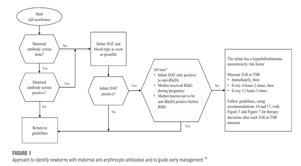
Figure-1: Approach to identify newborns with maternal anti-erythrocyte antibodies and to guide early management (6)
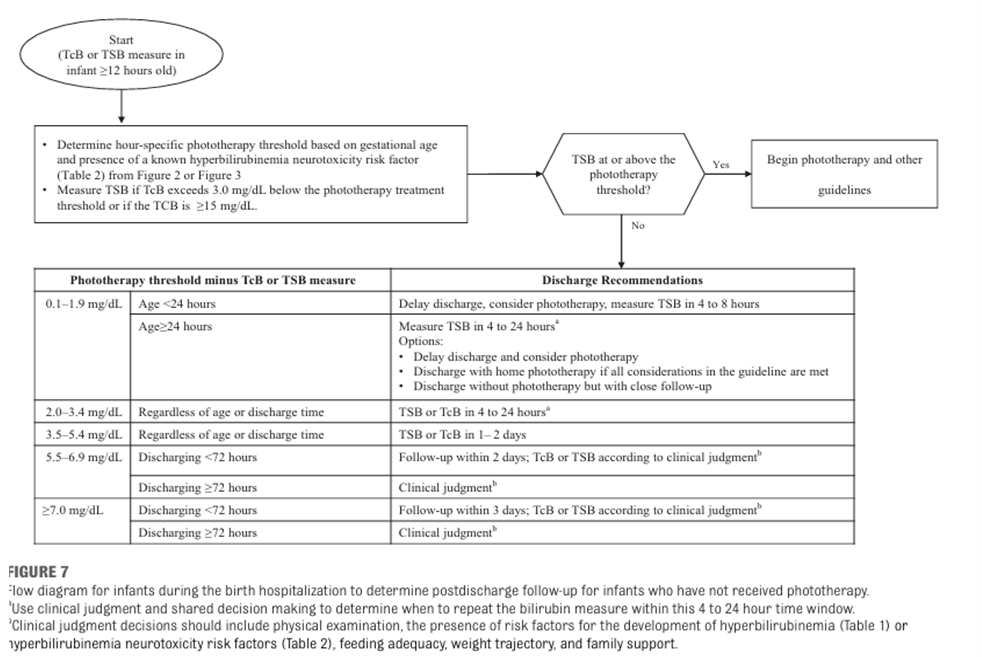
Figure-2: Flow diagram for infants during birth hospitalization to monitor jaundice follow up (6)
Table-6: Timing of follow up (unless seen earlier according to nomogram):
|
Infant Discharged |
Should be seen by Age |
|
Before age 24 hours |
72 hours |
|
Between 24-47.9 hours |
96 hours |
|
Between 48 and 72 hour |
120 hours |
(AAP guideline, Pediatrics 2004)(3)
Table-7: Risk Factors for Development of significant hyperbilirubinemia: (6)
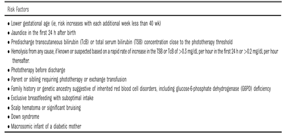
Table-8: Hyperbilirubinemia neurotoxicity risk factors: (6)
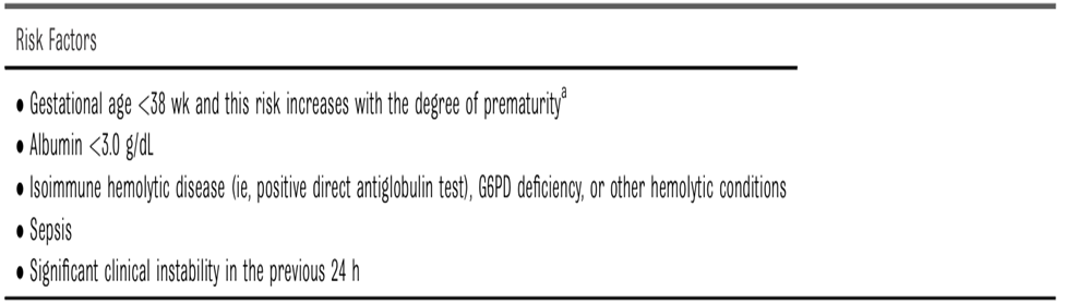
Table-9: Clinical assessment of neurotoxicity using the Modified BIND score: (51)
|
CLINICAL SIGN |
SCORE |
SEVERITY |
Date/Time |
|
MENTAL STATUS |
|||
|
-Normal |
0 |
None |
|
|
-Sleepy but arousable - Decreased feeding |
1 |
Mild |
|
|
- Lethargy - Poor suck and/or - Irritable/jittery with short-term strong suck |
2 |
Moderate |
|
|
-Semi-coma -Apnea -Seizures -Coma |
3 |
Severe |
|
|
MUSCLE TONE |
|||
|
- Normal |
0 |
None |
|
|
- Persistent mild hypotonia |
1 |
Mild |
|
|
- Moderate hypotonia - Moderate hypertonia - Increasing arching of neck and trunk on stimulation without spasms of arms and legs and without trismus |
2 |
Moderate |
|
|
- Persistent retrocollis - Opisthotonus - Crossing or scissoring of arms or legs but without spasms of arms and legs and without trismus |
3 |
Severe |
|
|
Total / 3 |
|||
|
CRY PATTERN |
|||
|
- Normal |
0 |
None |
|
|
- High pitched |
1 |
Mild |
|
|
- Shrill |
2 |
Moderate |
|
|
- Inconsolable crying or - Cry weak or absent in child with previous history of high pitched or shrill cry |
3 |
Severe |
|
|
Total / 3 |
|||
|
OCCULOMOTOR OR EYE MOVEMENTS |
|||
|
- Normal |
0 |
None, Mild |
|
|
- Sun-setting - Paralysis of Upward Gaze |
3 |
Severe |
|
|
Total / 3 |
|||
|
Total ABE Score / 12 |
|||
Choose one number for each domain. Final score out of 12 (zero: Normal, 1-4: mild encephalopathy, 5-6: moderate encephalopathy, 7-12: severe encephalopathy) (58)
Table-10: Laboratory Evaluation of the Jaundiced Infant of 35 or More Weeks’ Gestation: (3)
|
Indications |
Assessments |
|
Jaundice in first 24 hour |
Measure TcB and/or TSB and decide according to Table-7 |
|
Infant receiving phototherapy or TSB rising rapidly, crossing percentiles (Figure-6), and unexplained by history and physical examination
|
-Blood type and Coombs’ test. - Complete blood count and smear. - Reticulocyte count. - It is an option to perform G6PD (if retics are high and no incompatibility), and ETCO2, if available. - Measure (direct) conjugated bilirubin. - Repeat TSB in 4–24 hour depending on infant’s age and TSB level. |
|
TSB approaching exchange levels or not responding to phototherapy |
Perform reticulocyte count, G6PD, albumin, ETCO if available and consider exchange transfusion.
|
|
Elevated direct-reacting (or conjugated) bilirubin level |
Do urinalysis and urine culture. Evaluate for sepsis including TORCH screen if indicated by history and physical examination. |
|
Jaundice present at or beyond age 2 weeks, or sick infant |
- Total and direct-reacting (or conjugated) bilirubin level, if direct-reacting bilirubin elevated, evaluate for causes of cholestasis. - Check results of newborn thyroid and galactosemia screen, and evaluate infant for signs or symptoms of hypothyroidism or late onset sepsis. |
Table-11: Example of a Clinical Pathway for Management of the Newborn Infant Readmitted for Phototherapy or Exchange Transfusion: (3)
Treatment:
- Use intensive phototherapy and/or exchange transfusion
Laboratory tests:
- TSB and direct-reacting (or conjugated) bilirubin levels.
- Blood type (ABO, Rh).
- Direct antibody test (or Coombs’ test).
- Serum albumin.
- Complete blood cell count with differential and smear for red cell morphology.
- Reticulocyte count.
- ETCO (if available).
- G6PD if suggested by ethnic or geographic origin or if poor response to phototherapy.
- Urine for reducing substances.
- If history and/or presentation suggest sepsis, perform blood culture, urine culture, and cerebrospinal fluid for protein, glucose, cell count, and culture.
Interventions:
- If TSB ≥25 mg/dL (428 µmol/L) or ≥20 mg/dL (342 µmol/L) in a sick infant or infant ≤ 38 week gestation, obtain a type and cross match, and request blood in case an exchange transfusion is necessary.
- In infants with isoimmune hemolytic disease and TSB level rising in spite of intensive phototherapy or within 2-3 mg/dL (34-51 µmol/L) of exchange level (Figures-7 & 8), administer intravenous immunoglobulin 0.5–1 g/kg over 2 h, and repeat in 12 h if necessary.
- If infant’s weight loss from birth is 12% or there is clinical or biochemical evidence of dehydration, recommend expressed breast milk or formula. If oral intake is in question, give intravenous fluids.
- Parent Information leaflet
الصفراء في الأطفال حديثي الولادة:
ما هى الصفراء فى الأطفال حديثى الولادة ؟
هى عبارة عن حدوث اصفرار ببياض العين و الجلد عند المولود ، وهى حالة قد تتطلب عناية خاصة بحديثى الولادة.
كيف تحدث الصفراء فى حديثى الولادة ؟
تحدث نتيجة زيادة مستوى المادة المسببة للصفراء في الدم عن الحد الطبيعي. و هي مادة تُنتج عند:
· تكسير خلايا الدم الحمراء.
· مشاكل بالكبد (عدم اكتمال نمو الكبد أو وجود التهابات أو اعتلال بالكبد).
· انسداد القنوات المرارية.
ما هى أنواع الصفراء فى حديثى الولادة ؟
1- الصفراء الفسيولوجية (الطبيعية):
· هي عادة تظهر بنهاية اليوم الثالث أو الرابع من الولادة وتستمر لمدة عشرة أيام أو أسبوعين من يوم الولادة على الأكثر.
· وتحدث لأن إنزيمات الكبد المسئولة عن التخلص من المادة المسببة للصفراء فى الطفل حديث الولادة تكون غير مكتملة جزئيا .
2- الصفراء المرضية: وتحتاج لتدخل طبي
· من أشهر أسبابها: تكسير الدم نتيجة عدم توافق فصيلة دم الطفل مع دم الأم (إذا كان الطفل يحمل فصيلة موجبة والأم تحمل فصيلة سالبة والأب يحمل فصيلة موجبة).
· يحدث ذلك خلال 24 ساعة من الولادة.
· تحدث أيضا نتيجة وجود التهابات أو اعتلال بالكبد أو انسداد القنوات المرارية.
الأطفال الأكثر عرضة للإصابة بصفراء حديثي الولادة:
1- الأطفال المبتسرين (أقل من 9 شهور أو 36 أسبوع حمل).
2- وجود اخ او اخت عانى سابقا من صفراء حديثي الولادة.
3- ناقصي النمو.
4- إذا أصيبت الأم بأي حرارة أو طفح جلدي أثناء الحمل.
5- إذا كانت فصيلة الام 0 أو الأم تحمل فصيلة سالبة والأب يحمل فصيلة موجبة).
أعراض الصفراء في حديثي الولادة:
· اصفرار بشرة الطفل.
· اصفرار بياض العين.
علامات الخطر:
· ظهور الاصفرار في اليوم الأول من الولادة.
· تحول لون البراز إلى اللون الفاتح المقارب للبياض.
· وجود تشنجات أو تقوس بالضهر.
وعند ظهور أي من هذه العلامات يجب التوجه إلى الطبيب فوراً.
ما هي مضاعفات مرض الصفراء؟
ارتفاع المادة المسببة للصفراء بدم الطفل يؤدي الى وصولها الى أجزاء ومراكز هامة بالمخ ومن ثم حدوث مضاعفات خطيرة مثل:
الاعتلال الدماغي الحاد (الشلل الدماغي): وتكون أعراضه:
· الخمول أو صعوبة الاستيقاظ.
· البكاء عال النبرة.
· قلة الرّضاعة.
· تقوس الرقبة والجسم للوراء.
· تأخر تطور النمو العقلي.
· فقدان السمع.
كيف يتم تشخيص الصفراء في حديثي الولادة:
· قياس نسبة المادة المسببة للصفراء عبر الجلد.
· قياس نسبة المادة المسببة للصفراء في الدم.
علاج الصفراء في حديثي الولادة:
عند ملاحظة الاصفرار على بشرة الطفل يجب التوجه الى الطبيب فورا ويحدد الطبيب طريقة العلاج على حسب نسبة الصفراء في دم الطفل:
1- المتابعة: وتستمر كل 72 ساعة مع استمرار الرضاعة الطبيعية المنتظمة (8:12) مرة يوميا.
2- العلاج الضوئي في الحضانة.
3- تغيير دم الطفل في حالات الارتفاع الشديد لنسبة الصفراء في الدم.
4- فى حالة انسداد القنوات المرارية نتوجه للعلاج الجراحي.
ماذا تفعل بعد خروج الطفل من الحضانة؟
· يجب متابعة الصفراء بعد 24 ساعة.
· يجب عمل فحص للسمع لأي طفل عانى من نسبة صفرا مرتفعة.
معتقدات خاطئة:
· عدم عرض الطفل الأصفر على الطبيب.
· أن حالة الطفل ستتحسن تحت الإضاءة المنزلية المعتادة (اللمبة النيون). أو في الشمس.
· ان الفيتامينات أو الجلوكوز أو الاجار أو الاعشاب علاج للصفراء.
· ان الرضاعة الطبيعية لا يمكن ان تستمر في حالة وجود الصفراء او العلاج الضوئي.
Instructions to parents before discharge from maternity unit:
Parents should be provided with written information to facilitate post discharge care including the date, time and place of follow up appointment and when necessary prescription and appointment for a follow up TcB or TSB.
Birth hospitalization information including the last TcB or TSB and the age at which it was measured and DAT results (if any) should be documented.
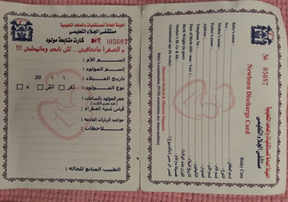
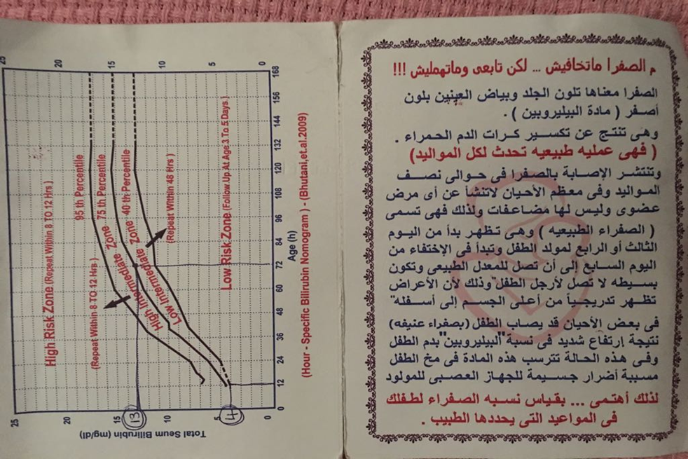
Figure-3: Example of parent card
- Appendix
Appendix 1: Phototherapy
Introduction:
Clinical trials have validated the efficacy of phototherapy in reducing excessive unconjugated hyperbilirubinemia, and its implementation has drastically curtailed the use of exchange transfusions. (3) The initiation and duration of phototherapy is defined by a specific range of total bilirubin values based on an infant’s postnatal age and the potential risk for bilirubin neurotoxicity. (3) Clinical response to phototherapy depends on the efficacy of the phototherapy device as well as the balance between an infant’s rates of bilirubin production and elimination. The active agent in phototherapy is light delivered in measurable doses, which makes phototherapy conceptually similar to pharmacotherapy.
When to start phototherapy (6):
· If TsB at/above PHOTOTHERAPY THRESHOLD
· If TsB less than 3 mg/dL below PHOTOTHERAPY THRESHOLD but with signs/symptoms of ACUTE HEMOLYSIS.
· Provider may start phototherapy if TsB < 3 mg/dL below threshold in order to prevent readmission.
Figure-4: Phototherapy thresholds for babies with no neurotoxicity risk factors (6)
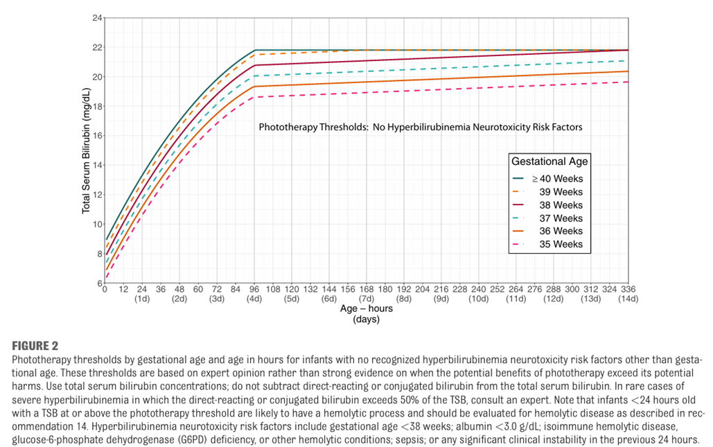
Figure-5: Phototherapy thresholds for babies with neurotoxicity risk factors (6)
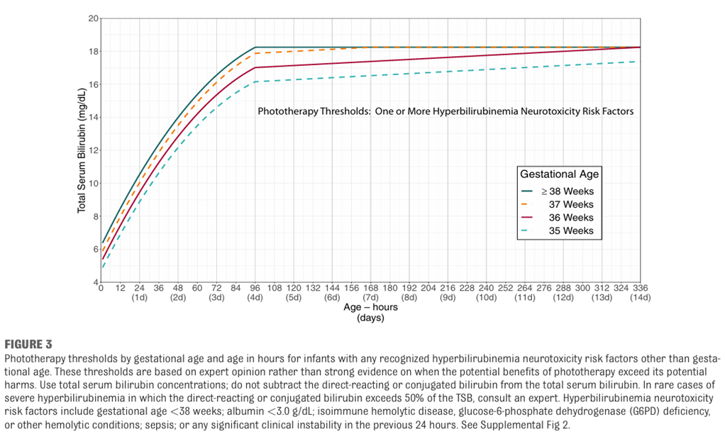
Commercial light sources:
A wide selection of commercial phototherapy devices is available in Egypt.
Phototherapy devices can be categorized according to their light source as follows:
· Fluorescent-tube devices that emit different colors (cool white daylight, blue [B], special blue [BB], turquoise, and green) and are straight (F20 T12, 60 cm, 20 W), U-shaped, or spiral-shaped;
· Metal halide bulbs, used in spotlights and incubator lights;
· Light-emitting diodes (LEDs) used with fiber-optic light guides in pads, blankets, or spotlights;
· High-intensity LEDs, used as over- and under-the-body devices. (5)
Standards for phototherapy devices:
Factors to consider in prescribing and implementing phototherapy are:
· Emission range of the light source,
· Light intensity (irradiance):
- Standard PT units deliver 8–10 W/ cm2 per nm.
- Intensive PT requires 30 W/cm2 per nm, if the level of bilirubin is approaching exchange transfusion only INTENTSIVE phototherapy will be effective.
· Exposed (“treatable”) body surface area illuminated
· Reduction in total bilirubin concentration. (5)
Table-12: Factors That Affect the Dose and Efficacy of Phototherapy (PT): (5)
PT: phototherapy; LE: light-emitting. *Olympic Bili Bassinet (Olympic Medical, Seattle, WA)
|
Table-13: Practice considerations for optimal administration of phototherapy: (5)
|
Checklist |
Recommendation |
Implementation |
|
|
Light source (nm) |
Wavelength spectrum in 460- 490-nm blue-green light region |
Know the spectral output of the light source. |
|
|
Light irradiance (W·cm2·nm1) |
Use optimal irradiance: 30 W·cm2·nm1 within the 460- to 490-nm waveband |
Ensure uniformity over the light footprint area. |
|
|
Body surface area (cm2) |
Expose maximal skin area |
Reduce blocking of light. |
|
|
Timeliness of implementation |
Urgent or “crash-cart” intervention for excessive hyperbilirubinemia |
May conduct procedures while infant is on phototherapy. |
|
|
Continuity of therapy |
Briefly interrupt for feeding, parental bonding, nursing care |
After confirmation of adequate bilirubin concentration decrease. |
|
|
Efficacy of intervention |
Periodically measure rate of response in bilirubin load reduction |
Degree of total serum/plasma bilirubin concentration decrease. |
|
|
Duration of therapy |
Discontinue at desired bilirubin threshold; be aware of possible rebound increase |
Serial bilirubin measurements based on rate of decrease. |
|
What decline in the Serum Bilirubin can you expect?
· With extremely high bilirubin (more than 30 mg/dL), and intensive phototherapy, a decline of as much as 10 mg/dL can occur within few hours, and a decrease of at least 0.5 to 1 mg/dL/ hour can be expected in the first 4 to 8 hours.
· On average, for infants >35 weeks’ gestation readmitted for phototherapy, intensive phototherapy can produce a decrement of 30% to 40% in the initial bilirubin level by 24 hours after initiation of phototherapy.
· The most significant decline will occur in the first 4 to 6 hours.
· With standard phototherapy systems, a decrease of 6- 20% of the initial bilirubin level can be expected in the first 24 hours.
Safety and protective measures:
· A clinician skilled in newborn care should assess the neonate’s clinical status during phototherapy to ensure adequate hydration, nutrition, and temperature control. Clinical improvement or progression of jaundice should also be assessed, including signs suggestive of early bilirubin encephalopathy (ABE).
· The distance of the phototherapy device from the infant should be safely minimized.
· It is important to note that the intensity of light decreases at the outer perimeter of the light footprint and to recognize the effects of physical factors that could impede or obstruct light exposure to have maximum exposure.
· Temperature should be monitored during phototherapy and a neutral thermal environment ensured.
· Baby should be monitored for adequate hydration by daily weighing and assessment of wet nappies.
· Phototherapy does not use ultraviolet light and exposure to lights is mostly harmless. Devices must comply with general safety standards.
· The genitalia should be protected from light during phototherapy.
· Give baby eye protection and routine eye care.
Other clinical considerations include:
· Phototherapy is contraindicated in infants with congenital porphyria or those treated with photosensitizing drugs.
· Prolonged phototherapy is associated with increased oxidant stress and lipid peroxidation and riboflavin deficiency.
When should phototherapy be stopped?
· There is no standard for discontinuing phototherapy.
· For infants who are readmitted after their birth hospitalization (usually for TSB levels of 18 mg/dL or higher), phototherapy may be discontinued when the serum bilirubin level falls below 13 to 14 mg/dL.
· Do not delay discharge to observe the infant for rebound.
· If phototherapy is used for infants with hemolytic diseases or is initiated early and discontinued before the infant is 3 to 4 days old, a follow-up bilirubin measurement within 24 hours after discharge is recommended.
Intermittent Versus Continuous Phototherapy:
· NO rationale for intermittent phototherapy exists.
· If the infant’s bilirubin level is approaching the exchange transfusion zone, intensive phototherapy should be administered continuously until a satisfactory decline in the serum bilirubin level occurs or exchange transfusion is initiated.
· Phototherapy may be interrupted for breastfeeding or brief parental visits as long as the level of bilirubin is not approaching the exchange zone.
Hydration:
· No evidence that excessive fluid administration affects the serum bilirubin concentration.
· If a baby with high bilirubin levels is also mildly dehydrated. This may need supplemental fluid intake to correct the dehydration.
· Encourage frequent breastfeeding but if baby is dehydrated despite frequent breastfeeding, supplement with a milk-based formula, because it inhibits the enterohepatic circulation of bilirubin and should help to lower the serum bilirubin level.
· Routine intravenous fluid or other supplementation (e.g., with dextrose water) of term and near-term infants receiving phototherapy is NOT required or indicated unless there is evidence of dehydration or escalation of care is required. (6)
Safety and state of the art use:
PHOTOTHERAPY IS A DRUG like Pharmacotherapy.(20)
In order to provide effective PT and improve outcomes, the following rules should be followed:
· Use photons as a drug when prescribing phototherapy.
· Use a narrow‐spectrum blue light (450 nm).
· Measure irradiance for specific wavelength.
· Use the least irradiance dose to achieve an efficient decrease in bilirubin load.
· Use phototherapy for the shortest possible time and only when indicated. (71)
Complications:
· Phototherapy in hospital separates mother and infant. Eye patching is disturbing to parents.
· Diarrhoea, intestinal hypermotility.
· Temperature instability.
· Phototherapy rash.
· Possible long term effects:
- Teratogenic effects, one study reported that phototherapy was associated with DNA damage. However, there is no evidence that this effect on DNA at a microscopic level can lead to long-term adverse effects in phototherapy-treated babies.
- Melanocytic nevus development.
· Infants with cholestatic jaundice may develop a dark, grayish-brown discoloration of the skin, serum, and urine (the bronze infant syndrome). This syndrome generally has had few deleterious consequences, and if there is a need for phototherapy, the presence of direct hyperbilirubinemia should not be considered a contraindication to its use. This is particularly important in sick neonates.
· Rarely, purpura and bullous eruptions have been described in infants with severe cholestatic jaundice receiving phototherapy and severe blistering and photosensitivity during phototherapy have occurred in infants with congenital erythropoietic porphyria.
· Congenital porphyria or a family history of porphyria is an absolute contraindication to the use of phototherapy, as is the concomitant use of drugs or agents that are photosensitizers.
Home phototherapy:
· Home phototherapy should not be used unless there is quality assurance regarding the phototherapy device, the implementation and the monitoring of bilirubin at home. Very close supervision of medical practitioners by a regulator of the process is required to avoid malpractice. Since that is currently not available, it is not recommended to have home phototherapy until these rules are fulfilled.
Escalation of care:
The intensive care that some infants with elevated or rapid increasing bilirubin concentration require to prevent the need for an exchange and possibly prevent kernicterus. (6)
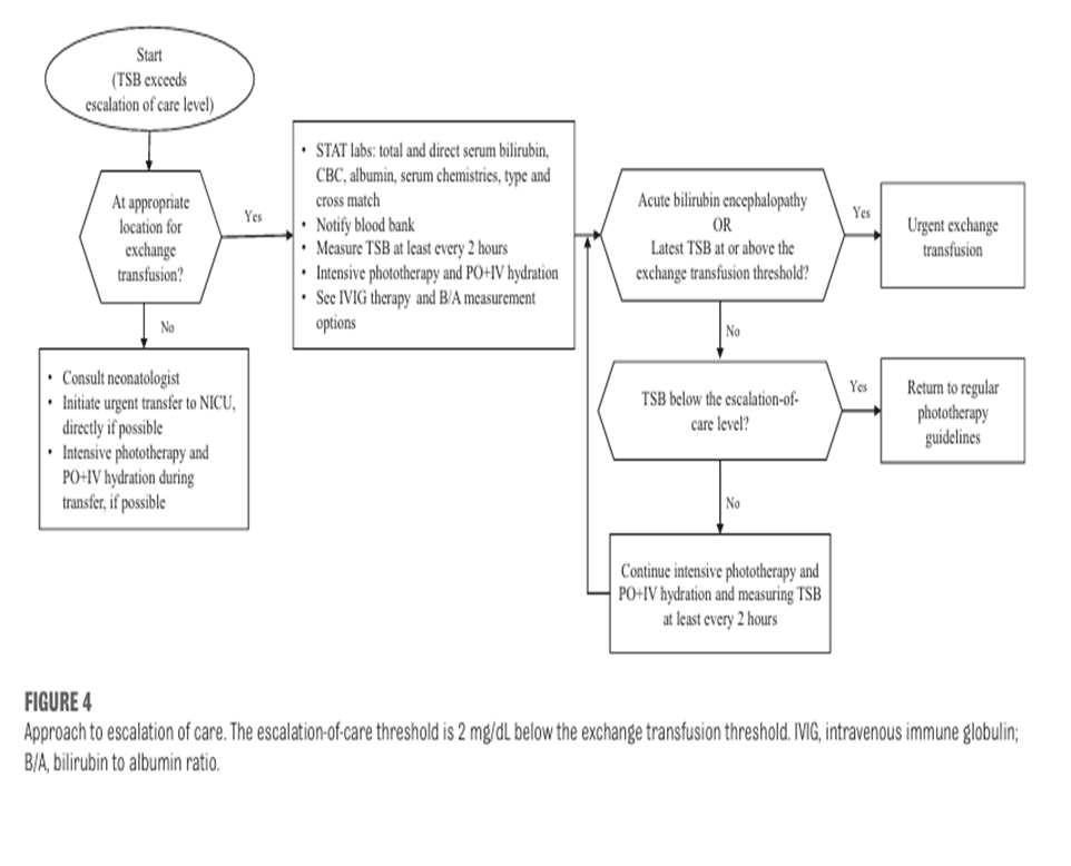
Figure-6: Approach to escalation of care
Appendix 2: Exchange Transfusion
· Exchange transfusion is an intensive invasive procedure and should not be performed in a Level 1 NICU.
· “Exchange transfusion must take place in an intensive care setting with intensive physiological and biochemical monitoring, carried out by staff trained in the procedure following written informed parental consent”, cited by British Committee for Standards in Hematology. (72)
· Exchange transfusion is usually carried out by repeatedly exchanging small samples (5-10 ml/kg) of blood through an umbilical venous catheter and replacing with same volumes by compatible blood.
· Neonatal exchange transfusion is becoming an infrequent procedure because severe Rh hemolytic disease of the newborn is becoming increasingly rare due to:
- Maternal anti-D therapy and intrauterine transfusion of affected fetuses.
- Maximizing phototherapy use prevents the need for exchange transfusion, even at very high level of TSB.
- Intravenous immunoglobulin (IVIG) acts by preventing the destruction of sensitized erythrocytes.
Benefits of Exchange Transfusion in severely jaundiced babies:
· Rapidly lowers the serum bilirubin level and reduce the risk of brain damage and kernicterus.
· Corrects anemia and increase the oxygen carrying capacity of the infant’s blood.
· Removes damaged and antibody coated red cells.
Indication for exchange transfusion in severe hyperbilirubinemia:
· Any baby showing signs of intermediate or advanced stages of acute bilirubin encephalopathy (e.g., hypertonia, arching, retrocollis, opisthotonus, high pitched cry, or recurrent apnea) irrespective of bilirubin level.
· If the TSB is at or above the exchange transfusion threshold.
· If intensive phototherapy is failing to lower TSB and it is approaching exchange level
· If the bilirubin /albumin ratio is:
- ≥8.0 If the gestational age is ≥38 weeks’ gestation and there are no hyperbilirubinemia neuro toxicity risk factors,
- ≥7.2 If the gestational age is ≥38 weeks’ gestation and there is at least 1 hyperbilirubinemia neurotoxicity risk factor,
- ≥7.2 If the gestational age is 35 through 37 weeks’ gestation with no hyperbilirubinemia neurotoxicity risk factor,
- 6.8 If the gestational age is 35 through 37 weeks’ gestation and at least 1 hyperbilirubinemia neurotoxicity risk factor. (62)
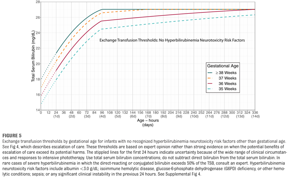
Figure-7: Exchange transfusion threshold by gestational age for infants with no recognized hyperbilirubinemia neurotoxicity risk factors
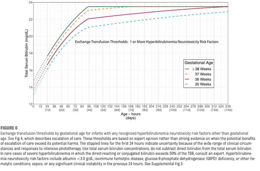
Figure-8: Exchange transfusion threshold by gestational age for infants with any recognized hyperbilirubinemia neurotoxicity risk factors(6)
Table-14: B/A Ratio at which Exchange Transfusion should be considered: (6)
· The bilirubin/ albumin ratio can be used with TSB level to help determine need for exchange transfusion.
· The bilirubin/ albumin ratio threshold for exchange transfusion, measured as TSB (measured in mg/ dL) divided by serum albumin (measured in g/dL), varies by gestational age and risk.
· An exchange transfusion may be considered if the bilirubin albumin ratio is:
- ≥8.0 if the gestational age is ≥38 weeks’ gestation and there are no hyperbilirubinemia neuro toxicity risk factors
- ≥7. 2 if the gestational age is ≥ 38 weeks’ gestation and there is at least 1 hyperbilirubinemia neurotoxicity risk factors
- ≥7.2 if the gestational age is 35 through 37 weeks’ gestation with no hyperbilirubinemia neurotoxicity risk factors
- ≥6.8 if the gestational age is 35 through 37 weeks’ gestation and at least 1 hyperbilirubinemia neurotoxicity risk factors.
Use of IVIG: (6)
Intravenous immunoglobulin (IVIG) 0.5-1 g/kg over 2 hours:
Provide to infants with isoimmune hemolytic disease (i.e. positive DAT) whose TSB reaches or exceeds the escalation of care threshold. This dose can be repeated in 12 hours.
Table-15: Procedures for Exchange Transfusion:
|
Steps |
Preparation for exchange transfusion |
|
1 |
Parent/care giver Information and consent Informed parental consent about the procedure and risks of exchange transfusion should be obtained and documented wherever possible. This should be a written consent. |
|
2 |
Order the blood : Which blood group to use? · Cross-matched washed packed red blood cells mixed with thawed adult fresh-frozen plasma to a hematocrit approximating 40% is preferred for exchange transfusions. The additional albumin-containing fresh frozen plasma that infants receive by keeping the hematocrit close to 40% will augment bilirubin removal. (72-74). · Use blood group O packed RBCs (with low titer hemolysins) Rh negative or Rh identical with the neonate suspended in AB plasma for exchange transfusion. · If not found then: if - Rh isoimmunization: use Rh negative and blood group O or blood group of the baby suspended in AB plasma. Cross matched with the baby’s and mother’s blood. - ABO incompatibility: Rh compatible and blood group O (not that of the baby), suspended in AB plasma, cross matched with baby’s and mother’s blood. - Other conditions: baby’s group and Rh type, cross matched with the baby’s and mother’s blood. Use a double-volume exchange transfusion: · Do not perform a single volume exchange. · A double volume exchange transfusion will exchange approximately 80% of the infant’s blood volume:(2 x 80 mls / Kg = 160 / per Kg = double volume blood exchange) Red Blood Cell Requirements: · 5 days old or less. · Negative for any antigens to which the mother or baby has antibodies. · Irradiated – use within 24 hours. Irradiation is a MUST if the infant has had a previous intrauterine transfusion and is preferable for all exchange transfusions if no delay will occur. · CMV negative. · Plasma reduced with HCT 40%. · It should be warmed to 37oc immediately before transfusion by a blood warmer. (Not to be transfused straight from 40C) |
|
3 |
Gain intravenous access: · Central via the umbilical vein. · Or a combination of central and peripheral lines. · Any central line should have its position confirmed by X-ray prior to procedure and checked for patency. |
|
4 |
Prepare equipment: Make a check list with the following before starting exchange: · Continuous cardio-respiratory monitoring equipment, including SpO2, and temperature should be available and resuscitation equipment. · Blood sugar monitoring available. · Exchange Transfusion tray. · Blood: do not collect blood until ready to begin exchange. · Protective clothing - Sterile gowns, gloves and masks. · Blood warmer and extension set. · Blood giving set with appropriate 170-200-micron filter. · Stand for blood. · Laboratory bottles = Full blood count x 2, urea and electrolytes x 2 including calcium and glucose. Blood culture x 2, capillary gas for pH x 2, (x 2 = pre and post specimens required ). · 2 ml, 5 ml, 10 ml and 60 ml syringes and needles. · Ventilator and intubation equipment, also suction equipment. (available if needed) · Sharps bin box. · High stools for doctor and nurse as the procedure up to 3 hours. · Timing device for timing infusion and withdrawal of aliquots. |
|
5 |
Ensure a neonatal screening blood spot has been taken prior to procedure. |
|
6 |
A minimum of 2 staff are required for the procedure (senior doctor and trained nurse). |
|
Steps |
Management prior to exchange transfusion |
|
1 |
· Nurse the infant in the radiant overhead heater if available, in the intensive care nursery with continuation of phototherapy. |
|
2 |
· Monitor heart rate, respiratory rate, oxygen saturation, blood pressure and temperature, a baseline set of observations including general color and tone and blood sugar should be documented before the procedure starts. |
|
3 |
· Stop enteral feeds one hour before the procedure, leaving the nasogastric tube on free drainage, (to reduce the risk of necrotizing enterocolitis). |
|
4 |
· Recheck TSB and check blood sugar (and effect of treatment) prior to picking up blood. |
|
5 |
· Set up the blood warmer. Using aseptic non touch technique when appropriate. |
|
6 |
· Collect and check the blood with another trained member of staff. · Warm the blood in a towel if warmer is not available. · Do not place blood on servo heater. Prime tubing. |
|
7 |
· Establish volume of aliquots prior to commencement · Aliquots usually tolerated for exchange transfusion: Ø <1500 grams: 5 ml Ø 1500-2490 grams :10 ml Ø 2500 grams – 3500 : 15 ml |
|
8 |
Bloods tests, blood sugar and a starting set of observations are recorded |
|
Steps |
Management during Exchange Transfusion |
|
1 |
Define responsibility: · Senior doctor (person A) is responsible for infusing and withdrawing an equal amount of blood. · Person B is responsible for the accurate recording of blood input and withdrawal on the Exchange Transfusion Fluid Chart (Table-17). |
|
2 |
· One member of staff (person A) flushes the input line with sodium chloride, connects the blood giving set, and connects the first syringe to the withdrawal line. Start by withdrawal of aliquot send to lab, and continue to infuse and withdraw equal amounts so that an equal rate is maintained. · Person B records this. ·
Small aliquots exchanged at a slower rate are usually better
tolerated by infants with cardiovascular instability. The time for each
in/out should be around 4-10 minutes (withdrawal and infusion of blood should
be performed at the same rate). In and
out aliquots are repeated sequentially until desired volume for exchange · A timer may be used for guiding speed of aliquots. |
|
3 |
· Whilst the exchange transfusion is in progress, the volume of blood exchanged in and out is recorded in the fluid balance chart with the Time. |
|
4 |
· The first aliquot of blood removed should be sent for estimation of full blood count, (FBC), differential, urea and electrolytes (U&Es), liver function tests, split bilirubin, Ca, hematocrit and glucose. · In severe cases consider: - coagulation screen, glucose 6-phosphate dehydrogenase (G6PD). |
|
5 |
· This process of administration and withdrawal continues until the agreed volume of blood has been exchanged or the condition of the baby determines cessation. · At the end of the procedure the infant’s fluid balance should be in deficit of one aliquot. |
|
6 |
· The last withdrawn blood should be sent for blood sugar, urea and electrolytes, bilirubin - total + direct, FBC, hematocrit, magnesium, calcium, blood gases and consider coagulation screen. |
|
7 |
· Continuously observation is required for Any signs of deterioration including cyanosis, pallor, abdominal distension, vomiting and blood in stools - Record heart rate, respirations, temperature, oxygen saturation and blood pressure every 15 minutes, any changes in these should be reported immediately. - Signs of hypocalcaemia - Tachycardia, irritability, seizures. - Monitor capillary blood glucose every 30 minutes. - All lines should be monitored for extravasation, blanching, erythema, leaking and edema. |
|
8 |
· Aim for procedure should take 2 hours (no longer than 3 hours). · On completion of the exchange transfusion, the lines are again flushed with NaCl. |
|
9 |
· Disposal of equipment –in accordance with local policy. |
|
10 |
· Documentation – ALL the procedure should be documented including: - All transfusion records, timing, accurate fluid balance, observation, samples taken and their results. - Also any untoward reaction, corrective measures taken and outcomes. |
|
11 |
· The baby should be left in a comfortable position and parents informed that the procedure has been completed and the condition of the baby. |
|
12 |
· Stop the procedure immediately and review if there are any concerns or untoward reactions. |
|
13 |
· Do NOT routinely administer intravenous calcium. (23) |
|
Steps |
Management following exchange transfusion |
|
1 |
· Continue intensive phototherapy after exchange, checking serum bilirubin as required. |
|
2 |
· Take blood pressure at completion of procedure. · Continue to monitor vital signs hourly for 6 hours or as the condition of the baby indicates. · After this time, and if stable and within normal limits routine observations may be recommenced. |
|
3 |
· Measure TSB level within 2 hours and manage according to threshold graphs (Figures-7 & 8). · Continue to monitor CBC and reticulocytes. |
|
4 |
· Monitor the blood sugar 1, 2 and 4 hours after exchange regularly according to condition for rebound hypoglycemia. |
|
5 |
· Observe catheter sites for bleeding/signs of infection. |
|
6 |
· Observe urine for blood, observe stools for blood. |
|
7 |
· Feeding: - Observe for abdominal distension, and for signs of feed intolerance, gastric aspirate and vomiting. - Commence feeds on medical advice, but not before 3 hours post-exchange transfusion. |
|
8 |
· Neonatal screening must not be taken until 4 days after the exchange as blood transfusion will invalidate all the tests. |
|
9 |
· All infants should receive regular audiology testing as they are at risk of hearing loss due to the severely raised serum bilirubin levels. |
Risks/Complications of exchange transfusion
There is a 5% risk of complication (3)
Exchange Transfusion Reaction:
· Tachycardia or bradycardia.
· Respiratory Distress.
· Dramatic changes in blood pressure.
· Temperature irregularity.
· Rash.
· Cyanosis or pallor.
· Hypoglycemia
· Hypocalcaemia (treatment see below)
· Cardiac Arrhythmias.
· Heart failure (fluid overload).
Other risk/complications:
· Administration of incorrect blood product.
· Volume overload.
· Hemodynamic instability.
· Acid base instability.
· Apnea.
· Electrolyte imbalances (hypocalcaemia, hypoglycemia, hypomagnesaemia).
· Convulsions.
· Hypotension (volume depletion).
· Embolism (air/thrombus).
· Graft versus host reaction.
· Infection.
· Necrotizing enterocolitis.
· Temperature instability.
· Thrombocytopenia.
· Thrombosis.
· Intraventricular hemorrhage.
Table-16: Exchange Transfusion Fluid Chart: (24)
|
Name |
|
|||||||
|
DOB |
|
|||||||
|
Hospital no |
|
|||||||
|
Aliquot Number |
Time |
IN |
OUT |
Total Balance (in/out) |
||||
|
aliquot mls |
Total mls |
aliquot mls |
Total mls |
|||||
|
1 |
|
|
|
|
|
|
|
|
|
2 |
|
|
|
|
|
|
|
|
|
3 |
|
|
|
|
|
|
|
|
|
4 |
|
|
|
|
|
|
|
|
|
5 |
|
|
|
|
|
|
|
|
|
6 |
|
|
|
|
|
|
|
|
|
7 |
|
|
|
|
|
|
|
|
|
8 |
|
|
|
|
|
|
|
|
|
9 |
|
|
|
|
|
|
|
|
|
10 |
|
|
|
|
|
|
|
|
|
11 |
|
|
|
|
|
|
|
|
|
12 |
|
|
|
|
|
|
|
|
|
13 |
|
|
|
|
|
|
|
|
|
14 |
|
|
|
|
|
|
|
|
|
15 |
|
|
|
|
|
|
|
|
|
16 |
|
|
|
|
|
|
|
|
|
17 |
|
|
|
|
|
|
|
|
|
18 |
|
|
|
|
|
|
|
|
|
19 |
|
|
|
|
|
|
|
|
|
20 |
|
|
|
|
|
|
|
|
|
21 |
|
|
|
|
|
|
|
|
|
22 |
|
|
|
|
|
|
|
|
|
23 |
|
|
|
|
|
|
|
|
|
24 |
|
|
|
|
|
|
|
|
|
25 |
|
|
|
|
|
|
|
|
- Glossary
1. Acceptability
Is the extent to which the users are likely to adopt a recommendation, based on internal qualities such as clarity, comprehensiveness, and logical reasoning and on external factors such as the burden imposed on the process and system of care, patient and providers attitudes and beliefs, and patients’ needs, expectations, and preferences.
2. Adaptation (of guidelines)
Is the systematic approach to considering the use and/or modification of (a) guidelines(s) produced in one cultural and organizational setting for application in different context? Adaptation can be used as an alternative to de novo guideline development or for customizing (an) existing guideline(s) to suit the local context.
3. Adoption (of a guideline)
Is the acceptance of a guideline as a whole after the assessment of its quality, currency, and content? When health care providers (or other users of recommendations) adopt a guideline, they feel committed to change their practices in accordance with the recommendations of the guideline.
4. Applicability
Is the extent to which the users are able to put a recommendation into practice, based on internal qualities such as a clearly defined eligible patient population that matches the population to which the intervention is targeted in the local setting and external factors such as the availability of the necessary knowledge, skills, provider time, staff, equipment, and other resources.
Applicability is sometimes taken as a synonym for feasibility:
· Feasibility of the acquisition of necessary skills and knowledge.
· Feasibility of the necessary increase in provider time, staff, equipment, and so on.
5. Culture
Culture represents the norms and values of a specific group, community, or population.
6. Diffusion
Is a passive means of transferring knowledge; it is not directed towards a target audience (e.g. publication of articles in medical journals).
7. Dissemination
Is more active than diffusion in that it targets a specific audience and involve tailoring the information for that audience (e.g. of dissemination strategies include targeted mailings, presentations, and press conferences.
8. Evidence-based principles
Evidence-Based Medicine (EBM) has been defined as ― the conscientious, explicit, and judicious use of current best evidence in making decisions about the care of individual patients. The practice of EBM means integrating individual clinical expertise with the best available external clinical evidence from systematic research.
9. Evidence tables
Are summaries of the most salient information from studies identified in the systematic review. The elements of evidence tables are dependent on the types of information in studies related to a particular topic but might include information such as the article reference, the study type (e.g. RCT or Cohort), the number of patients and their characteristics, and the intervention, comparison arms, outcome measures, and effect sizes.
10. Guideline or Clinical Practice Guideline (CPG)
Systematically developed statements about specific health problems, intended to assist practitioners and patients in making decisions about appropriate health care.
11. Guideline consistency
Agreement between the evidence and the recommendations, based on the:
§ Comprehensiveness of the study search and selection process,
§ Coherence between the results of the studies and their interpretation by the guideline authors, and
§ Transparency between interpretation and recommendations.
12. Guideline content
In the ‘ADAPTE Manual and Resource Toolkit for Guideline Adaptation’ document, guideline content refers to the recommendations in the source guidelines.
13. Guideline currency
A CPG may be considered up to date ―when (no) new information on interventions, outcomes, and performance justifies updating (it).
14. Guideline quality
By quality of clinical practice guidelines, we mean the confidence that the potential biases of guideline development addressed adequately and that the recommendations are both internally and externally valid and are feasible for practice. This process involves taking into account the benefits, harms and costs of the recommendations, as well as the practical issues attached to them. Therefore, the assessment (of quality) includes judgments about the methods used for developing the guidelines, the content of the final recommendations, and the factors linked to their uptake.
15. Guideline topic
In the ADAPTE Manual and Resource Toolkit for Guideline Adaptation' document, the topic refers to the theme of the guideline, as described in the guideline title, for a targeted population (disease and patients) and intervention. The purpose, the audience, and the setting intended for the guideline, although not necessarily explicitly stated in the title, are also part of the topic. A guideline on a given topic may contain more than one health question.
16. Health question or clinical question or key question
Is a precisely described health issue (e.g. clinical, professional practice or public health) relating to the topic of the guideline? Guideline may include one or more questions.
17. Implementation
Implementation includes methods to promote the uptake of research findings into routine healthcare in both clinical and policy contexts and hence to improve the quality and effectiveness of healthcare. It includes the study of influences on healthcare professional and organizational behavior.
18. Intra-class correlations
Intra-class correlations provide a measurement of the extent to which two or more raters agree when rating the same set of things. It is a reliability index and is typically a ratio of the variance of interest over the sum of the variance of interest plus error.
19. Recommendation
Any statement that promote or advocate a particular course of action in clinical care.
20. Stakeholder
A stakeholder is an individual, group and/or organization with a stake in your decision to implement a guideline. Stakeholders include individuals or groups who will be directly or indirectly affected by the implementation of a guideline.
21. Source guideline
In the ADAPTE Manual and Resource Toolkit for Guideline Adaptation' document, source guideline refers to those guidelines selected to undergo assessments of quality, currency, content, consistency, and acceptability/applicability and upon which an adapted guideline may be based.
- Limitation of the guidelines
· Most of the recommendation statements from one source & many recommendations of low strength of evidence (GPS)
· No data registry in Egypt about neonatal jaundice.
· The guidelines directed only for term and late preterm newborns
Suggestions:
· It is essential to have data registry about neonatal jaundice cases and cases with BIND.
- Further research needs
· To study the actual effectiveness of IVIG in arresting hemolysis and progression of severe hyperbilirubinemia
· To prepare guidelines for premature babies.
· To study the safety, practicality and efficacy of home phototherapy remain a research priority.
- Monitoring and evaluating the impact of the guideline
· Research should be directed toward methods for disseminating the information contained in this guideline to increase awareness on the part of physicians, residents, nurses, and parents concerning the issues of neonatal hyperbilirubinemia and strategies for its management.
· In addition, monitoring systems should be established to identify the impact of these guidelines on the incidence of acute bilirubin encephalopathy and kernicterus and the use of phototherapy and exchange transfusions.
- REFERENCES
1. The ADAPTE Collaboration (2009). The ADAPTE Process: Resource Toolkit versions for Guideline Adaptation. Version 2.0 Available from: https://www.g-i-n.net/ 2009.
2. Amer YS, Elzalabany MM, Omar TI, Ibrahim AG, Dowidar NL. The 'Adapted ADAPTE': an approach to improve utilization of the ADAPTE guideline adaptation resource toolkit in the Alexandria Center for Evidence-Based Clinical Practice Guidelines. Journal of evaluation in clinical practice. 2015;21(6):1095-106.
3. American Academy of Pediatrics Subcommittee on Hyperbilirubinemia. Management of hyperbilirubinemia in the newborn infant 35 or more weeks of gestation. Pediatrics. 2004;114(1):297-316.
4. Maisels MJ, Bhutani VK, Bogen D, Newman TB, Stark AR, Watchko JF. Hyperbilirubinemia in the newborn infant > or =35 weeks' gestation: an update with clarifications. Pediatrics. 2009;124(4):1193-8.
5. Bhutani VK, Committee on F, Newborn, American Academy of P. Phototherapy to prevent severe neonatal hyperbilirubinemia in the newborn infant 35 or more weeks of gestation. Pediatrics. 2011;128(4):e1046-52
6. Kemper AR, Newman TB, Slaughter JL et al. Clinical practice guideline revision: Management of hyperbilirubinemia in the newborn infant 35 or more weeks of gestation. Pediatrics.2022; 150 (3): e2022058859.
7. Ayyappan S, Philip S, Bharathy N, Ramesh V, Kumar CN, Swathi S, et al. Antioxidant status in neonatal jaundice before and after phototherapy. Journal of pharmacy & bioallied sciences. 2015;7(Suppl 1):S16-21.
8. Bhutani VK, Johnson LH, Keren R. Diagnosis and management of hyperbilirubinemia in the term neonate: for a safer first week. Pediatric clinics of North America. 2004;51(4):843-61.
9. Maisels MJ. Epidemiology of neonatal jaundice. In: Maisels MJ, Watchko JF, editors. Neonatal jaundice. Amsterdam: Harwood Academic Publishers; 2000. p. 37–49.
10. Ebbesen F, Andersson C, Verder H, Grytter C, Pedersen-Bjergaard L, Petersen JR, et al. Extreme hyperbilirubinaemia in term and near-term infants in Denmark. Acta paediatrica. 2005;94(1):59-64.
11. Slusher TM, Zamora TG, Appiah D, Stanke JU, Strand MA, Lee BW, et al. Burden of severe neonatal jaundice: a systematic review and meta-analysis. BMJ paediatrics open. 2017;1(1):e000105.
12. Basheer H, Makhlouf M, El Halawany F, Fahmy N, Iskander I. Screening for neonatal jaundice in El Galaa Teaching Hospital: A Egyptian Maternity Hospital – Can the model be replicated? Journal of Clinical Neonatology. 2017;6(2):128-33.
13. Gamaleldin R, Iskander I, Seoud I, Aboraya H, Aravkin A, Sampson PD, et al. Risk factors for neurotoxicity in newborns with severe neonatal hyperbilirubinemia. Pediatrics. 2011;128(4):e925-31.
14. El Houchi SZ, Iskander I, Gamaleldin R, El Shenawy A, Seoud I, Abou-Youssef H, et al. Prediction of 3- to 5-Month Outcomes from Signs of Acute Bilirubin Toxicity in Newborn Infants. The Journal of pediatrics. 2017;183:51-5 e1.
15. Mostafa MA et al. Knowledge of Neonatal Hyperbilirubinemia Among Primary Health Care Physicians: A Single-Center Experience. Clinical Medicine Insights: Pediatrics. 2019; 13: 1–5.
16. Iskander I, Gamaleldin R, Kabbani M. Root causes for late presentation of severe neonatal hyperbilirubinaemia in Egypt. Eastern Mediterranean health journal = La revue de sante de la Mediterranee orientale = al-Majallah al-sihhiyah li-sharq al-mutawassit. 2012;18(8):882-7.
17. Shapiro SM. Definition of the clinical spectrum of kernicterus and bilirubin-induced neurologic dysfunction (BIND). Journal of perinatology : official journal of the California Perinatal Association. 2005;25(1):54-9.
18. Amin S.B., Bhutani V.K., Watchko J.F. Apnea in acute bilirubin encephalopathy. Semin. Perinatal. 2014;38:407–411. doi: 10.1053/j.semperi.2014.08.003
19. Shaprio S.M. Chronic bilirubin encephalopathy: Diagnosis and outcome. Semin. Fetal Neonatal Med. 2010;15:157–163
20. Valinjkar SJ et al, Rebound hyperbiliurbinemia in neonates after phototherapy and factors affecting it. JMSCR 2017;5(3):19003-19014
21. Taylor JA, Stout JW, de Greef L, Goel M, Patel S, Chung EK, et al. Use of a Smartphone App to Assess Neonatal Jaundice. Pediatrics. 2017;140(3).
22. Hansen TW. The role of phototherapy in the crash-cart approach to extreme neonatal jaundice. Seminars in perinatology. 2011;35(3):171-4.
23. Singla DA, Sharma S, Sharma M, Chaudhary S. Evaluation of Risk Factors for Exchange Range Hyperbilirubinemia and Neurotoxicity in Neonates from Hilly Terrain of India. International journal of applied & basic medical research. 2017;7(4):228-32.]\
24. National Institute for Health and Care Excellence (NICE): Jaundice in the newborn under 28 days [CG98.1]; 2010.
25. National Institute for Health and Care Excellence (NICE): Addendum to Jaundice in newborn babies under 28 days. London:; 2016 May. (Clinical Guideline, No. 98.1.) Available from: https://www.ncbi.nlm.nih.gov/books/NBK550520/
26. Barrington KJ, Sankaran K, Canadian Paediatric Society, Fetus and Newborn Committee. Guidelines for detection, management and prevention of hyperbilirubinemia in term and late preterm newborn infants | Canadian Paediatric Society: Canadian Paediatric Society; 2018 [updated 28/2/2018; cited 2018 15/6/2018]. Available from: https://www.cps.ca/en/documents/position/hyperbilirubinemia-newborn.
27. Queensland Clinical Guidelines. Neonatal jaundice clinical guideline education presentation E22.7-1-V3-R27. Queensland Health. 2018.
28. Brouwers MC, Kho ME, Browman GP, Burgers JS, Cluzeau F, Feder G, et al. AGREE II: advancing guideline development, reporting and evaluation in health care. Canadian Medical Association Journal. 2010;182(18):E839-E42.
29. Alshehri A, Almazrou S, Amer Y. Methodological frameworks for adapting global practice guidelines to national context int eh Eastern Mediterranean region. EMHJ, 2023;1:29(7):541-549
30. Practice bulletin No. Practice bullitin no 181summary: prevention of RHD alloimmunization Obstet gynecol.2017;130(2): 481-483
31. Vats K, Watchco JF. Coordinating care across the perinatal continuum in hemolytic disease of the fetus and newborn: the timely handoff of a postive maternal antierythrocyte antibody screen. J Pediatr. 2019; 214: 212-216
32. Erdeve O. Management of neonatal jaundice in low-income and middle-income countries. BMJ Paediatr Open. 2020; 2;4(1):e000845
33. Chen Yj, Yeh TH, Chen CM. Effect of breast feeding frequency on hyperbilirubinemia in breast fed term neonates. Pediatr Int. 2015;57(6):1121-1125
34. Porter MLand Dennis BL.Hyperbilirubinemia in the term newborn. Am Fam Phys 2002;65(4):599-606
35. Kaplan M, Kaplan E, Hammerman C et al, POst photherapy neonatal bilirubin rebound : a potential cause of significant hyperbiliurbinemia . Arch dis child 2006; 9(1):31-34
36. Maayan-Metzger A, Schwartz T, Sulkes K, Merlob P. Maternal antiD prophylaxis during pregnancy does not cause neonatal hemolysis. Arch Dis Child Fetal Neonatal Ed. 2001; 84(1):F60–F62
37. Flaherman VJ, Maisels MJ; Academy of Breastfeeding Medicine. ABM clinical protocol #22: guidelines for management of jaundice in the breastfeeding infant 35 weeks or more of gestation — revised. Breastfeed Med. 2017;12 (5): 250-257
38. Maisels MJ, Clune S, Coleman K et al. The natural history of jaundice in predominantly breastfed infants. Pediatrics. 2014;134(2):e340-345
39. Meek JY, Noble L. American Academy of Pediatrics, Section on breastfeeding. Breast feeding and use of human milk. Pediatrics 2022; 150(1): e2022057989
40. Flaherman VJ, Schaefer EW, Kuzniewicz MW, Li SX, Walsh EM, Paul IM. Early weight loss nomograms for exclusively breastfed newborns. Pediatrics 2015; 135 (1) :e16-e23
41. Newman TB, Liljestrand P, Escobar GJ. Jaundice noted in the first 24 hours after birth in a managed care organization. Arch Pediatr Adolesc Med. 2002;156(12):1244–1250
42. Hulzebos CV, Vitek L, Coda Zabetta CD, et al. Screening methods for neonatal hyperbilirubinemia: benefits, limitations, requirements, and novel developments. Pediatr Res. 2021;90(2):272–276
43. Wainer S, Parmar SM, Allegro D, Rabi Y, Lyon ME. Impact of transcutaneous bilirubinometry program on resource utilization and severe hyperbilirubinemia. Pediatr 2012; 129(1): 77-86.
44. Konana OS, Bahr TM, Strike HR, Coleman J, Snow GL, Christensen RD. Decision accuracy and safety of transcutaneous bilirubin screening at intermountain healthcare. J Pediatr. 2021;228:53–57
45. Kuzniewicz MW, Park J, Niki H, Walsh EM, McCulloch CE, Newman TB. Predicting the need for phototherapy after discharge. Pediatrics. 2021;147(5): e2020019778
46. Maisels MJ, Deridder JM, Kring EA, Balasubramaniam M. Routine transcutaneous bilirubin measurements combined with clinical risk factors improve the prediction of subsequent hyperbilirubinemia. J Perinatol. 2009; 29(9):612–61733.
47. Elsaie AL, Taleb M, Nicosia A et al. Comparison of end tidal cardon monoxide measurements with direct antiglobin tests in the managmeent of neonatal hyperbilirurbinemia . J Perinatol. 2020; 40(10): 1513-1517.
48. Amegan-Aho KH, Segbefia CI, Glover NDO, Ansa GA, Afaa TJ. Neonatal Janudice: awareness , perception and preventive practices in expectant mothers. Ghana Med J.2019; 53(4):267-272.
49. Olusanya BO, Imosemi DO, Emokpae AA. Differences between transcutaneous and serum bilirubin measurements in Black African neonates. Pediatrics. 2016;138(3):e20160907
50. Engle WD, Jackson GL. Transcutaneous bilirubinometry. Semin Perinatol.2014; 34(7) :438-451.
51. Radmacher PG, Groves FD, Owa JA, Ofovwe GE, Amuabunos EA, Olusanya BO, et al. A modified Bilirubin-induced neurologic dysfunction (BIND-M) algorithm is useful in evaluating severity of jaundice in a resource-limited setting. BMC pediatrics. 2015;15:28
52. Watchko JF. Bilirubin-Induced Neurotoxicity in the Preterm Neonate. Clinics in perinatology. 2016;43(2):297-311
53. Johnson L, Brown AK, Bhutani VK. BIND-a Clinical Score For Bilirubin Induced Neurologic Dysfunction In Newborns. Pediatrics. 1999;104(3):746-7.
54. Ahlfors CE. Criteria for exchange transfusion in jaundiced newborns. Pediatrics. 1994;93(3):488–494
55. Wallenstein MB, Bhutani VK. Jaundice and kernicterus in the moderately preterm infant. Clinics in perinatology. 2013;40(4):679-88.
56. Amin SB, Saluja S, Kler N. Unbound bilirubin and acute bilirubin encephalopathy in infants born late preterm and term with significant hyperbilirubinemia. J Pediatrics 2024;266:113880.
57. Noorulla F, Dedon R, Maisels MJ. Association of early direct bilirubin levels and biliary atresia among neonates. JAMA Netw Open. 2019;2(10):e1913321
58. Fawaz R, Baumann U, Ekong U, et al. Guideline for the evaluation of cholestatic jaundice in infants: joint recommendations of the North American Society for Pediatric Gastroenterology, Hepatology, and Nutrition and the European Society for Pediatric Gastroenterology, Hepatology, and Nutrition. J Pediatr Gastroenterol Nutr. 2017;64(1):154–168
59. Tridente A, De Luca D. Efficacy of light-emitting diode versus other light sources for treatment of neonatal hyperbilirubinemia: a systematic review and meta-analysis. Acta Paediatr. 2012; 101(5):458–465
60. Sachdeva M, Murki S, Oleti TP, Kandraju H. Intermittent versus continuous phototherapy for the treatment of neonatal non-hemolytic moderate hyperbilirubinemia in infants more than 34 weeks of gestational age: a randomized controlled trial. Eur J Pediatr. 2015;174(2):177–181
61. Grabenhenrich J, Grabenhenrich L, B€ uhrer C, Berns M. Transcutaneous bilirubin after phototherapy in term and preterm infants. Pediatrics. sive phototherapy. Arch Pediatr Ado lesc Med. 2002;156(7):669–672 119.
62. Ahlfors CE. Criteria for exchange transfusion in jaundiced newborns.Pediatrics. 1994;93(3):488–494
63. Louis D, More K, Oberoi S, Shah PS. Intravenous immunoglobulin in isoimmune haemolytic disease of newborn: an updated systematic review and meta-analysis. Archives of disease in childhood Fetal and neonatal edition. 2014;99(4):F325-31.
64. Singh M. Vitamin K during infancy: current status and recommendations. Indian Pediatr. 1997 Aug;34(8):708-12. PMID: 9492399
65. Drew JH, Wells R. The effect of drungs on albumin binding of bilirubin and on free bilirubin concentration as determined by flurescence quenching. Annals of pharmacology 1979;(13)9:486-560.
66. Ahlfors CE, Shwer ML, Ford KB. Bilirubin-albumin binding in neonatal salicylate intoxication. Dev Pharmacol Ther. 1982;4(1-2):47-60.
67. Shankaran S, Poland RL. The displacement of bilirubin from albumin by furosemide. J Pediatr. 1977 Apr;90(4):642-6.
68. Pacifici GM, Marcini G. Clinical Pharmacology of Ceftriaxone in Neonates and Infants: Effects and Pharmacokinetics Systematic Review;2017 5751-5777
69. Zecca E, Romagnoli C, De Carolis MP, Costa S, Marra R, De Luca D. Does Ibuprofen increase neonatal hyperbilirubinemia? Pediatrics. 2009 Aug;124(2):480-4. doi: 10.1542/peds.2008-2433.
70. Martínez-Cruz CF, García Alonso-Themann P, Poblano A, Cedillo-Rodríguez IA. Hearing and neurological impairment in children with history of exchange transfusion for neonatal hyperbilirubinemia. Int J Pediatr. 2014;2014:605828.
71. Ree, I. M. C., Smits-Wintjens, V. E. H. J., van der Bom, J. G., van Klink, J. M. M., Oepkes, D., & Lopriore, E. (2017). Neonatal management and outcome in alloimmune hemolytic disease. Expert Review of Hematology, 10(7), 607–616
72. Watchko JF. Emergency release uncross-matched packed red blood cells for immediate double volume exchange transfusion in neonates with intermediate to advanced acute bilirubin encephalopathy: timely but insufficient? J Perinatol. 2018; 38(8):947–953
73. Sproul A, Smith L. Bilirubin equilibra tion during exchange transfusion in hemolytic disease of the newborn. J Pediatr. 1964;65:12–26
74. Valaes T. Bilirubin distribution and dynamics of bilirubin removal by exchange transfusion. Acta Paediatr (Stockh). 1963;52(suppl 149):57–69
- Annexes
Declaration of Conflict of Interests:
The members of the guideline development/ adaptation group and the external review group have no academic, financial, or competing interests to declare and none of them were involved in the development of the original source guideline (s).
Annex Table 17: Results of the AGREE II assessment of the source guidelines:
|
AGREE II/ CPGs |
Percentage |
|
Domain 1 (Scope) |
81 % |
|
Domain 2 (Stakeholder) |
76 % |
|
Domain 3 (Rigour) |
83 % |
|
Domain 4 (Clarity) |
88 % |
|
Domain 5 (Applicability) |
68 % |
|
Domain 6 (Independence) |
80 % |
|
Overall assessment. |
78 % |
|
Recommend for use (Overall assessment) |
Yes with modifications |