Most common fish viral diseases
| Site: | EHC | Egyptian Health Council |
| Course: | Aquatic animal medicine Guidelines |
| Book: | Most common fish viral diseases |
| Printed by: | Guest user |
| Date: | Sunday, 20 September 2026, 6:51 PM |
Description
"last update: 7 July 2026" Download Guideline
Table of contents
- - Acknowledgment
- - Aim
- - Scope
- - Introduction
- - Viral Nervous Necrosis (VNN)
- - Infectious Pancreatic Necrosis (IPN)
- - Viral Encephalitis of Tilapia Larvae
- - Spring Viremia of Carp (SVC)
- - Koi Herpesvirus (KHV) disease or Carp Interstitial Nephritis and Gill Necrosis Virus (CNGV)
- - Diagnosis of viral diseases
- - Prevention and control of viral diseases
- - Treatment Using Antiviral drugs
- - References
- Acknowledgment
We would like to acknowledge the committee of National Egyptian Guidelines for Veterinary Medical Interventions, Egyptian Health Council for adapting this guideline.
Executive Chief of the Egyptian Health Council: Prof. Mohamed Mustafa Lotief.
Head of the Committee: Prof. Ahmed M Byomi
The rapporteur of the Committee: Prof. Mohamed Mohamedy Ghanem.
Scientific Group Members: Prof. Gamal A. Sosa., Prof. Nabil Yassien, Prof. Ashraf Aldesoky Shamaa, Prof. Amany Abbass, Prof. Dalia Mansour, Dr. Essam Elmarakby, Dr. Mohamed Elsharkawy, Dr. Naglaa Radwan, Dr. Hend El Sheikh
Authors: Mohamed Faisal1,2; Adel A. Shaheen1, Amany A. Abbass1; Amel M. El Asely1; Eman A. Abd El-Gawad1; Hiam S. Elabd1; Aya F. Matter1; Hadeer A. Youssef1, and Amira M. El-Daim1.
1Department of Aquatic Animal Medicine, Faculty of Veterinary Medicine, Benha University, Egypt.
2College of Veterinary Medicine, Michigan State University, USA.
- Aim
The key objective is to present concise and thorough information on the important viral diseases affecting fish in aquaculture systems. It aims to highlight the variety of fish viruses, their epidemiology, pathophysiology, and diagnostic techniques, while stressing their economic, ecological, and societal impacts. In addition, it critically assesses current preventative and control methods.
- Scope
The most important emerging fish viruses, including tilapia lake virus, viral nervous necrosis, infectious pancreatic necrosis, viral encephalitis of tilapia larvae, Spring viremia of carp, Koi Herpesvirus. In this respect and to provide a better understanding of the variety of viruses that pose a threat for cultured fish species, topics such as epidemiology, global distribution, conventional and molecular methods of diagnosis are included. These elements provide a framework for advancing knowledge and guiding for long-term prevention strategies.
- Introduction
Aquaculture has become one of the most rapidly expanding food production industries globally, significantly contributing to the increasing demand for economical protein and enhancing global food security. Tilapia is known as fundamental species in aquaculture because to its flexibility, fast growth, and economic significance (Ashouri et al., 2023). The intensification of aquaculture methods has led to promote susceptibility to infectious diseases, with viral infections posing significant and unpredictable risks (Uma, 2025; Combe et al., 2023).
Numerous viruses have been considered as significant obstacles to the sustainability of aquaculture. Tilapia Lake Virus (TiLV) has attracted international concern owing to its elevated mortality rates and extensive prevalence (Shafi, 2025; Aich et al., 2022), while other pathogens such as Nervous Necrosis Virus (NNV), Infectious Pancreatic Necrosis Virus (IPNV), Viral Encephalitis of Tilapia Larvae, Spring Viremia of Carp Virus (SVCV), and Koi Herpesvirus (KHV) highlight the diversity and complexity of viral infections impacting cultured fish (John et al., 2023; Machimbirike et al., 2019; Sano et al., 2006). These viruses are associated diseases that affect fish livestock health and aquicultural output while also imposing considerable economic, ecological, and social impacts (Jennings et al., 2016).
Studying the epidemiology, pathophysiology, and diagnostic techniques of various viral diseases is crucial for optimal management (Admasu & Wakjira, 2021; Noga, 2010; Georgiadis et al., 2001). Conventional diagnostic methods, enhanced by sophisticated molecular techniques, have augmented detection and monitoring (MacAulay et al., 2022); yet deficiencies persist in recognizing viral reservoirs and predicting outbreak patterns (Grubaugh et al., 2019). Furthermore, existing preventative and control techniques which range from biosecurity measures to experimental vaccines necessitate additional refining to guarantee the long-term sustainability of aquaculture systems (Can et al., 2023).
- Viral Nervous Necrosis (VNN)
Definition:
Nervous necrosis virus (NNV), also known as Viral Encephalopathy and Retinopathy (VER). It is a highly devastating disease affects marine and freshwater fishes characterized clinically by abnormal swimming behavior and congestion and edema in brain and retina. Fry and juveniles fish are highly susceptible with mortalities ranged 80-100% due to destruction of central nervous system and brain.
Etiology:
Nervous necrosis virus (NNV) is the causative agent of VNN, it is a betanodavirus classified within the family Nodaviridae. NNV is a small naked virus whose genome is organized into two molecules of positive sense and single – strand RNA (RNA1 and RNA2). RNA1 codes for the RNA-dependent RNA-polymerase (RdRp), whereas RNA2 encodes the capsid protein (Cp) (Mori et al., 1992 ). The NNV classification is based on a partial sequence of the RNA2 that differentiates 4 genotypes: Stripped Jack Nervous Necrosis Virus (SJNNV). Red-Spotted Grouper Nervous Necrosis Virus (RGNNV), Tiger Puffer Nervous Necrosis Virus (TPNNV) and Barfin Flounder Nervous Necrosis Virus (BFNNV) (Bandín & Souto, 2020).
Mode of transmission:
The Nervous necrosis virus (NNV) is transmitted through two primary pathways: horizontally (fish -to- fish via water) through cohabitation with infected fish and vertically (spawners- to -offspring through eggs) from the broodstock to offspring as virus particles released during spawning (Munday et al., 2002).
NB. Outbreak survivors act as asymptomatic carriers shed virus into water or via eggs.
Susceptible host:
Marine and freshwater fishes are susceptible to infection, such as European Sea bass, gilthead Sea bream, Groupers, Barfin flounder, Striped Jack and Tiger puffer, Eel and Tilapia.
Clinical signs:
Nervous manifestation in the form of abnormal swimming behavior (spiral, whirling, and side swimming), exophthalmia, skin erosions and change in pigmentation
PM lesions:
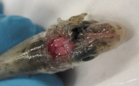
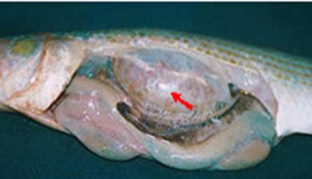
Swim bladder inflamed, hemorrhagic or edematous brain, popeye, blindness and abdominal distension are observed in affected fish.
Some fish undergo a subclinical course exhibiting no clinical signs or lesions.
|
|
|
|
A sea bass showing with chronic VNN showed hemorrhagic or edematous brain thefishsite.com/articles/farmers-sought-for-betanodavirus-forum |
Swim bladder showed inflammation |
Histopathological lesions:
Large vacuolated areas, gliosis, perivascular cuffing, the cell bodies of neurons showed pyknosis and lysis, and inclusion bodies in the nervous tissue in Brain.
Massive vacuolar degeneration and necrosis of the inner and outer nuclear layers of retina
Hyperplasia and necrotic changes in gas glandular epithelia and cellular vacuolation in swim bladder
- Infectious Pancreatic Necrosis (IPN)
Definition:
It is highly contagious viral disease of salmonid fish especially fry and fingerlings characterized clinically by high mortality rate, abnormal swimming behaviors and pancreatic necrosis.
Etiology:
Infectious pancreatic necrosis virus (IPNV) has hexagonal profile without envelopes and a diameter approximately 60 nm in diameter whose genome is bi-segmented double-stranded RNA (dsRNA) belonging to Aquabirnavirus subgroup of the Birnaviridae (Munro & Midtlyng, 2011).
Mode of Transmission:
The virus is efficiently transmitted horizontally through cohabitation with infected fish and vertically by shedding virus via eggs.
Infection survivors act as asymptomatic carriers shed virus into water for years, serving as reservoirs and disseminating it through water via feces, particularly during stress, and the breeders through their reproductive products (Reno, 1999).
|
|
Salmonids are highly susceptible - other fish species like common carp and tilapia are affected and serve as asymptomatic carriers.
|
|
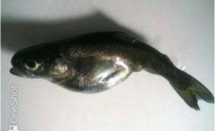
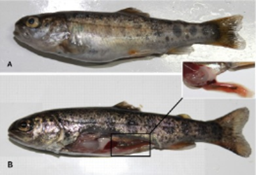
Clinical signs: Abnormal swimming behavior Anorexia, Darkened skin. Exophthalmia, Hemorrhages in the ventral surfaces and Abdominal distension. High mortality reached 100% in fry and young fingerlings.
PM lesions:
Congestion and hemorrhagic areas in the pancreas, intestine and stomach. Abnormal pale gills, spleen and liver and ascetic fluid in the abdominal cavity.
|
|
|
Histopathological lesions:
Necrosis, Vacuolation and Pyknotic nuclei in liver cells.
Necrosis of the pancreatic acinar cells and inflammatory infiltration early stage.
Necrosis in the hematopoietic tissues of the kidney and spleen.
Desquamation and sloughing of intestinal mucosa.
- Viral Encephalitis of Tilapia Larvae
Definition:
It is an acute highly contagious viral disease affecting tilapia larvae characterized clinically by high mortalities, erratic swimming behavior, and exophthalmia and eye cataract.
Etiology:
Viral encephalitis of tilapia larvae, Alpha-herpesvirus, icosahedral hexagonal, double strand (dsDNA) virus (Shlapobersky et al., 2010). It primarily attacks the central nervous system, causing extremely high mortality rates among larvae in hatcheries
Mode of Transmission:
It is transmitted both horizontally through cohabitation with infected fish and vertically from the spawner to offspring by viral particles shed during spawning (Machimbirike et al., 2019).
Susceptible host: Tilapia larvae and fry
Clinical Signs:
Abnormal swimming behavior "whirling syndrome", dark pigmentation, skin erosion, discoloration, and loss of scales, exophthalmia, abdominal distension, gill paleness and loss of appetite. High mortality rates, sometimes exceeding 90%.
PM Lesions:
Paleness of liver and kidney, enlarged spleen and gall bladder. Mild abdominal distention with ascitic fluid, and exophthalmia. Intestinal tract empty or filled with fluid accumulation.
Histopathological lesions
Foci of gliosis and perivascular cuffing of lymphocytes in the brain cortex; severe necrosis and vacuolation in brain tissue ‘encephalon’, and specific inclusion bodies within the affected neuron. Brain hemorrhage also observed
Syncytial cells formation (fused multinucleated cells), inclusion bodies and increased melanomacrophages centers in Liver
Necrosis, lymphoid depletion and increased melanomacrophages centers of spleen.
severe necrosis in renal tissues.
- Spring Viremia of Carp (SVC)
Definition:
It is an acute highly contagious viral disease affecting common carp, koi and other cyprinid fish characterized clinically by high mortalities, abdominal distention, and hemorrhaging on the skin and gills.
Etiology:
Spring viremia of carp, it is caused by spring viraemia of carp virus (SVCV), a rhabdovirus of the type species, Carp sprivivirus, in the genus Sprivivirus (Dixon & Stone, 2017).
Mode of transmission:
The transmission of SVCV is horizontal (Fijan, 1988) through direct contact with infected fish, contaminated water with shedding virus. The virus has been retrieved from ovarian fluid (Békési and Csontos, 1985), however vertical transmission remains unproven.
Susceptible host:
Cyprinid species as common carp and koi carp
Other fish species as Nile tilapia and rainbow trout.
Clinical Signs:
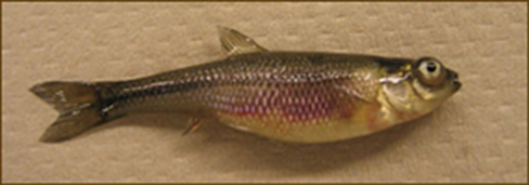
loss of equilibrium, dark coloration, exophthalmia, hemorrhages on the skin, fins and vent, abdominal distension or dropsy and a protruding vent often with trailing mucoid fecal casts.
PM lesions:
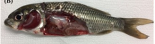
Enlargement of spleen and kidneys, petechial hemorrhages in internal organs and muscles, adhesin of internal organs my occurred, presence of ascetic fluid in the abdominal cavity, degeneration of gills, congestion of intestine and focal hemorrhage of swim bladder. Intestine inflamed and filled with mucous,
|
|
|
Histopathological lesions
Necrosis of the internal organs, enlargement of melanomacrophage centers in spleen, lymphocyte infiltration in the swim bladder. Renal tubules are clogged with casts, and the cells undergo hyaline degeneration and vacuolation.
- Koi Herpesvirus (KHV) disease or Carp Interstitial Nephritis and Gill Necrosis Virus (CNGV)
Definition:
It is an acute highly contagious viral disease affecting common carp and koi characterized clinically by high mortalities, signs of hypoxia, enophthalmia and hemorrhagic spots on the body surface and erratic swimming behavior.
Etiology:
The etiological agent of Koi Herpesvirus (KHV) or Carp Interstitial Nephritis and Gill Necrosis Virus (CNGV), is a third Cyprinid herpesvirus-3 (CyHV-3) a spherical to pleomorphic envelope with linear double-stranded DNA (Gotesman et al., 2013).
Mode of transmission:
Horizontal transmission through cohabitation with infected fish, CyHV-3 may penetrate via the skin, and the gill (Costes et al. 2009). Ingesting CyHV-3-positive material offers an alternative route for CyHV-3 to enter via the pharyngeal periodontal mucosa, where virus proliferates extensively in the gut (Fournier et al. 2012).
Susceptible host:
All varieties and subspecies of common carp and common carp hybrids
Clinical signs:
Skin had patches of pale coloration or reddening, excessive or reduced mucous secretion and sandpaper-like skin texture, enophthalmia (sunken eyes), hemorrhagic spots on the skin and base of the fins, and fin erosion, Signs of hypoxia, loss of equilibrium and disorientation,
PM lesions:
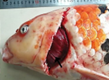
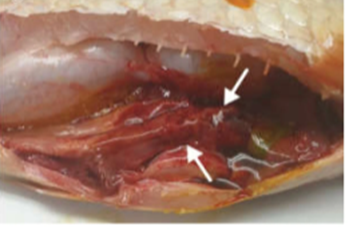
Gills had excess mucus, paleness and mottled with red and grayish-white patches, adhesion of abdominal cavity, enlarged kidney and liver, and petechial hemorrhages on the internal organs.
|
|
|
Histopathological lesions:
Necrosis in primary and secondary lamellae, fusion of secondary lamellae and adhesion of gill filaments, and hyperplasia and hypertrophy of branchial epithelium.
Necrosis of Kidneys hematopoietic tissues, severe interstitial nephritis with heavy infiltration with inflammatory cells and destruction of renal tubules.
Inflammation and necrosis in liver and spleen
Intranuclear inclusions in the cells of gills and kidneys.
Signet ring’ appearance of branchial epithelial cells (chromatin margins to the edges of the nucleus creating a distinct visual ring.)
- Diagnosis of viral diseases
A: Field-level / Presumptive methods:
· Case history and clinical signs
· Postmortem lesions
· Rapid test Kits (Antigen: KHV, SVCV, VNN)
· Water quality parameters.
· New trend ‘New Field-lab’
- Portable qPCR: Run WOAH assay in 1 hr at hatchery.
- Digital PCR: quantification for carrier fish with very low load of virus.
- eDNA qPCR: environmental DNA qPCR used to detect virus from pond water.
B: Laboratory methods / Confirmatory methods
· WOAH - World Organization for Animal Health – requires 2 lab methods for official confirmation of listed virus diseases. the laboratory methods include:
1-Virus Isolation in Cell Culture
2-Molecular Methods:
· Conventional PCR
· Real-time qPCR
· LAMP - Loop-mediated isothermal amplification
· Sequencing
3-Serological Methods:
· Serum Neutralization.
· IFAT - Indirect fluorescent antibody techniques.
4. Histopathology study
· Histopathology
· Immunohistochemistry IHC
5. Fluorescence In-Situ Hybridization FISH
6. Electron Microscopy
- Prevention and control of viral diseases
➡️ Diseases prevention and control using biosecurity measures
- Application of biosecurity measures in the hatchery
i. Periodical screening to the broodstock and eggs
To prevent vertical transmission of infections, parent stocks must be regularly screened using laboratory assays to detect and eliminate infected fish, ensuring that only disease-free fish can be used for egg production in aquaculture. Individual fish from the parent stock should be tagged and examined for viral infections using sperm, ovarian fluid, and other materials for virus isolation and identification through cell culture and/or reverse transcription polymerase chain reaction (RT-PCR) (Watanabe et al., 2000).
ii. Application of standard operating procedures Standard Operating Procedures (SOPs) for Brood Stock Stations
According to Palić et al. (2015) SOPs should integrate biosecurity into daily operations, covering:
• Facility design & flow: Clear separation of restricted and non-restricted areas, with defined movement paths for personnel and stock.
• Disinfection protocols: Procedures for personnel, equipment, and vehicles to prevent pathogen transfer.
• Water management: Treatment of inflow water and safe handling of wastewater.
• Feeding & husbandry: Documented feeding regimes, verified feed sources, and use of immunostimulants or vaccines.
• Routine screening: Protocols for sample collection, diagnostic testing, and scheduled health checks for viral diseases.
• Outbreak response: Isolation of affected stock, disinfection, hygienic disposal of dead fishes, and mandatory reporting of notifiable diseases to national veterinary authorities.
• Record keeping: Standardized forms capturing health status, disinfection logs, egg production, disease screening, diagnostic results, feed use, growth performance,
· Implementation of Biosecurity on Aquaculture Farms
- The biosecurity in the fish farms includes, newly acquired stock should be held in a separate facility for 4–6 weeks, with no sharing of equipment or personnel with resident fish. Vaccination before integration is recommended. Key transmission indices include regulating water temperature to reduce disease occurrence and enhance immune response, maintaining appropriate stocking densities to minimize stress and transmission, and treating inflow water with filtration and ultraviolet (UV) irradiation to prevent viral entry.
- Chemical disinfectants (Timsen (n- Alkyl dimethyl benzyl ammonium chloride. It is broad-spectrum quaternary ammonium compound with powerful bactericide, fungicide, viricide and algicide activity), iodophores and H2O2) are essential for inactivating viral pathogens in aquaculture. Effective selection should consider efficacy, environmental impact, human safety, and effects on cultured species.
-Routine disinfection of equipment, facilities, and personnel is critical, and fish eggs should be treated with iodophor (25 ppm for 20 minutes or 50 ppm for 15 minutes) or sodium hypochlorite (4–6% for 5 minutes) (Russell Danner & Merrill, 2005).
-Eco-friendly antiviral compounds in aquaculture are big focus right now because the chemicals compounds hurt fish immunity, water and environment.
- Water quality must also be monitored, as high ammonia, nitrate, or low pH levels stress fish and increase susceptibility to infection. Adequate aeration is equally important to prevent hypoxia, which can be fatal to fish eggs.
Research by Amend & Pietsch (1972) further demonstrated that iodophors possess strong virucidal activity against major salmonid viruses, including IHNV, IPNV, and VHSV.
- Biological Biosecurity Measures
Biological processes are vital for protecting aquaculture species against viral infections. Key strategies include vaccination of fish, use of certified disease-free eggs or livestock, and adoption of disease-resistant strains. Policies such as “eggs only” combined with certified eggs have successfully prevented viral introductions in salmon farms, highlighting the higher risk of disease transmission through live fish transfers (Mweemba et al., 2024).
National biosecurity programs coordinate farm-level and brood-station disease control with international standards. Guidelines should cover outbreak management, eradication compliance, and restrictions on the movement of eggs, live fish, postlarvae shrimp, frozen shrimp, and other aquatic organisms (Scarfe, 2003). Authorities are responsible for routine surveillance, providing diagnostic services, supervising eradication programs, and regulating imports and exports of aquaculture products.
Although the profile of aquatic viral diseases varies among countries, many pathogens are transboundary, requiring common strategies for control. To coordinate these efforts, international organizations such as the Food and Agriculture Organization (FAO) and the World Organization for Animal Health (WOAH) provide global frameworks. The FAO issues guidelines for disease prevention in food animals, while the WOAH develops science-based standards for all animal health, including aquatic species. Through the Aquatic Animal Health Code and Manual of Diagnostic Tests for Aquatic Animals, the OIE ranks diseases by risk level and provides guidance on diagnosis, surveillance, disinfection, prevention, eradication, and restocking. These frameworks also support national biosecurity programs and ensure compliance with health certification requirements for international trade. In principle, both FAO and WOAH not only address transboundary diseases but also empower countries to establish their own effective biosecurity measures (Subasinghe et al., 2023).
➡️ Breeding resistant strains.
- Selective breeding of diseases resistance
Efforts to breed fish resistant to viral diseases have gained momentum only in recent decades, as many viral pathogens were identified during this period. In Norway, selective breeding has been a key component of salmon programs since 1993, leading to the identification of strains highly susceptible to infectious pancreatic necrosis virus (IPNV). These strains have been valuable not only for developing resistant lines but also for optimizing challenge models in vaccine efficacy trials.
- Genetic Selection of Disease-Resistant Strains
Advances in high-throughput genome mining have enabled the identification of genetic markers linked to disease resistance in aquaculture species. The most widely used approach is quantitative trait loci (QTL) analysis “ it is a statistical method used to identify specific region of the genome (DNA segments) associated with phenotypic variation in quantitative complex traits like height, yield or disease susceptibility” (Buchmann, 2022), which statistically links phenotypic traits such as post-challenge survival to specific genomic regions. By crossing fish with different susceptibility traits and exposing them to viral pathogens, researchers can identify QTLs associated with resistance or susceptibility. Common markers include single nucleotide polymorphisms (SNPs) and microsatellites, both of which have been successfully applied in salmonids to identify resistance to infectious pancreatic necrosis virus (IPNV).
➡️ Vaccination
Vaccination is one of the most effective ways to prevent viral diseases, as immunized fish are less likely to become infected. Successful vaccines depend on identifying pathogen surface antigens that trigger protective immune responses. Delivery systems may use inactivated or live attenuated viruses, or molecular methods that express immunogenic proteins. The choice of system depends on safety, cost, efficacy, and the type of immune response required.
➡️ Immunostimulants
Immunostimulants are widely used in aquaculture eco-friendly strengthen the immune system of fish and reduce the impact of viral infections. They do not directly destroy viruses, but they enhance both innate and adaptive immunity, making fish less susceptible to outbreaks.
Natural compounds such as β-glucans, chitosan, and plant extracts (garlic, turmeric, neem) stimulate macrophages and improve antiviral defense. Probiotics like Lactobacillus and Bacillus enhance gut immunity and help lower viral loads. Vitamins and minerals including vitamin C, vitamin E, selenium, and zinc act as antioxidants, supporting immune cell function. Synthetic agents such as levamisole and CpG oligonucleotides directly modulate immune responses and trigger antiviral signaling pathways.
The benefits of immunostimulants include reduced mortality during viral outbreaks such as Koi Herpesvirus (KHV), Infectious Pancreatic Necrosis (IPN), and Viral Hemorrhagic Septicemia (VHS). They also improve vaccine efficacy when used as adjuvants and provide a sustainable alternative to antibiotics, avoiding resistance and chemical residues.
- Treatment Using Antiviral drugs
Are there antiviral drugs actually approved for fish or aquatic animals?
United States: No antiviral drug is currently approved for food fish.
European Union: No Direct viral inhibitors approved
World Organization for Animal Health: No list of approved antiviral therapeutics
Reasons causing Limitation of using antiviral in aquaculture include:
· Viruses caused rapid onset of disease and kill fish fast. Drugs are given to fish incorporated in diet so they take several days to reach desirable level in tissue.
· Fish are poikilothermic and drug metabolism depends on water temp, so dosing unreliable.
· Experimental trials resulted in accumulation of antivirals in fish flesh.
· Viruses’ mutation and drugs not effective in open pond.
· Limitation of the drug delivery as injection of fish is not practical as well as bath treatments.
- References
1. Admasu, F., & Wakjira, M. (2021). Pathology of epizootic-infectious diseases of fishes in aquaculture. Biomedical Journal of Scientific & Technical Research, 40(2), 31984-31995.
2. Aich, N., Paul, A., Choudhury, T. G., & Saha, H. (2022). Tilapia lake virus (TiLV) disease: Current status of understanding. Aquaculture and fisheries, 7(1), 7-17.
3. Ashouri, G., Hoseinifar, S. H., El-Haroun, E., Imperatore, R., & Paolucci, M. (2023). Tilapia fish for future sustainable aquaculture. In Novel approaches toward sustainable tilapia aquaculture (pp. 1-47). Cham: Springer International Publishing.
4. Bandín, I., & Souto, S. (2020). Betanodavirus and VER Disease: A 30-year Research Review. Pathogens, 9(2), 106. https://doi.org/10.3390/pathogens9020106
5. Békési, L., & Csontos, L. (1985) Isolation of spring viraemia of carp virus from asymptomatic broodstock carp, Cyprinus carpio L. Journal of Fish Diseases, 8, 471–472.
6. Buchmann, K. (2022). Genetic breeding, disease resistance and immunity. In Principles of Fish Immunology: From Cells and Molecules to Host Protection (pp. 657-670). Cham: Springer International Publishing.
7. Can, E., Austin, B., Steinberg, C., Carboni, C., Sağlam, N., Thompson, K., ... & Ergün, S. E. B. A. H. A. T. T. İ. N. (2023). Best practices for fish biosecurity, well-being and sustainable aquaculture. Sustainable Aquatic Research, 2(3).
8. Combe, M., Reverter, M., Caruso, D., Pepey, E., & Gozlan, R. E. (2023). Impact of global warming on the severity of viral diseases: a potentially alarming threat to sustainable aquaculture worldwide. Microorganisms, 11(4), 1049.
9. Costes, B., Raj, V. S., Michel, B., Fournier, G., Thirion, M., Gillet, L., ... & Vanderplasschen, A. (2009). The major portal of entry of koi herpesvirus in Cyprinus carpio is the skin. Journal of virology, 83(7), 2819-2830.
10. Dixon, P., & Stone, D. A. V. I. D. (2017). Spring viraemia of carp. In Fish viruses and bacteria: pathobiology and protection (pp. 79-90). Wallingford UK: CABI.
11. Eyngor, M., Zamostiano, R., Kembou Tsofack, J. E., Berkowitz, A., Bercovier, H., Tinman, S., ... & Eldar, A. (2014). Identification of a novel RNA virus lethal to tilapia. Journal of clinical microbiology, 52(12), 4137-4146.
12. Fijan, N. (1988) Vaccination against spring viraemia of carp. In: Ellis, A.E. (ed.) Fish Vaccination. Academic Press, London, pp. 204–215.
13. Fournier, G., Boutier, M., Stalin Raj, V., Mast, J., Parmentier, E., Vanderwalle, P., ... & Vanderplasschen, A. (2012). Feeding Cyprinus carpio with infectious materials mediates cyprinid herpesvirus 3 entry through infection of pharyngeal periodontal mucosa. Veterinary Research, 43(1), 6.
14. Georgiadis, M. P., Gardner, I. A., & Hedrick, R. P. (2001). The role of epidemiology in the prevention, diagnosis, and control of infectious diseases of fish. Preventive Veterinary Medicine, 48(4), 287-302.
15. Gotesman, M., Kattlun, J., Bergmann, S. M., & El-Matbouli, M. (2013). CyHV-3: the third cyprinid herpesvirus. Diseases of aquatic organisms, 105(2), 163-174.
16. Grubaugh, N. D., Ladner, J. T., Lemey, P., Pybus, O. G., Rambaut, A., Holmes, E. C., & Andersen, K. G. (2019). Tracking virus outbreaks in the twenty-first century. Nature microbiology, 4(1), 10-19.
17. Jennings, S., Stentiford, G. D., Leocadio, A. M., Jeffery, K. R., Metcalfe, J. D., Katsiadaki, I., ... & Verner‐Jeffreys, D. W. (2016). Aquatic food security: insights into challenges and solutions from an analysis of interactions between fisheries, aquaculture, food safety, human health, fish and human welfare, economy and environment. Fish and Fisheries, 17(4), 893-938.
18. John, K. R., Sivasankar, P., & George, M. R. (2023). Status of emerging and reemerging fish viral infections in India. In Viral Outbreaks-Global Impact and Newer Horizons. IntechOpen.
19. MacAulay, S., Ellison, A. R., Kille, P., & Cable, J. (2022). Moving towards improved surveillance and earlier diagnosis of aquatic pathogens: From traditional methods to emerging technologies. Reviews in Aquaculture, 14(4), 1813-1829.
20. Machimbirike, V. I., Jansen, M. D., Senapin, S., Khunrae, P., Rattanarojpong, T., & Dong, H. T. (2019). Viral infections in tilapines: More than just tilapia lake virus. Aquaculture, 503, 508-518.
21. Mori K.-I., Nakai T., Muroga K., Arimoto M., Mushiake K., Furusawa I. Properties of a New Virus Belonging to Nodaviridae Found in Larval Striped Jack (Pseudocaranx dentex) With Nervous Necrosis. Virology . 1992;187(1):368–371. doi: 10.1016/0042-6822(92)90329-N.
22. Munday B.L., Kwang J. & Moody N. (2002) Review article Betanodavirus infections of teleost fish: a review. Journal of Fish Diseases 25, 127–142.
23. Munro, E. S., & Midtlyng, P. J. (2011). Infectious pancreatic necrosis and associated aquatic birnaviruses. In Fish diseases and disorders. Volume 3: viral, bacterial and fungal infections (pp. 1-65). Wallingford UK: CABI.
24. Mweemba, H., Mutoloki, S., & Evensen, Ø. (2024). Prevention and control of viral diseases in aquaculture. In Aquaculture Virology (pp. 91-108). Academic Press.
25. Noga, E. J. (2010). Fish disease: diagnosis and treatment. John Wiley & Sons.
26. Sano, M., Nakai, T., & Fijan, N. (2006). 5 Viral Diseases and Agents. Fish Diseases and Disorders, 3, 166.
27. Palić, D., Scarfe, A. D., & Walster, C. I. (2015). A standardized approach for meeting national and international aquaculture biosecurity requirements for preventing, controlling, and eradicating infectious diseases. Journal of Applied Aquaculture, 27(3), 185-219.
28. Reno, P.W. Infectious pancreatic necrosis virus and associated aquatic birnavirus. In Fish Diseases and Disorders, Vol. 3: Viral, Bacterial and Fungal Infections; Woo, P.T.K., Bruno, D.W., Eds.; CAB International: New York, NY, USA, 1999; pp. 1–55.
29. Russell Danner, G., & Merrill, P. (2005). Disinfectants, disinfection, and biosecurity in aquaculture. Aquaculture biosecurity: prevention, control, and eradication of aquatic animal disease, 91-128.
30. Scarfe, A. D. (2003). State, regional, national, and international aquatic animal health policies: focus for future aquaculture biosecurity. Biosecurity in aquaculture production systems: exclusion of pathogens and other undesirables. The World Aquaculture Society, Baton Rouge, Louisiana, 233-262.
31. Shafi, M. E. (2025). A comprehensive review of summer mortality syndrome in fish: Causes, climate impact, and mitigation strategies. Journal of Advanced Veterinary and Animal Research, 12(3), 890.
32. Shlapobersky, M., Sinyakov, M. S., Katzenellenbogen, M., Sarid, R., Don, J., & Avtalion, R. R. (2010). Viral encephalitis of tilapia larvae: primary characterization of a novel herpes-like virus. Virology, 399(2), 239-247.
33. Subasinghe, R., Alday‐Sanz, V., Bondad‐Reantaso, M. G., Jie, H., Shinn, A. P., & Sorgeloos, P. (2023). Biosecurity: Reducing the burden of disease. Journal of the World Aquaculture Society, 54(2), 397-426.
34. Uma, A. (2025). Pathogen and Disease Transmission in Aquatic Animals. In Management of Fish Diseases (pp. 19-55). Singapore: Springer Nature Singapore.
35. Watanabe, K. I., Nishizawa, T., & Yoshimizu, M. (2000). Selection of brood stock candidates of barfin flounder using an ELISA system with recombinant protein of barfin flounder nervous necrosis virus. Diseases of aquatic organisms, 41(3), 219-223.
36. Wood, E. M., Snieszko, S. F., & Yasutake, W. T. (1955). Infectious pancreatic necrosis in brook trout. Archives of Pathology, 60, 26-