the Management of Adrenal Lesions
| Site: | EHC | Egyptian Health Council |
| Course: | General surgery Guidelines |
| Book: | the Management of Adrenal Lesions |
| Printed by: | Guest user |
| Date: | Sunday, 20 September 2026, 5:52 PM |
Description
"last update: 3 August 2026" Download Guideline
- Executive summary
This guideline provides healthcare professionals with evidence based recommendations on the management of adrenal lesions.
· We recommend that patients with adrenal incidentalomas are discussed in a multidisciplinary expert team meeting, (a radiologist, an endocrinologist, and a surgeon), if at least one of the following criteria is met: 1) Imaging is not consistent with a benign lesion, (2) there is evidence of hormone excess (including mild autonomous cortisol secretion [MACS] in patients with clinically relevant comorbidities potentially attributable to cortisol), (3) adrenal surgery is considered and/or (4) evidence of significant tumor growth during follow-up imaging, (Good practice statement).
· We recommend using CT as the first imaging modality if not yet performed to establish with the highest possible certainty if an adrenal mass is benign or malignant at the time of initial detection, (Strong recommendation).
· We advise that if the CT is consistent with a benign adrenal mass, no further imaging is required, (Conditioned recommendation).
· We advise performing an immediate additional imaging, if CT is equivocal with a tumor size < 4 cm, and the results of the hormonal work-up do not indicate significant hormone excess. Alternatively, interval imaging in 12 months by CT (or MRI) could be performed, (Conditional recommendation).
· If the adrenal mass is ≥ 4 cm and CT suggests that there is a relevant risk that this lesion is malignant, we advise discussing such cases in a multidisciplinary team meeting prior to surgery, (Good Practice Statement).
· We suggest completely staging the patient (including at least thoracic CT and/or FDG-PET/CT, (Conditional recommendation).
· We recommend against the use of an adrenal biopsy in the diagnostic work-up of patients with adrenal masses unless there is a history of extra-adrenal malignancy, (Strong recommendation).
· We advise measurement of sex steroids and precursors of steroidogenesis (ideally using multi-steroid profiling by tandem mass spectrometry) in patients in whom by imaging or clinical features an adrenocortical carcinoma is suspected, (Conditional recommendation).
· We recommend that patients with adrenal incidentalomas undergo a 1-mg overnight dexamethasone suppression test to exclude autonomous cortisol secretion, (Strong recommendation).
· We recommend using post dexamethasone serum cortisol levels ≤50 nmol/L (≤1.8 µg/dL) as a diagnostic criterion for the exclusion of autonomous cortisol secretion, (Strong recommendation).
· We recommend that in patients without signs and symptoms of overt Cushing’s syndrome a post-dexamethasone serum cortisol concentration above 50 nmol/L (> 1.8 µg/dL) should be considered as MACS. In these patients, we recommend that ACTH-independency should be confirmed, (Strong recommendation).
· We recommend against considering patients with MACS (per definition without specific clinical signs of Cushing’s syndrome) as being at high risk for development of overt Cushing’s syndrome, (Strong recommendation).
· We recommend screening patients with adrenal incidentaloma and MACS for hypertension and type 2 diabetes mellitus, (Strong recommendation).
· We recommend discussing the option of surgery with the patient who has MACS in addition to relevant comorbidities and a unilateral adrenal mass. The proposal to perform surgery should be established within an expert multi-disciplinary group, (Strong recommendation).
· We advise excluding pheochromocytoma by measurement of plasma free metanephrines or urinary fractionated metanephrines in all patients with adrenal lesions, (Conditional recommendation).
· We advise to measure VMA in urine to diagnose pheochromocytoma as an alternative to metanephrine, (Good practice statement).
· In patients with concomitant hypertension or un-explained hypokalemia, we recommend use of the aldosterone/renin ratio to evaluate primary aldosteronism, (Strong recommendation).
· We recommend adrenalectomy as the standard of care for unilateral adrenal tumors with clinically significant hormone excess. In patients with MACS, surgery can be considered in patients with relevant co-morbidities, (Strong recommendation).
· We recommend against performing surgery in patients with an asymptomatic, non-functioning unilateral adrenal mass and obvious benign features on imaging studies, (Strong recommendation).
· If surgery is indicated for a benign adrenal mass causing hormone excess (including MACS), we advise that a minimally invasive approach is used, (Conditional recommendation).
· We advise performing minimally invasive adrenalectomy by an expert high-volume adrenal surgeon in patients with unilateral adrenal masses with radiological findings suspicious of malignancy and a diameter ≤6 cm, but without evidence of local invasion, (Conditional recommendation).
· We advise open adrenalectomy for unilateral adrenal masses with radiological findings suspicious of malignancy and signs of local invasion, (Conditional recommendation).
· We recommend perioperative glucocorticoid treatment at surgical stress doses in all patients undergoing surgery and a preoperative morning serum cortisol >50 nmol/L (1.8 µ/dL) after a 1 mg overnight dexamethasone test, (Strong recommendation).
· We advise that patients with MACS (similarly to patients with adrenal Cushing's syndrome) that underwent surgery be followed by an endocrinologist until recovery of hypothalamic-pituitary-adrenal axis function has been documented, (Conditional recommendation).
· We advise against further imaging during follow-up in patients with an adrenal lesion with clear benign features on imaging studies, (Conditional recommendation).
· In patients with an indeterminate adrenal mass (by imaging), opting not to undergo adrenalectomy following initial assessment, we advise repeating CT or MRI after 6-12 months to exclude significant growth. We advise surgical resection if the lesion enlarges by more than 20% in maximum diameter, (Conditional recommendation).
· We recommend against repeated hormonal work-up in patients with hormonal work-up results within the reference range at initial evaluation unless new clinical signs of endocrine activity appear, (Strong recommendation).
· In patients with MACS, who do not undergo an adrenalectomy, we recommend only annual reassessment of comorbidities potentially attributable to cortisol, (Strong recommendation).
- Recommendations
Section 1: General remarks:
· We recommend that patients with adrenal incidentalomas are discussed in a multidisciplinary expert team meeting, (a radiologist, an endocrinologist, and a surgeon), if at least one of the following criteria is met: 1) Imaging is not consistent with a benign lesion, (2) there is evidence of hormone excess (including mild autonomous cortisol secretion [MACS] in patients with clinically relevant comorbidities potentially attributable to cortisol), (3) adrenal surgery is considered and/or (4) evidence of significant tumor growth during follow-up imaging, (Good practice statement).
Section 2: Assessment of the risk of malignancy:
· We recommend using CT as the first imaging modality if not yet performed to establish with the highest possible certainty if an adrenal mass is benign or malignant at the time of initial detection, (Strong recommendation, moderate certainty evidence, (1)).
· We advise that if the CT is consistent with a benign adrenal mass, no further imaging is required, (Conditioned recommendation, moderate certainty evidence, (1)).
· We advise performing an immediate additional imaging, if CT is equivocal with a tumor size < 4 cm, and the results of the hormonal work-up do not indicate significant hormone excess. Alternatively, interval imaging in 12 months by CT (or MRI) could be performed, (Conditional recommendation, very low certainty evidence, (1)).
· If the adrenal mass is ≥ 4 cm and CT suggests that there is a relevant risk that this lesion is malignant, we advise discussing such cases in a multidisciplinary team meeting prior to surgery, (Good Practice Statement).
· We suggest completely staging the patient (including at least thoracic CT and/or FDG-PET/CT, (Conditional recommendation, very low certainty evidence, (1)).
· We recommend against the use of an adrenal biopsy in the diagnostic work-up of patients with adrenal masses unless there is a history of extra-adrenal malignancy, (Strong recommendation, moderate certainty evidence, (1)).
· We advise measurement of sex steroids and precursors of steroidogenesis (ideally using multi-steroid profiling by tandem mass spectrometry) in patients in whom by imaging or clinical features an adrenocortical carcinoma is suspected, (Conditional recommendation, low certainty evidence, (1)).
Section 3: Assessment for hormone excess:
• We recommend that patients with adrenal incidentalomas undergo a 1-mg overnight dexamethasone suppression test to exclude autonomous cortisol secretion, (Strong recommendation, moderate certainty evidence, (1)).
• We recommend using post dexamethasone serum cortisol levels ≤50 nmol/L (≤1.8 µg/dL) as a diagnostic criterion for the exclusion of autonomous cortisol secretion, (Strong recommendation, low certainty evidence, (1)).
• We recommend that in patients without signs and symptoms of overt Cushing’s syndrome a post-dexamethasone serum cortisol concentration above 50 nmol/L (> 1.8 µg/dL) should be considered as MACS. In these patients, we recommend that ACTH-independency should be confirmed, (Strong recommendation, low certainty evidence, (1)).
• We recommend against considering patients with MACS (per definition without specific clinical signs of Cushing’s syndrome) as being at high risk for development of overt Cushing’s syndrome, (Strong recommendation, moderate certainty evidence, (1)).
• We recommend screening patients with adrenal incidentaloma and MACS for hypertension and type 2 diabetes mellitus, (Strong recommendation, low certainty evidence, (1)).
• We recommend discussing the option of surgery with the patient who has MACS in addition to relevant comorbidities and a unilateral adrenal mass. The proposal to perform surgery should be established within an expert multi-disciplinary group, (Strong recommendation, very low certainty evidence, (1)).
• We advise excluding pheochromocytoma by measurement of plasma free metanephrines or urinary fractionated metanephrines in all patients with adrenal lesions, (Conditional recommendation, moderate certainty evidence, (1)).
• We advise to measure VMA in urine to diagnose pheochromocytoma as an alternative to metanephrine, (Good practice statement).
• In patients with concomitant hypertension or un-explained hypokalemia, we recommend use of the aldosterone/renin ratio to evaluate primary aldosteronism, (Strong recommendation, moderate certainty evidence, (1)).
Section 4: Surgical treatment:
• We recommend adrenalectomy as the standard of care for unilateral adrenal tumors with clinically significant hormone excess. In patients with MACS, surgery can be considered in patients with relevant co-morbidities, (Strong recommendation, moderate certainty evidence, (1)).
• We recommend against performing surgery in patients with an asymptomatic, non-functioning unilateral adrenal mass and obvious benign features on imaging studies, (Strong recommendation, low certainty evidence, (1)). If surgery is indicated for a benign adrenal mass causing hormone excess (including MACS), we advise that a minimally invasive approach is used, (Conditional recommendation, very low certainty evidence, (1)).
• We advise performing minimally invasive adrenalectomy by an expert high-volume adrenal surgeon in patients with unilateral adrenal masses with radiological findings suspicious of malignancy and a diameter ≤6 cm, but without evidence of local invasion, (Conditional recommendation, very low certainty evidence, (1)).
• We advise open adrenalectomy for unilateral adrenal masses with radiological findings suspicious of malignancy and signs of local invasion, (Conditional recommendation, very low certainty evidence, (1)).
• We recommend perioperative glucocorticoid treatment at surgical stress doses in all patients undergoing surgery and a preoperative morning serum cortisol >50 nmol/L (1.8 µ/dL) after a 1 mg overnight dexamethasone test, (Strong recommendation, moderate certainty evidence, (1)).
• We advise that patients with MACS (similarly to patients with adrenal Cushing's syndrome) that underwent surgery be followed by an endocrinologist until recovery of hypothalamic-pituitary-adrenal axis function has been documented, (Conditional recommendation, low certainty evidence, (1)).
Section 5: Follow-up of patients not undergoing adrenal surgery after initial assessment:
• We advise against further imaging during follow-up in patients with an adrenal lesion with clear benign features on imaging studies, (Conditional recommendation, moderate certainty evidence, (1)).
• In patients with an indeterminate adrenal mass (by imaging), opting not to undergo adrenalectomy following initial assessment, we advise repeating CT or MRI after 6-12 months to exclude significant growth. We advise surgical resection if the lesion enlarges by more than 20% in maximum diameter, (Conditional recommendation, very low certainty evidence, (1)).
• We recommend against repeated hormonal work-up in patients with hormonal work-up results within the reference range at initial evaluation unless new clinical signs of endocrine activity appear, (Strong recommendation, low certainty evidence, (1)).
• In patients with MACS, who do not undergo an adrenalectomy, we recommend only annual reassessment of comorbidities potentially attributable to cortisol, (Strong recommendation, low certainty evidence, (1)).
- Acknowledgement
We would like to acknowledge the Guideline Development Group, (GDG) committee for developing this guideline.
Chair of GDG: Mostafa Abdel-Hamed Soliman, Professor of Surgery, Cairo University.
Members of GDG (Alphabetically):
Abdel-Wahab Mohamed Ezzat, Professor of Surgery, Ain -Shams University.
Ahmed Abdel-Raouf Elgeidie, Professor of Digestive Surgery, Mansoura University.
Ahmed Rifat Mosaeed Mohamed Elsherif, Assistant Professor of Surgery, Cairo University.
Alaa Abdallah, Professor of Surgery, Ain -Shams University.
Atef Abdel-Ghani Salem, Professor of Surgery, Benha University.
Hesham Abdel-Raouf El-Akkad, Professor of Surgery, Ain-Shams University.
Ibrahim El-Zayat, Head of Surgery Department, Aswan University.
Khaled Abdallah El-Fiky, Professor of Surgery, Ain-Shams University.
Khaled Amer, Professor of Surgery, Military Medical Academy.
Khaled Safwat, Professor of Surgical Oncology and Endoscopy, Zagazig University.
Mohamed Ibrahim Abdel-Hamed Al-Said, Professor of Surgery, Zagazig University.
Mohamed Saber Abd Elkhaliq Mostafa, Assistant Professor of Surgery, Cairo University.
Tarek Ibrahim, Professor of Surgery, National Liver Institute, Menofia University.
- Abbreviations
ADPKD |
Autosomal Dominant Polycystic Kidney Disease |
AGREEII |
Appraisal of Guidelines for Research and Evaluation II |
CT |
Computed Tomography |
FDG/Pet/CT |
Flurodeoxyglucose Positron Emission Tomography |
GDG |
Guidelines Development Group |
GRADE |
Grading of Recommendations, Assessment, Development and Evaluation |
MACS |
Mild Autonomous Cortisol Secretion |
MRI |
Magnetic Resonance Imaging |
PCLD |
Polycystic Liver Disease |
VMA |
Valynile Mandelic Acid |
- Glossary
Adrenal incidentaloma:
An adrenal incidentaloma is an adrenal mass detected on imaging not performed for a suspected adrenal disease. By this strict definition, the imaging study is not done for signs and symptoms potentially indicative of adrenal hormone excess (eg, pheochromocytoma, Cushing’s, or Conn’s syndrome) or an otherwise suspected adrenal disease, but rather for the evaluation of symptoms that are not suggestive of an adrenal problem, such as investigations for abdominal or back pain, (1).
Functioning tumor:
It is an adrenal tumor that secretes hormones in an autonomous/unregulated manner, (1).
- Introduction
Adrenal incidentalomas are adrenal masses
detected on imaging performed for reasons other than suspected adrenal disease.
In most cases, adrenal incidentalomas are nonfunctioning adrenocortical
adenomas but may also require therapeutic intervention including that for
adrenocortical carcinoma, pheochromocytoma, hormone-producing adenoma, or
metastases, (1).
In recent years,
several studies have investigated steroid
profiling as a tool to discriminate benign
from malignant adrenal tumors, (2).
For the differentiation
of malignant from benign adrenal tumors there are still 3 main imaging techniques in mainstream
clinical use: CT, MRI, and Pet/CT, (3 &4). The conservative treatments for the group
of patients ranged from pharmacotherapeutic interventions for comorbidities to watchful waiting only were studied. The quality of evidence
from these cohort studies is low to
very low, mainly due to confounding and
the lack of a standardized protocol, (5).
Etiology of adrenal tumors
presented as adrenal incidentaloma, is summarized in table 1.
Minimally invasive
surgery was comparable to open surgery in most of the studies, (6).
- Scope and purpose
The scope of this guideline is to set recommendations for the diagnosis and treatment of adrenal lesions. The main purpose of these guidelines is to minimize malpractice and poor surgical decision, to improve the quality of medical care and surgical service, to provide the good surgical practice to our patients, and finally to be cost effective.
- Target audience
The principle targeted candidates are the practicing surgeons, however endocrinologist, urologists and radiologists and all specialists involved in the treatment of adrenal lesions are also included.
- Methods
A comprehensive search for guidelines was
undertaken to identify the relevant guidelines to consider for adaptation.
Inclusion/ exclusion criteria followed in
the search and retrieval of guidelines to be adapted:
Selecting only evidence-based guidelines
(guideline must include a report on systematic literature searches and explicit
links between individual recommendations and their supporting evidence)
Selecting
only national and/or international guidelines
Specific
range of dates for publication (using Guidelines published or updated within the last five years)
Selecting
peer reviewed publications only.
Selecting
guidelines written in English language.
Excluding guidelines written by a single
author, not on behalf of an organization to be valid and comprehensive, a
guideline ideally requires multidisciplinary input
Excluding guidelines published without
references as the panel needs to know whether a thorough literature review was
conducted and whether current evidence was used in the preparation of the
recommendations
The following characteristics of the retrieved
guidelines were summarized in:
Developing organization/authors
Date of publication, posting, and release
Country/language of publication
Date of posting
and/or release
Dates of the search used by the source guideline developers
All retrieved Guidelines were screened and
appraised using AGREE II instrument (www.agreetrust.org) by at least three members. The panel
decided on a cut-off point (any guideline scoring above 50% on the rigor dimension was retained). The GDG decided to adapt
the European Society of Endocrinology clinical practice guidelines on the
management of adrenal incidentalomas, in collaboration with the European
Network for the Study of Adrenal Tumors, 2023,(1).
Evidence assessment
According to WHO Handbook for Guidelines,
we used the GRADE (Grading of Recommendations, Assessment, Development and
Evaluation) approach to assess the quality of a body of evidence, develop
and report recommendations. GRADE methods are used by WHO because these represent
internationally agreed standards for
making transparent recommendations. Detailed GRADE information is available on
the following sites:
•
GRADE
working group: https://www.gradeworkinggroup.org/
•
GRADE
online training modules: http://cebgrade.mcmaster.ca/
Specifically, the quality of evidence was
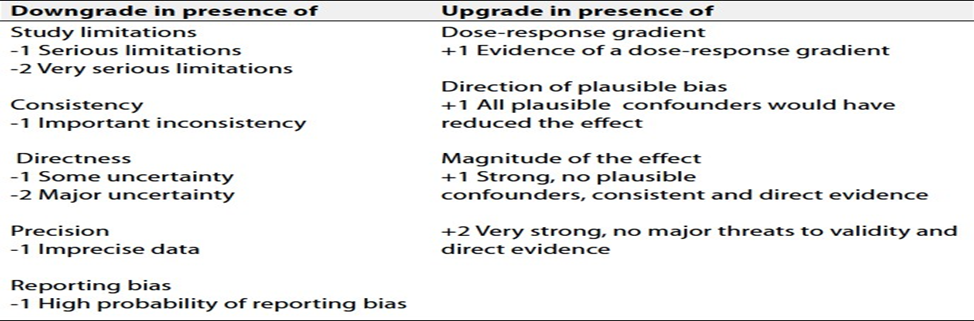
graded as ‘High’, ‘Moderate’, ‘Low’ or ‘Very low’, (table 2& 3).
The strength of the
recommendation
The strength of a recommendation
communicates the importance of adherence to the recommendation.
Strong recommendations
With strong recommendations, the guideline
communicates the message that the desirable effects of adherence to the
recommendation outweigh the undesirable effects. This means that in most
situations the recommendation can be adopted as policy.
Conditional recommendations
These are made when there is greater
uncertainty about the four factors above or if local adaptation has to account
for a greater variety in values and preferences, or when resource use makes the intervention suitable for some, but
not for other locations. This means that there is a need for substantial debate
and involvement of stakeholders before this recommendation can be adopted as
policy.
When not to make recommendations
When there is lack of evidence on the
effectiveness of an intervention, it may be appropriate not to make a recommendation.
- Research needs
1. The fracture risk in
incidentalomas associated with MACS.
2. Minimally invasive and/or
robotic vs open surgery in patients with potentially malignant adrenal mass.
3. Quality of life, mental health,
cognition, and frailty in patients with adrenal incidentalomas, both
non-functioning and the ones with MACS.
4. New biomarkers to identify
patients with clinically relevant cortisol excess.
- Clinical indicators for monitoring
1. Adrenal imaging, (CT, MRI or Pet/CT).
2.
Dexamethasone suppression test.
3. Plasma free
metanephrines, urinary fractionated metanephrines, or VMA in urine. Aldosterone/renin
ratio.
4. Documentation of management of adrenal
lesion.
- Updating of the guideline
The GDG committee for guidelines development is responsible for the continuous evaluation of evidence available about adrenal lesions. The present guidelines will be updated in case of significant changes based on new evidence.
- Annexes
Table 1: Etiology of adrenal tumors presented as adrenal incidentaloma.
|
Etiology |
Prevalence of the different entities among adrenal incidentalomas |
|
Adrenocortical adenoma or macronodular bilateral adrenal hyperplasia |
80%-85% |
|
Nonfunctioning |
40%-70% |
|
Mild autonomous cortisol secretion |
20%-50% |
|
Primary aldosteronism |
2%-5% |
|
Overt Cushing's syndrome |
1%-4% |
|
Other benign mass: Myelolipoma |
3%-6% |
|
Cyst and pseudocyst |
1% |
|
Ganglioneuroma |
1% |
|
Schwannoma |
<1% |
|
Hemorrhage |
<1% |
|
Pheochromocytoma |
1%-5% |
|
Adrenocortical carcinoma |
0.4%-4% |
|
Other malignant mass (mostly adrenal metastases): |
3%-7% |
Table 2: Quality and Significance of the Four Levels of Evidence in GRADE
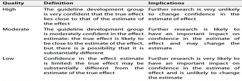
Table 3: Factors that Determine How to Upgrade or Downgrade the Quality of Evidence