BURN INJURIES
| Site: | EHC | Egyptian Health Council |
| Course: | Emergency Medicine Guidelines |
| Book: | BURN INJURIES |
| Printed by: | Guest user |
| Date: | Sunday, 20 September 2026, 9:43 PM |
Description
"last update: 10 August 2026" Download Guideline
- Executive Summary
This guideline provides standardized, evidence-based recommendations for the recognition and management of Burn.
1-) Burn injury First aid
Recommendation 1: (First Aid Education)
We recommend educating both the public and first responders is essential for enhancing awareness and ensuring optimal first responder performance.
· Strength of recommendation: Strong
Recommendation 2: (First Aid Safety)
We recommend that first responder must ensure the removal of the individual from all potential hazards, including heat or flame, live electrical sources, and chemicals. It is essential that the responder prioritises the safety of themselves, the patient, and any bystanders.
· Strength of recommendation: Strong
Recommendation 3: (Cooling Burns)
We recommend in the event of heat or flame injuries, to cool the burn wound with clean, running water at a temperature that is comfortable for the patient for 15–20 minutes, ideally within three hours of the injury. Following the cooling process, the individual should be kept warm, and primary medical attention should be sought promptly.
· Strength of recommendation: Strong
Recommendation 4: (Chemical Injuries)
We recommend in case of chemical injuries, the first responder should promptly remove and safely dispose of all contaminated clothing and materials, followed by thorough irrigation with water for up to 45 minutes. Additionally, it is important to identify the chemical agent involved and adhere to established protocols specific to that substance, if applicable.
· Strength of recommendation: Strong
Recommendation 5: (Electrical Injuries)
We recommend when providing first aid for electrical injuries, it is essential to prioritise safety by deactivating the electrical source or isolating the victim using a nonconductive material. Initiate cardiopulmonary resuscitation (CPR) if required, and subsequently manage burns by applying appropriate cooling measures.
· Strength of recommendation: Strong
2-) Resuscitation in Burn Patient
a-) Fluid resuscitation in burn patient:
Recommendation 6: Indication to start Iv fluids:
We recommend Initiating intravenous fluid resuscitation with salt‑containing solutions in:
-Adults with burns involving ≥20% of total body surface area (TBSA), or earlier if there are clinical signs of shock or significant comorbidities.
-Children with burns involving ≥10% TBSA.
· Strength of recommendation: Strong
Recommendation 7: Timing
We recommend initiating intravenous fluid resuscitation as early as possible, ideally within 2 hours of injury, for all patients who meet the TBSA thresholds (≥20% in adults, ≥10% in children). Do not delay resuscitation until arrival at a burn center—start at the first point of care once criteria are met.
· Strength of recommendation: Strong
Recommendation 8: Initial Fluid Type:
We recommend using a balanced crystalloid solution, such as Lactated Ringer’s (LR) or acetated Ringer’s, as the primary fluid for initial burn resuscitation. Avoid using normal saline as the sole resuscitation fluid due to the risk of hyperchloremic metabolic acidosis.
· Strength of recommendation: Strong
Recommendation 9: Initial 24-hour Volume:
We recommend using a validated formula (such as the Parkland formula) to estimate the initial 24-hour fluid requirement but emphasize that this is only a starting point. The calculated volume should be adjusted frequently based on urine output and clinical endpoints, not given as a fixed amount.
· Strength of recommendation: Strong
Recommendation 10: Titration Targets:
We recommend adjusting fluid administration based on physiological endpoints, primarily urine output, rather than relying solely on calculated formula volumes. Continuous reassessment is essential to avoid both under- and over-resuscitation.
· Strength of recommendation: Strong
b-) Inhalation burn:
Recommendation11: Clinical Indicators for Airway Burns
We recommend using clinical indicators, such as injury circumstances (closed-space exposure, inhalation of hot vapour/liquid) and physical findings (soot in mouth, singed nasal hairs, facial burns), rather than using invasive diagnostic tools (e.g., laryngoscopy, bronchoscopy), as findings that suggest airway burns.
· Strength of recommendation: Strong
Recommendation 12: Radiographic Monitoring
We recommend perform serial plain chest radiography in the acute period for the early diagnosis of respiratory disorders.
· Strength of recommendation: Strong
Recommendation 13: Early Preventive Intubation
We suggest performing early preventive intubation if inhalation injury is suspected and airway oedema is likely.
· Strength of recommendation: Conditional
c-) Pain management and sedation in burn patients.
Recommendation14: Pain Assessment
We recommend that the adequacy of pain control be routinely assessed using standardized scoring systems. Whenever possible, self-report measures should be utilized as the preferred method of pain assessment. In situations where patients are unable to self-report such as young children or individuals with impaired mental status validated behavioural pain assessment scales should be employed to ensure accurate and effective pain management.
Inhalation injury can cause rapid and progressive airway oedema, making delayed intubation difficult or impossible once swelling develops. Early (preventive) endotracheal intubation ensures a secure airway before obstruction occurs, reducing the risk of respiratory failure and mortality.
Although not all patients with suspected inhalation injury will require intubation, the potential harm from a lost airway outweighs the risks of early intervention when airway oedema is likely. Therefore, early intubation is conditionally recommended in such cases.
· Strength of recommendation: Strong
Recommendation 15: Multimodal analgesia
We recommend that analgesia should be used as an
essential component in the management of burn patients. A multimodal approach
is preferred, combining agents with different mechanisms of action; however,
single-agent therapy may be appropriate in selected cases.
Recommended options include opioids and non-opioid agents such as acetaminophen
(paracetamol), nonsteroidal anti-inflammatory drugs (NSAIDs), ketamine,
dexmedetomidine, gabapentin, and pregabalin.
· Strength of recommendation: Strong
Recommendation 16: Assessment of agitation before sedation
We recommend timely identification and treatment of any underlying causes of agitation including pain, delirium, hypoxemia, hypoglycaemia, and hypotension before sedative administration.
· Strength of recommendation: Strong
d-) Sedation During Tracheal Intubation in Burn Patients
Recommendation 17: Multimodal Sedation Strategy Egyptian National Burn Guidelines
We recommend that sedation during tracheal intubation in burn patients be guided by a multimodal strategy that prioritizes patient safety, minimizes adverse effects, and supports optimal outcomes. This approach should be tailored to the clinical condition, burn severity, and ICU status of each patient.
· Strength of recommendation: Strong
e-) Recommendation regarding antibiotic administration / tetanus vaccination:
Recommendation 18: Systemic Antibiotic Administration
We advise against use uniform systemic administration of antibiotics except in immunocompromised patients with contaminated wounds.
Routine preventive systemic antibiotic administration is not currently recommended due to insufficient evidence supporting its effectiveness. However, in patients with contaminated wounds, immunocompromised individuals (such as those with diabetes), children, and perioperative patients, targeted antibiotic prophylaxis may be considered. In such cases, the selection of antibiotics should be guided by local microbial patterns, facility-specific characteristics, and wound culture results.
· Strength of recommendation: Conditional
Recommendation 19: Tetanus Prophylaxis
We recommend the administration of tetanus toxoid (TT) or human tetanus immunoglobulin (TIG) for contaminated burns
It is recommended to manage contaminated burns in the same manner as other wounds. Although no clear national guideline exists in Egypt regarding anti-tetanus treatment for burn patients, the committee reached a strong consensus on its necessity, given the potentially fatal nature of tetanus and existing recommendations supporting prophylaxis for contaminated burns. Clinically, it is often difficult to clearly differentiate between “tetanus-prone” and “non-tetanus-prone” wounds, as tetanus can develop even from minor injuries such as small scratches, burns involving less than 1% TBSA, or sometimes without any apparent wound. Therefore, administration of tetanus toxoid (Tt) or tetanus immune globulin (TIG) is recommended for patients with incomplete or uncertain primary immunization, and for those with contaminated burns occurring more than 5–10 years after their last vaccination, depending on the level of wound contamination.
· Strength of recommendation: Strong
3- Burn Wound care management
Recommendation
20: - Blister Management
We suggest intact blisters should generally be left undisturbed, while large, tense, or contaminated blisters may require aspiration or deroofing
Strength of recommendation: conditional
Recommendation 22: -Use of Antimicrobial Ointments for Superficial Burns
We consider the use of topical antimicrobial ointments (e.g., bacitracin, polymyxin B, mupirocin) for small, superficial burns, particularly on sensitive areas such as the face.
Strength of recommendation: Conditional
Recommendation 23: - Dressing Technique Selection
We consider starting with conventional dressings (e.g., gauze with antimicrobial ointment) for heavily exudative or contaminated wounds, then transitioning to advanced or biologic occlusive dressings based on wound characteristics and healing progression. Closed dressing techniques are preferred over open techniques for most wounds.
Strength of recommendation: good practise statement
Recommendation 24: Management of raw burnt areas for deep partial-thickness and full-thickness burns.
We recommend immediate or early burn wound excision (within 24–72 hours) over conservative care for deep partial-thickness and full-thickness burns.
Strength of recommendation: strong
Recommendation 25: -Duration and Frequency of Dressing Changes
We recommend minimizing the frequency of dressing changes and maintaining long-lasting occlusive dressings whenever possible.
Strength of recommendation: Strong
4- Referral Guidelines for Burn Injuries
Recommendation 26: -Referral Based on Burn Size
We advise referring patients to a designated burn center if they have:
Partial-thickness burns >10% TBSA (any age), or
Full-thickness burns of any size.
· Strength of recommendation: good practice statement
Recommendation 27: -Referral Based on Anatomic Location:
· Strength of recommendation: good practice statement
Recommendation 28: -Referral Based on Mechanism of Injury:
We recommend referral to designated burn center for:
- Electrical burns (including lightning)
- Chemical burns
- Suspected or confirmed inhalation injury
· Strength of recommendation: good practice statement
Recommendation 29: - Referral Based on Patient Factors:
We advise referral to a designated burn center for:
ü Burns in patients with significant comorbidities (e.g., diabetes, cardiac disease, immunosuppression)
ü Burns with concomitant trauma where burns pose the greatest risk
ü Children in facilities without paediatric burn expertise
· Strength of recommendation: good practice statement
Recommendation 30: -Referral and Transfer Arrangements:
We suggest the referring facility establish communication with the receiving burn center prior to transfer, confirm bed availability, and provide comprehensive patient information. A structured handover and safe transport with adequate monitoring and personnel are essential for optimal continuity of care.
· Strength of recommendation: good practice statement
- Recommendations
1-) Burn injury First aid
Recommendation 1: (First Aid Education)
We recommend educating both the public and first responders is essential for enhancing awareness and ensuring optimal first responder performance.
· Strength of recommendation: Strong
· Level of evidence: moderate (11-34)
· Remarks:
First aid following a burn injury is typically administered initially by laypersons with limited skill sets until trained responders become available. This immediate intervention is critical, as it mitigates the severity of burn injuries and contributes to improved patient outcomes. However, first aid provided by either trained caregivers or bystanders is frequently inadequate. Both resource-rich and resource-limited settings exhibit shortcomings in the quality of burn first aid, highlighting the essential need for education among caregivers, the public, and first responders to ensure effective and appropriate initial treatment.
Statistical evidence demonstrates that burn injuries occur most frequently among toddlers younger than four years old, highlighting the necessity of educating both parents and caregivers. Incorporating first aid training into school curricula ensures broad educational outreach, effectively engaging diverse age groups, including young children. Furthermore, implementing such training in workplace settings and across the broader community is vital to achieving widespread dissemination of essential first aid knowledge.
First aid is a crucial component of secondary prevention and represents a responsibility for health systems to ensure the presence of qualified first responders, as well as to educate the public in order to decrease the impact of injury and improve outcomes. Conversely, improper first aid measures—such as using substances like toothpaste or butter instead of appropriate burn cooling techniques—can be detrimental.
Recommendation 2: (First Aid Safety)
We recommend that first responder must ensure the removal of the individual from all potential hazards, including heat or flame, live electrical sources, and chemicals. It is essential that the responder prioritises the safety of themselves, the patient, and any bystanders.
· Strength of recommendation: Strong
· Level of evidence: moderate (35-42)
· Remarks:
Individuals providing first aid should prioritise their own safety by avoiding flames, smoke, falling debris, and compromised structures. Securing personal safety is essential before assisting victims away from the source of injury, particularly in cases involving electrical or chemical burns. After ensuring the fire is extinguished, all burned clothing should be gently removed. If jewellery cannot be safely taken off, this should be clearly communicated to the hospital emergency department for appropriate management.
The application of ointments, lotions, or creams is not recommended, as these substances are generally ineffective, difficult to remove, and may be detrimental to patient care. Blisters should remain intact, with decisions regarding their management deferred to healthcare professionals
Recommendation 3: (Cooling Burns)
We recommend in the event of heat or flame injuries, to cool the burn wound with clean, running water at a temperature that is comfortable for the patient for 15–20 minutes, ideally within three hours of the injury. Following the cooling process, the individual should be kept warm, and primary medical attention should be sought promptly.
· Strength of recommendation: Strong
· Level of evidence: low (38,43_57)
· Remarks:
Cooling burn wounds is widely recognised as appropriate first aid by numerous regulatory bodies. Nevertheless, the optimal methodology and application of this intervention remains inconclusive. Current evidence primarily derives from prospective and retrospective cohort studies, as well as animal and simulation models.
A large prospective cohort study of 2,897 patients in Australia and New Zealand found that water-cooling as first aid substantially reduces the need for surgical intervention, shortens hospital stays, and lowers ICU admission rates, particularly when applied for up to 20 minutes. Animal research further demonstrated that 20 minutes of cooling accelerates complete re-epithelialization (4.0 weeks) compared to cases without cooling. Nevertheless, evidence on the optimal duration of cooling is currently insufficient to make definitive recommendations. Despite this limitation, international experts widely acknowledge cooling as an effective first-aid measure and generally recommend its application for up to 20 minutes. Prolonged cooling beyond this duration increases the risk of hypothermia. Furthermore, some researchers suggest that burn cooling may still offer benefits if performed up to three hours’ post-injury.
Global expert opinion supports cooling burns with water for up to 20 minutes to mitigate the risk of hypothermia associated with longer durations. The optimal temperature should be adjusted according to patient comfort.
The optimal method for applying cool water—whether by running water, compresses soaked in water, or water sprays—remains uncertain. Nevertheless, due to its cost-effectiveness and accessibility, including in low-resource settings, the majority of global experts recommend the use of running water. The application of ice is discouraged because of the associated risk of hypothermia.
There is substantial literature supporting cold water as a first aid measure, though robust evidence remains limited. Since 1965, UK ambulance services have advised using cold water and avoiding lotions in burn management.
Recommendation 4: (Chemical Injuries)
We recommend in case of chemical injuries, the first responder should promptly remove and safely dispose of all contaminated clothing and materials, followed by thorough irrigation with water for up to 45 minutes. Additionally, it is important to identify the chemical agent involved and adhere to established protocols specific to that substance, if applicable.
· Strength of recommendation: Strong
· Level of evidence: moderate (58_71)
· Remarks:
Chemical burns commonly result from exposure to industrial or household cleaning agents, through direct contact with acids or alkalis, ingestion, or inhalation of fumes. First aid may be administered by the affected individual or a bystander; it is essential that anyone providing assistance wears appropriate protective equipment such as gloves, masks, and eye protection. The severity of injury is determined by the duration of exposure, therefore, prompt and safe removal of the chemical is crucial. Removal of contaminated clothing and thorough irrigation of the affected area for 30 minutes to 2 hours is recommended. Irrigation should be performed with running water that drains away, rather than sitting in a tub, to prevent spreading the chemical to unaffected areas.
Some chemicals react exothermically when exposed to water, so these substances must be brushed off prior to washing to avoid further complications.
Recently, Diphoterine—a polyvalent, amphoteric, chelating, hypertonic solution—has been introduced for decontamination of both acidic and alkaline burns. It is available as a spray for cutaneous burns and as an eye wash, with a specific formulation for hydrofluoric acid burns. Diphoterine has been employed in industrial settings as a first aid measure for several decades and has only recently become utilized in hospital care over the past 15 years. However, cost considerations may limit its availability in resource-constrained environments, and current evidence supporting its efficacy remains limited.
Recommendation 5: (Electrical Injuries)
We recommend when providing first aid for electrical injuries, it is essential to prioritise safety by deactivating the electrical source or isolating the victim using a nonconductive material. Initiate cardiopulmonary resuscitation (CPR) if required, and subsequently manage burns by applying appropriate cooling measures.
· Strength of recommendation: Strong
· Level of evidence: moderate (72-79)
· Remarks:
According to published literature, electric burns constitute approximately 5.8% of all burn cases. However, in limited-resource settings, the incidence may rise to between 21% and 27%. Electric burns are classified as either high or low voltage; burns resulting from currents exceeding 1000 volts are considered high voltage. These injuries can occur through direct contact with the source or via arching, where electrical discharge between the source and the victim generates enough heat to ignite clothing, leading to additional flame burns.
High voltage electricity may induce tetanic muscle contraction, rendering the individual unable to release the cable, which can result in significant skeletal injury or death. Consequently, individuals exposed to high voltage electric burns should receive trauma life support measures and extended cardiopulmonary resuscitation (CPR) if cardiac arrest occurs. Low voltage injuries, typically caused by currents of 120–230 volts, generally produce minor burns at the points of current entry and exit.
The primary objective of first aid is to secure the area, as the victim may act as a conductor and pose an electrocution risk to rescuers. The power source must be disconnected, and rescuers must exercise caution, as residual electricity may persist in components such as capacitors or condensers. Victims should be removed using non-conductive materials (such as dry wood), and standing on wet surfaces should be strictly avoided during rescue efforts.
Lightning commonly causes superficial burns that often display a characteristic spidery pattern. However, the magnitude of the electrical charge can result in catastrophic injuries. Immediate CPR should be initiated and continued until first responders arrive on the scene.
2-) Resuscitation in Burn Patient
a-) Fluid resuscitation in burn patient:
Recommendation 6: Indication to start Iv fluids:
We recommend Initiating intravenous fluid resuscitation with salt‑containing solutions in:
-Adults with burns involving ≥20% of total body surface area (TBSA), or earlier if there are clinical signs of shock or significant comorbidities.
-Children with burns involving ≥10% TBSA.
· Strength of recommendation: Strong
· Level of evidence: low (80_92)
· Remarks:
These thresholds prevent burn shock while limiting over-resuscitation. They align with ISBI and ABLS standards and are supported by international consensus, though comparative trials are limited. Early initiation within these TBSA ranges reduces mortality and organ failure.
Remarks:
1. In adults with burns of 15–20% TBSA, resuscitation may be started earlier based on overall clinical condition, presence of inhalation injury, or hemodynamic instability.
2. Requirements should be calculated using body weight and %TBSA, and fluids should be titrated to urine output and clinical endpoints, not fixed volumes alone.
3. Early initiation (within 2 hours of injury) is associated with improved outcomes, especially in pediatric patients.
4. These thresholds align with ISBI and ABLS guidance and are consistent with Japanese and JDA recommendations, which allow earlier initiation in selected cases.
5. In resource‑limited or mass‑casualty settings, oral resuscitation protocols may be used temporarily if IV access is delayed.
We recommend initiating intravenous fluid resuscitation as early as possible, ideally within 2 hours of injury, for all patients who meet the TBSA thresholds (≥20% in adults, ≥10% in children). Do not delay resuscitation until arrival at a burn center—start at the first point of care once criteria are met.
· Strength of recommendation: Strong
· Level of evidence: low (5,81,85,92_97,114-117)
· Remarks:
Delays >2 h in children with large burns are associated with higher sepsis, renal injury, and mortality; early start reduces hypoperfusion and organ injury.
Remarks:
· Early initiation reduces the risk of hypovolemic shock, renal failure, and mortality, particularly in paediatric patients with large burns.
· If transfer to a higher-level facility is required, begin resuscitation before transport.
· In delayed presentations, do not give the entire missed volume as a bolus; instead, start at a slightly higher rate and titrate to urine output and clinical endpoints.
· Oral or enteral resuscitation may be used temporarily in resource-limited or mass-casualty settings if IV access is not immediately available.
Recommendation 8: Initial Fluid Type:
We recommend using a balanced crystalloid solution, such as Lactated Ringer’s (LR) or acetated Ringer’s, as the primary fluid for initial burn resuscitation. Avoid using normal saline as the sole resuscitation fluid due to the risk of hyperchloremic metabolic acidosis.
· Strength of recommendation: Strong
· Level of evidence: low (8,81,84,85,89,96)
· Remarks:
All guidelines favour balanced crystalloids as standard; no outcome advantage for early colloids over crystalloids; saline associated with acidosis/AKI risk.
Remarks:
· Why balanced crystalloids?
LR and acetated Ringer’s are closer to plasma composition, reducing the risk of hyperchloremic metabolic acidosis and renal complications compared to normal saline.
· Glucose considerations:
Do not add glucose to resuscitation fluids for adults. For children, provide a separate maintenance infusion with 5% dextrose to prevent hypoglycaemia.
· Colloids:
Not recommended in the first 24 hours for adults. In children, selective early albumin use may be considered after 8–12 hours in large burns to reduce fluid creep (see separate recommendation).
· Avoid hypertonic saline, HES, and high-dose vitamin C:
These are not recommended for routine use due to safety concerns (renal failure, mortality risk) and lack of proven survival benefit.
· Resource-limited settings:
If LR acetated Ringer’s is unavailable, normal saline can be used temporarily, but switch to balanced crystalloids as soon as possible.
Recommendation 9: Initial 24-hour Volume:
We recommend using a validated formula (such as the Parkland formula) to estimate the initial 24-hour fluid requirement but emphasize that this is only a starting point. The calculated volume should be adjusted frequently based on urine output and clinical endpoints, not given as a fixed amount.
· Strength of recommendation: Strong
· Level of evidence: moderate (8,81,85,85,89,96,118)
· Remarks:
Parkland/modified Brooke are widely used starts; multiple sources warn that sticking to formula without titration causes over‑resuscitation (oedema, “intra-abdominal hypertension/abdominal compartment syndrome, acute respiratory distress syndrome IAH/ACS, ARDS). JDA cites a meta‑analysis supporting Parkland but grades it lower due to fluid‑creep risk.
Remarks:
· Parkland formula:
4 mL × body weight (kg) × %TBSA burned for the first 24 +hours.
Give half of the calculated volume in the first 8 hours from the time of burn, and the remaining half over the next 16 hours.
· Modified Brooke formula:
2 mL × body weight (kg) × %TBSA burned (used in some centers to reduce risk of over-resuscitation).
· Titration is essential:
Monitor urine output (adults: 0.3–0.5 mL/kg/h; children: ~1 mL/kg/h) and clinical signs (mental status, perfusion, lactate).
· Avoid “fluid creep” (excessive fluid leading to oedema, abdominal compartment syndrome, and respiratory compromise).
Do not use formulas as rigid prescriptions, they are guides for initial rate setting only.
Delayed presentation: Do not give the entire missed volume as a bolus; increase the rate moderately and titrate to endpoints.
Recommendation 10: Titration Targets:
We recommend adjusting fluid administration based on physiological endpoints, primarily urine output, rather than relying solely on calculated formula volumes. Continuous reassessment is essential to avoid both under- and over-resuscitation.
· Strength of recommendation: Strong
· Level of evidence: low (8,81,84,85,89,96)
· Remarks:
Burn guidelines converge on Urine Output (UOP) as the primary bedside endpoint; invasive strategies have not improved outcomes over UOP‑guided care in early burn shock. (1,83,87)
· Urine output targets:
Adults: 0.3–0.5 mL/kg/hour (≈30–50 mL/hour)
Children: ~1.0 mL/kg/hour
· Additional indicators:
Mental status, heart rate, blood pressure, capillary refill, and serum lactate/base deficit.
· Avoid “fluid creep”:
Excessive fluid can cause pulmonary oedema, extremity compartment syndrome, and abdominal compartment syndrome.
· Invasive monitoring:
Routine use of central venous pressure or advanced hemodynamic monitoring is not recommended for initial resuscitation, no proven benefit over urine output and clinical endpoints.
· Delayed presentation:
Do not give the entire missed volume as a bolus; increase infusion rate moderately and titrate to urine output.
b-) Inhalation burn:
Recommendation11: Clinical Indicators for Airway Burns
We recommend using clinical indicators, such as injury circumstances (closed-space exposure, inhalation of hot vapour/liquid) and physical findings (soot in mouth, singed nasal hairs, facial burns), rather than using invasive diagnostic tools (e.g., laryngoscopy, bronchoscopy), as findings that suggest airway burns.
· Strength of recommendation: Strong
· Good practice (103,104)
· Remarks:
Clinical indicators such as closed-space exposure, inhalation of hot vapour, soot in the mouth, singed nasal hairs, and facial burns are reliable early predictors of airway injury, as they directly reflect exposure to heat and smoke. These signs can be assessed immediately and guide timely airway protection before oedema develops. Invasive tools like laryngoscopy or bronchoscopy, though more specific, may delay intervention and require specialized resources. Using clinical findings therefore provides a practical and evidence-based approach for predicting airway burns and deciding on early intubation.
Recommendation 12: Radiographic Monitoring
We recommend perform serial plain chest radiography in the acute period for the early diagnosis of respiratory disorders.
· Strength of recommendation: Strong
· Level of evidence: moderate (123,124)
· Remarks:
Serial plain chest radiography allows early detection of evolving respiratory complications such as pulmonary oedema, atelectasis, or pneumonia following inhalation injury. Although initial chest X-rays may appear normal, repeated imaging helps monitor progression and guides timely respiratory support.
Compared with more complex imaging modalities, plain radiography is quick, widely available, low-cost, and non-invasive—making it suitable for repeated use in the acute period. This supports its routine use for early diagnosis and monitoring of respiratory disorders.
Recommendation 13: Early Preventive Intubation
We suggest performing early preventive intubation if inhalation injury is suspected and airway oedema is likely
· Strength of recommendation: Conditional
· Level of evidence: moderate (123,124)
· Remarks:
Inhalation injury can cause rapid and progressive airway oedema, making delayed intubation difficult or impossible once swelling develops. Early (preventive) endotracheal intubation ensures a secure airway before obstruction occurs, reducing the risk of respiratory failure and mortality.
Although not all patients with suspected inhalation injury will require intubation, the potential harm from a lost airway outweighs the risks of early intervention when airway oedema is likely. Therefore, early intubation is conditionally recommended in such cases.
c-) Pain management and sedation in burn patients.
Recommendation14: Pain Assessment
We recommend that the adequacy of pain control be routinely assessed using standardized scoring systems. Whenever possible, self-report measures should be utilized as the preferred method of pain assessment. In situations where patients are unable to self-report such as young children or individuals with impaired mental status validated behavioural pain assessment scales should be employed to ensure accurate and effective pain management.
Inhalation injury can cause rapid and progressive airway oedema, making delayed intubation difficult or impossible once swelling develops. Early (preventive) endotracheal intubation ensures a secure airway before obstruction occurs, reducing the risk of respiratory failure and mortality.
Although not all patients with suspected inhalation injury will require intubation, the potential harm from a lost airway outweighs the risks of early intervention when airway oedema is likely. Therefore, early intubation is conditionally recommended in such cases.
· Strength of recommendation: Strong
· Level of evidence: moderate (97-113)
· Remarks:
Accurate and consistent pain assessment is fundamental for effective burn pain management.
Self-report tools (e.g., Visual Analog Scale [VAS], Numeric Rating Scale [NRS], Faces Pain Scale-Revised [FPS-R]) are the gold standard when feasible, providing direct input from the patient.
Validated behavioural tools are essential alternatives for non-communicative patients (e.g., FLACC scale for infants/children, COMFORT scale and Behavioural Pain Scale [BPS] for critically ill patients).
Routine use of standardized scales reduces under-treatment and supports individualized analgesic strategies across care teams.
Recommendation 15: Multimodal analgesia
We recommend that analgesia should be used as an
essential component in the management of burn patients. A multimodal approach
is preferred, combining agents with different mechanisms of action; however,
single-agent therapy may be appropriate in selected cases.
Recommended options include opioids and non-opioid agents such as acetaminophen
(paracetamol), nonsteroidal anti-inflammatory drugs (NSAIDs), ketamine,
dexmedetomidine, gabapentin, and pregabalin.
· Strength of recommendation: Strong
· Level of evidence: moderate (97-113)
· Remarks:
- Burn pain is complex and dynamic, encompassing background, procedural, breakthrough, and neuropathic components.
- Opioids remain a cornerstone for severe acute burn pain but should not be used in isolation whenever possible due to risk of tolerance, dependence, and side effects.
- Evidence supports the use of multimodal analgesia: acetaminophen and NSAIDs reduce opioid requirements; ketamine provides analgesia and opioid-sparing effects; dexmedetomidine offers sedation and analgesia without respiratory depression; gabapentinoids and pregabalin are effective for neuropathic pain.
- A multimodal regimen maximizes analgesic efficacy while minimizing adverse effects from any single drug class.
Recommendation 16: Assessment of agitation before sedation
We recommend timely identification and treatment of any underlying causes of agitation including pain, delirium, hypoxemia, hypoglycaemia, and hypotension before sedative administration.
· Strength of recommendation: Strong
· Level of evidence: moderate (99,100)
· Remarks:
- Other than sedation required for endotracheal intubation, which is not the target of this recommendation. Sedation is needed to alleviate agitation and anxiety, which are common in burn patients and may result from pain, fear, delirium, metabolic disturbances, hypoperfusion or hypoxia
- Uncontrolled agitation increases oxygen consumption, and may lead to accidental self-extubation or interference with lines and dressings.
- Sedation can improve comfort and safety but carries risks such as delirium, hemodynamic compromise, airway and ventilatory problems.
- Therefore, clinicians must first correct reversible causes of agitation, as possible, if there is (e.g., pain, hypoxia, metabolic derangements,) before giving sedatives.
- Non-pharmacologic measures such as reassurance, reorientation, environmental control, and family involvement should be implemented initially, especially in resource-limited settings.
- Sedation should only be used when non-pharmacologic and/or analgesic strategies are insufficient to control agitation and anxiety.
d-) Sedation During Tracheal Intubation in Burn Patients
Recommendation 17: Multimodal Sedation Strategy Egyptian National Burn Guidelines
We recommend that sedation during tracheal intubation in burn patients be guided by a multimodal strategy that prioritizes patient safety, minimizes adverse effects, and supports optimal outcomes. This approach should be tailored to the clinical condition, burn severity, and ICU status of each patient.
· Strength of recommendation: Strong
· Level of evidence: moderate (97,111)
(Grade B supported by systematic reviews and cohort studies; consensus in international guidelines).
· Remarks:
Burn patients undergoing tracheal intubation require effective sedation and analgesia to manage pain and anxiety. Excessive use of opioids and benzodiazepines can lead to complications such as respiratory depression, ileus, delirium, and immunosuppression. A multimodal approach combining opioids with non-opioid analgesics and safer sedative agents can improve clinical outcomes.
e-) Recommendation regarding antibiotic administration / tetanus vaccination:
Recommendation 18: Systemic Antibiotic Administration
We advise against use uniform systemic administration of antibiotics except in immunocompromised and patients with contaminated wounds.
Routine preventive systemic antibiotic administration is not currently recommended due to insufficient evidence supporting its effectiveness. However, in patients with contaminated wounds, immunocompromised individuals (such as those with diabetes), children, and perioperative patients, targeted antibiotic prophylaxis may be considered. In such cases, the selection of antibiotics should be guided by local microbial patterns, facility-specific characteristics, and wound culture results.
· Strength of recommendation: Conditional
· Level of evidence: moderate (112,113)
Recommendation 19: Tetanus Prophylaxis
We recommend the administration of tetanus toxoid (TT) or human tetanus immunoglobulin (TIG) for contaminated burns
It is recommended to manage contaminated burns in the same manner as other wounds. Although no clear national guideline exists in Egypt regarding anti-tetanus treatment for burn patients, the committee reached a strong consensus on its necessity, given the potentially fatal nature of tetanus and existing recommendations supporting prophylaxis for contaminated burns. Clinically, it is often difficult to clearly differentiate between “tetanus-prone” and “non-tetanus-prone” wounds, as tetanus can develop even from minor injuries such as small scratches, burns involving less than 1% TBSA, or sometimes without any apparent wound. Therefore, administration of tetanus toxoid (Tt) or tetanus immune globulin (TIG) is recommended for patients with incomplete or uncertain primary immunization, and for those with contaminated burns occurring more than 5–10 years after their last vaccination, depending on the level of wound contamination.
· Strength of recommendation: Strong
· Level of evidence: low (114_119)
3- Burn Wound care management
Recommendation 20: - Blister Management
We suggest intact blisters should generally be left undisturbed, while large, tense, or contaminated blisters may require aspiration or deroofing
· Strength of recommendation: conditional
· Level of evidence: moderate (120-125)
Remarks:
Intact blisters act as a natural biologic barrier, maintaining a sterile,
moist environment that reduces pain and may decrease infection risk compared to
early de-roofing. Their fluid contains inflammatory mediators, but the blister
roof protects the delicate epidermal layer underneath.
Deroofing is indicated for blisters that have ruptured, are grossly contaminated, are excessively large/tensive (causing pain or circulatory compromise), or are located over joints where they impede movement.
Recommendation 21: -Use of Topical Antimicrobial Agents and Antiseptics
We recommend the selective use of topical antimicrobial agents (e.g., silver-based dressings) and antiseptic solutions (e.g., diluted chlorhexidine) to reduce infection risk in burn wounds, balancing their antimicrobial benefits against potential cytotoxicity.
· Strength of recommendation: Strong
· Level of evidence: moderate (96,125-132)
Remarks:
Burn wounds are highly susceptible to colonization and infection, which can
convert partial-thickness injuries to full-thickness and cause sepsis.
Silver-based agents (e.g., silver sulfadiazine, nanocrystalline silver) provide broad-spectrum antimicrobial activity against common burn pathogens, including Pseudomonas aeruginosa and MRSA. However, silver ions can be cytotoxic to fibroblasts and keratinocytes, potentially delaying healing in clean, superficial burns.
Antiseptic solutions (e.g., 0.05% chlorhexidine, dilute acetic acid) are effective for managing colonized or infected wounds with a lower risk of developing bacterial resistance compared to topical antibiotics. They should be used at low concentrations to minimize tissue toxicity.
The decision should be guided by wound depth, signs of infection/local colonization, and the goal of therapy (prophylaxis vs. treatment).
Recommendation 22: -Use of Antimicrobial Ointments for Superficial Burns
We consider the use of topical antimicrobial ointments (e.g., bacitracin, polymyxin B, mupirocin) for small, superficial burns, particularly on sensitive areas such as the face.
· Strength of recommendation: Conditional
· Level of evidence: low (147,148)
Remarks:
For minor, clean superficial burns, the primary goals are to prevent infection
and support re-epithelialization with minimal interference.
Antimicrobial ointments provide effective prophylaxis against common skin pathogens (e.g., Staphylococcus aureus, Streptococcus pyogenes) and maintain a moist wound interface. They are generally less cytotoxic than silver-based agents, making them preferable for wounds expected to heal primarily within 7–14 days.
Their use on the face is favoured due to better cosmetic outcomes and reduced risk of silver-induced hyperpigmentation.
Recommendation 23: - Dressing Technique Selection
We consider starting with conventional dressings (e.g., gauze with antimicrobial ointment) for heavily exudative or contaminated wounds, then transitioning to advanced or biologic occlusive dressings based on wound characteristics and healing progression. Closed dressing techniques are preferred over open techniques for most wounds.
· Strength of recommendation: good practise statement
Remarks:
Conventional dressings (e.g., petrolatum gauze, wet-to-dry) are effective
for initial management to control exudate and debride non-viable tissue in
"dirty" wounds. Wet-to-dry dressings provide mechanical debridement
but should be discontinued once a clean granulating bed is established to avoid
damaging new tissue.
Advanced dressings (e.g., hydrocolloids, foams, hydrofibers) and biologic dressings (e.g., allograft, xenograft, biosynthetic skin substitutes) provide a superior moist environment, reduce pain, and can accelerate healing in clean partial- and full-thickness burns when available.
Closed dressings protect the wound from environmental contamination, reduce evaporative water and heat loss, and decrease pain. Open techniques (exposure method) may be considered temporarily in resource-limited settings or for specific anatomical areas, but they increase pain, risk of contamination, and metabolic demand.
Recommendation 24: Management of raw burnt areas for deep partial-thickness and full-thickness burns.
We recommend immediate or early burn wound excision (within 24–72 hours) over conservative care for deep partial-thickness and full-thickness burns.
· Strength of recommendation: strong
· Level of evidence: moderate (149-153)
Remarks:
The evidence strongly favors immediate or ultra-early burn wound
excision (within 24–72 hours) over conservative care for deep
partial-thickness and full-thickness burns. Immediate excision mitigates the
systemic inflammatory and hypermetabolic responses, reduces the risk of life-threatening
infections, and improves survival rate. While conservative care remains
appropriate for superficial burns, its application in deeper injuries is
associated with inferior outcomes, including higher mortality, prolonged
hospitalization, and increased scarring. Modern burn management protocols
should prioritize early surgical intervention, supported by aggressive
resuscitation and multidisciplinary care, to optimize patient outcomes.
Recommendation 25: -Duration and Frequency of Dressing Changes
We recommend minimizing the frequency of dressing changes and maintaining long-lasting occlusive dressings whenever possible.
· Strength of recommendation: Strong
· Level of evidence: moderate (154-157,160)
Remarks:
Frequent dressing changes disrupt the fragile wound bed, increase pain, and risk infection.
Occlusive dressings maintain a moist wound environment, which is critical for epithelial cell migration and faster re-epithelialization in superficial partial-thickness burns.
Evidence supports that leaving an appropriate occlusive dressing in place for at least 5–7 days significantly improves healing rates and reduces procedural pain and healthcare costs.
For contaminated or late-presenting wounds, initial frequent changes (e.g., every 48 hours) are necessary to monitor for infection before transitioning to a prolonged wear schedule.
4- Referral Guidelines for Burn Injuries
Recommendation 26: -Referral Based on Burn Size
We advise referring patients to a designated burn center if they have:
Partial-thickness burns >10% TBSA (any age), or
Full-thickness burns of any size.
· Strength of recommendation: good practice statement
Remarks:
Burn size is a primary determinant of mortality, morbidity, and need for specialized care.
The >10% TBSA threshold aligns with modern burn care standards (ABA/ISBI) and ensures early intervention for patients at risk of systemic complications such as burn shock, infection, and prolonged healing.
Full-thickness burns involve destruction of all skin layers and require surgical management (e.g., excision and grafting) best provided in a burn center.
Early referral improves outcomes by facilitating timely resuscitation, wound care, nutritional support, and rehabilitation.
Recommendation 27: -Referral Based on Anatomic Location:
We advise referral to a designated burn center for burns involving: Face, hands, feet, genitalia, perineum, or major joints.
· Strength of recommendation: good practice statement
Remarks:
Burns in these functionally and cosmetically critical areas are prone to poor outcomes if not managed with specialized expertise.
Facial burns risk airway compromise, inhalation injury, and significant scarring affecting vision, breathing, and appearance.
Hands, feet, and major joints require meticulous surgical and rehabilitative care to preserve mobility and function.
Genitalia and perineal burns present high risks of infection, urinary complications, and psychological impact.
Burn centers offer multidisciplinary teams including plastic surgeons, occupational therapists, and psychologists to optimize recovery.
Recommendation 28: -Referral Based on Mechanism of Injury:
- We recommend referral to a designated burn center for:
- Electrical burns (including lightning)
- Chemical burns
- Suspected or confirmed inhalation injury
· Strength of recommendation: good practice statement
Remarks:
Electrical injuries can cause deep tissue necrosis, compartment syndrome, cardiac arrhythmias, and delayed complications not apparent on initial exam.
Chemical burns require specific decontamination, neutralization, and toxicology management to prevent ongoing tissue damage and systemic toxicity.
Inhalation injury is associated with high mortality and requires advanced airway management, bronchoscopy, and ventilatory support often available only in specialized centers.
These injuries are complex and benefit from a team experienced in managing their systemic and local effects.
Recommendation 29: - Referral Based on Patient Factors:
We advise referral to a designated burn center for:
- Burns in patients with significant comorbidities (e.g., diabetes, cardiac disease, immunosuppression)
- Burns with concomitant trauma where burns pose the greatest risk
- Children in facilities without paediatric burn expertise
· Strength of recommendation: good practice statement
Remarks:
Comorbid conditions increase the risk of complications, delay healing, and require coordinated medical management.
Concomitant trauma (e.g., fractures, traumatic brain injury) complicates resuscitation and care; burn centers can collaborate with trauma services for integrated management.
Paediatric burns require age-specific fluid resuscitation, pain management, nutritional support, and psychosocial care best delivered in a paediatric burn program.
When immediate transfer is not possible, tele-consultation with a burn center is recommended to guide initial stabilization.
Recommendation 30: -Referral and Transfer Arrangements:
We suggest the referring facility establish communication with the receiving burn center prior to transfer, confirm bed availability, and provide comprehensive patient information. A structured handover and safe transport with adequate monitoring and personnel are essential for optimal continuity of care.
· Strength of recommendation: good practice statement
Remarks:
Pre-transfer communication ensures the receiving team is prepared for the patient's specific needs (e.g., airway management, resuscitation status) and confirms resource availability, preventing delays in care.
Structured handover, including mechanism of injury, burn size/depth, treatments given, vital signs, fluid resuscitation details, and comorbidities, reduces errors and supports informed decision-making upon arrival.
Safe transport by trained personnel with appropriate monitoring (e.g., vital signs, pulse oximetry) and the ability to manage emergencies (e.g., airway compromise, hypotension) is critical, especially for unstable patients or long distances.
Documentation and legal readiness, including completed transfer forms, consent, and contact information for both teams, facilitate a smooth transition and medico-legal protection.
These arrangements align with major trauma and burn care protocols, which emphasize that outcomes are improved by coordinated, systematic transfers rather than urgent, unstructured referrals.
1. National Burn Epidemiology and
Registry
Current Gap
Current evidence on burn epidemiology in Egypt remains sparse. Existing studies are predominantly single-centre and retrospective in design, with limited sample sizes, making it difficult to accurately assess the national burden and characteristics of burn injuries.
Research Priorities
National data on burn injuries epidemiology in Egypt are insufficient. Existing evidence is largely derived from single-centre studies with limited generalisability, constraining efforts to accurately assess the burden of burns and inform policy and service development, specially in conducting burn prevention intervention studies.
A national burn registry would provide robust data on the burden of burn injuries in Egypt, identify regional variations, monitor mortality trends, and enable outcome benchmarking across centres. This would support evidence-based decision-making and continuous improvement in burn care.
Expected impact
Robust epidemiological data will support evidence-informed policymaking, optimize resource distribution, and facilitate benchmarking to improve the quality of burn care.
2. Burn Prevention Research
Current Gap
Research Priorities
Available evidence and clinical experience suggest that paediatric scald injuries and flame burns associated with LPG use are among the most common causes of burn injuries in Egypt. Targeted prevention initiatives, particularly those delivered through schools, community settings and social media, may offer an effective means of reducing their occurrence. To maximize their impact, these interventions should be guided by high-quality epidemiological data that identify high-risk groups and geographic areas with the greatest burden of injury.
Expected Impact
Reduction in burn incidence and associated healthcare costs.
3. Burn Infection and
Antimicrobial Resistance
Current Gap
The increasing prevalence of multidrug-resistant organisms in Egypt highlights the need for comprehensive antimicrobial resistance surveillance. A national system for monitoring and reporting resistant pathogens would form a cornerstone of antimicrobial stewardship efforts, enable evidence-based antibiotic prescribing and strengthen infection prevention and control strategies.
Research Priorities
Systematic collection of multidrug-resistant organism data is needed to establish a national picture of the prevalence and distribution of resistant pathogens in burn care settings. Such surveillance would support the development of evidence-based guidelines and provide a framework for a national antibiotic stewardship programme tailored to burn care.
Another important research priority is enhancing rapid point-of-care diagnostic technologies. These tools could significantly reduce the time required to identify pathogens and antimicrobial susceptibility patterns compared with conventional culture-based methods. Earlier diagnosis would support more timely and targeted antimicrobial therapy, improve patient outcomes, and contribute to antimicrobial stewardship efforts.
Expected Impact
Enhanced infection control and antimicrobial stewardship.
4. Health Economics and Resource
Utilization
Current Gap
The economic burden of burn injuries in Egypt remains poorly understood and under-researched. Generating evidence in this area is essential to inform policy development, support resource allocation, and guide investment in burn prevention and care services. As burn injuries disproportionately affect children and young adults, they can result in substantial long-term losses in productivity and economic contribution. The broader social and economic impact of burns on patients, families, and society has received limited attention and warrants further investigation.
Research Priorities
Understanding the economic impact of burn injuries is critical for evidence-informed policymaking and healthcare planning. Future research should quantify both the direct and indirect costs of burns, including acute care, rehabilitation, loss of productivity, and the long-term physical and psychological effects on patients and their families. Such analyses should account for variations in burn size, severity, and cause to better inform resource allocation and prevention strategies.
Expected Impact
Health economic and resource utilization studies are essential to inform evidence-based policymaking and strategic investment in burn care services. These studies can demonstrate the substantial costs associated with burn injuries and reinforce the importance of prevention initiatives as the most effective and cost-efficient means of reducing the overall burden of burns on society.
5. Digital Health and Artificial
Intelligence
Current Gap
Research Priorities
The development of an integrated
digital burn care system in Egypt has the potential to significantly enhance
equity and access to specialist burn services, particularly in remote and
underserved areas where healthcare resources are limited. Digital tools that
support the assessment of burn size and depth could facilitate timely referral
of patients to the appropriate level of care, improving clinical
decision-making and patient outcomes.
In addition, telemedicine-based
outpatient services could support long-term follow-up, rehabilitation, and scar
management, ensuring continued access to specialist care for patients who might
otherwise face barriers to treatment. Such a digital infrastructure would also
provide the foundation for a national electronic burn registry, enabling
standardized data collection, surveillance, research, and quality improvement
initiatives. Establishing this capability should be considered a key priority
for advancing burn care in Egypt.
Expected Impact
- Acknowledgements
We would like to acknowledge the authors and contributors of the Egyptian National Burn Team for developing these guidelines.
Chair of the GDG: Prof. Dr. Jehan ElKholy
Members of this Guideline Development Group (GDG):
· Prof. Ahmed Hussien Mahmoud
· Prof. Ahmed Samy Saad
· Prof. Amr ElHasany
· Prof. Dalia Saad Abd-el-Kader
· Prof. Doaa Mansour
· Prof. Heba Ismail Ahmed Nagy
· Dr. Hend Essam Hasby
· Dr.Islam Mohamed ElSaeed
· Prof. Mohamed Ismail Mousa
· Prof. Mohamed Naeem Ramadan
· Dr. Mohamed Seif Elden
· Prof. Naiem Moiemen
· Prof. Roeya Aboelnasr
Methodologists:
1. Dr. Athar Reda
Ibrahim Mohamed
2. Dr. Heba Ali Abdelaziz Eldash
It was developed under Egypt's National Guideline Development and Adaptation Program, led by the Egyptian Health Council (EHC), the Ministry of Health and Population (MOHP), and the WHO Country Office Egypt.
- Abbreviations
· ABA: American Burn Association
· ABLS: Advanced Burn Life Support
· ACS: Acute Compartment Syndrome
· ARDS: Acute Respiratory Distress Syndrome
· AKI: Acute Kidney Injury
· CPR: Cardiopulmonary Resuscitation
· EHC: Egyptian Health Council
· HES: Hydroxyethyl Starch
· ICU: Intensive Care Unit
· ISBI: International Society for Burn Injuries
· JSBI: Japanese Society for Burn Injuries
· LR: Lactated Ringer's
· WHO: World Health Organization
· MoD: Ministry of Defence
· MOHP: Ministry of Health and Population
· MRSA: Methicillin-Resistant Staphylococcus aureus
· PPE: Personal Protective Equipment
· TBSA: Total Body Surface Area
· UOP: Urine Output
- Glossary
Burn: Damage to the skin or underlying tissues caused by thermal, chemical, electrical, or radiation energy.
Burn Shock: A type of hypovolemic shock resulting from massive fluid shifts and capillary leak following severe burns.
Debridement: Removal of non-viable tissue to reduce infection risk and promote healing.
Eschar: Dry, thick, leathery necrotic tissue formed after deep burns.
Escharotomy: A surgical incision through the eschar to relieve pressure and restore circulation or ventilation.
Fluid Resuscitation: Controlled IV fluid replacement to maintain organ perfusion after major burns.
Full-Thickness Burn: Burn extending through all skin layers and typically requiring surgical management.
Inhalation Injury: Respiratory tract damage caused by smoke or toxic gas inhalation.
Hypermetabolic State: Increased metabolic demand occurring after major burns, requiring enhanced nutritional support.
Parkland Formula: A formula used to calculate initial fluid requirements in burn patients (4 mL x body weight x TBSA).
Partial-Thickness Burn: Burn involving the epidermis and part of the dermis; may heal without surgery.
TBSA (Total Body Surface Area): The percentage of body surface affected by burns, used to determine severity and guide fluid resuscitation.
- Introduction
Burn injuries are complex and they necessitate vigilance in the clinical management, due to the multitude of related problems. Establishing the early steps of the management lays the foundation of all the treatment efforts that will ensue. Moreover, the initial steps shape the outcome profile of the patients. Hence the importance of the guidelines of the early management of burn patients. This helps quality assurance, reduces risks, and optimizes the cost-benefit relationship, while remaining adaptable to patient factors and local requirements.
The guidelines address the early phase of management. This includes among others, the airway, respiratory, pain and sedation requirements, wound care and fluid management. (1) Standardized management during the first 24-48 hours after burn injury is critical because these early decisions—airway protection, burn shock resuscitation, wound care, analgesia, and triage—directly influence survival and long-term outcomes. (1-3) Evidence-based protocols reduce variability in practice, minimize complications, and optimize resource utilization. (1,2)
This local guideline has been developed by adapting nationally recognized standards to our healthcare context, ensuring applicability within available resources while maintaining alignment with international best practices. (1-3) Credible guidelines follow transparent, multidisciplinary processes grounded in evidence-based medicine principles. The WHO Handbook for Guideline Development and the Institute of Medicine standards emphasize validity, clarity, and scheduled updates to maintain trustworthiness. These processes integrate high-quality research where available and expert consensus where evidence is limited—an inherent reality in burn care—while considering feasibility, equity, and patient values. (4,5)
International frameworks inform this national adaptation. The American Burn Association (ABA) provides detailed recommendations on burn shock resuscitation, electrical injuries, pain management, and infection control, complemented by the Advanced Burn Life Support (ABLS) manual for emergency implementation. (3,6) Similarly, the International Society for Burn Injuries (ISBI) and Japanese Society for Burn Injuries (JSBI) (7) promote globally applicable, resource-sensitive standards under the vision 'One World, One Standard of Care,' while Interburns operemarks standards guide tiered service delivery in diverse health systems. (1,2,8)
By adopting these principles, this guideline aims to harmonize care delivery, enhance quality assurance, and support clinicians in providing safe, effective, and timely interventions for burn patients. (1-8) This guideline has been developed within the framework of Egypt's National Guideline Development and Adaptation Program, jointly led by the Egyptian Health Council (EHC), the Ministry of Health and Population (MOHP), and the World Health Organization (WHO) Country Office Egypt. It follows the internationally endorsed GRADE-ADOLOPMENT methodology to ensure methodological rigor, transparency, and contextual relevance. (9,10)
- Scope and Purpose
This guideline focuses exclusively on the initial emergency management of burn patients, starting from first contact in prehospital or emergency settings through stabilization and preparation for definitive care. It addresses essential priorities such as airway security, breathing support, circulatory stabilization, pain and sedation strategies, wound coverage, and fluid resuscitation. The recommendations are designed for frontline healthcare providers, including emergency physicians, anaesthesiologists, critical care physicians, burn surgeons, plastic surgeons, general surgeons, ICU staff, emergency nurses, primary care physicians, and medical trainees (interns and residents), and apply to all levels of care, from basic emergency facilities to specialized burn centers. The scope deliberately excludes long-term rehabilitation, reconstructive surgery, and outpatient follow-up, concentrating instead on interventions that determine survival and early functional outcomes. These guidelines are adaptable to varying resource environments and emphasize practical steps that can be implemented safely and effectively within local constraints. (1-5,8,9)
The purpose of this guideline is to provide clear, evidence-informed recommendations for the early management of burn injuries in emergency settings. It aims to standardize clinical practice, improve patient outcomes, and reduce variability in care delivery. By integrating international best practices with local healthcare realities, this guideline supports clinicians in making timely, effective decisions during the critical initial phase of burn care. (1-5,9,10)
- Methodology
A comprehensive search for guidelines was undertaken to identify the most relevant guidelines to consider for adaptation.
Inclusion/ exclusion criteria were followed in the search and retrieval of guidelines to be adapted:
- Selecting only evidence-based guidelines (guidelines must include a report on systematic literature searches and explicit links between individual recommendations and their supporting evidence)
· Selecting only national and/or international guidelines
· Selecting peer-reviewed publications only
· Selecting guidelines written in the English language
· Excluding guidelines written by a single author, not on behalf of an organization, to be valid and comprehensive, a guideline ideally requires multidisciplinary input
· Excluding guidelines published without references, as the panel needs to know whether a thorough literature review was conducted and whether current evidence was used in the preparation of the recommendations
The following characteristics of the retrieved guidelines were summarized in:
· Developing organization/authors
· Date of publication, posting, and release
· Country/language of publication
· Date of posting and/or release
· Dates of the search used by the source guideline developers
All retrieved Guidelines were screened and appraised using AGREE II instrument (www.agreetrust.org) by at least three members. The panel decided on a cut-off point or ranked the guidelines (any guideline scoring above 50% on the rigor dimension was retained). The GDG decided to adapt the Guidelines for the management of conditions specifically related to stress by the WHO and the Guidelines of the International Society for the Study of Trauma and Dissociation (ISSTD).
- Target Audience
This guideline targets emergency physicians, general practitioners, policy makers, as well as non-governmental organizations (NGOs) and other stakeholders to offer the most appropriate treatment guidelines for trauma patients with massive bleeding.
· Emergency physicians
· Anesthesiologists and critical care physicians
· Burn surgeons and plastic surgeons
· General surgeons
· Intensive care unit (ICU) staff
· Emergency nurses
· Primary care physicians
· Medical trainees (interns and residents)
· policy makers
· non-governmental organizations (NGOs)
· Other stakeholders to offer the most appropriate treatment guidelines for Burn patients
➡️Evidence assessment
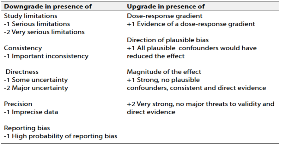
According to WHO Handbook for Guidelines, we used the GRADE (Grading of
Recommendations, Assessment, Development and Evaluation) approach to assess the quality of a body of evidence, develop and report recommendations. GRADE methods are used by WHO because they represent internationally agreed standards for making transparent recommendations. Detailed GRADE information is available on the following sites:
• GRADE working group: https://www.gradeworkinggroup.org/
• GRADE online training modules: http://cebgrade.mcmaster.ca/
Table 1 Quality and Significance of the four levels of evidence in GRADE:
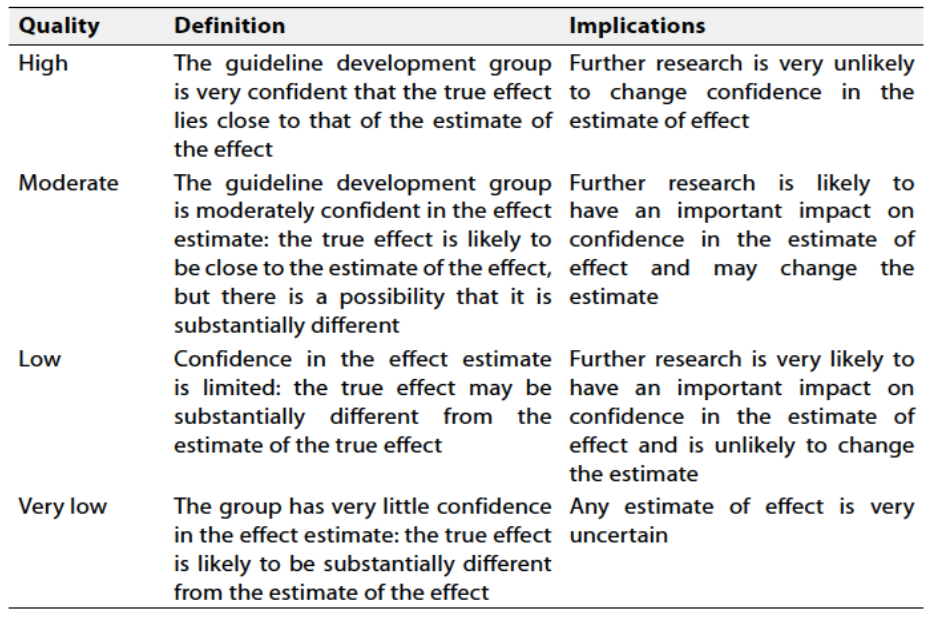
Table 2 Factors that determine How to upgrade or downgrade the quality of evidence
➡️The strength of the recommendation
The strength of a recommendation communicates the importance of adherence to the recommendation.
➡️Strong recommendations
With strong recommendations, the guideline communicates the message that the desirable effects of adherence to the recommendation outweigh the undesirable effects. This means that in most situations the recommendation can be adopted as policy.
➡️Conditional recommendations
These are made when there is greater uncertainty about the four factors above or if local adaptation should account for a greater variety in values and preferences, or when resource use makes the intervention suitable for some, but not for other locations. This means that there is a need for substantial debate and involvement of stakeholders before this recommendation can be adopted as policy.
➡️When not to make recommendations
When there is lack of evidence on the effectiveness of an intervention, it may be appropriate not to make a recommendation.
- Update of the guideline
This guideline will be updated whenever there is new evidence.
- References
1. Mosier MJ, Pham TN. American Burn Association practice guidelines: Burn shock resuscitation. J Burn Care Res. 2008;29:257–66.
2. Institute of Medicine. Clinical practice guidelines we can trust. Washington, DC: The National Academies Press; 2011.
3. Potokar T, Moghazy A, Peck M, Bendell R, Fanstone R. Setting standards for burn care services in low and middle income countries. Interburns; 2013.
4. World Health Organization. WHO handbook for guideline development. 2nd ed. Geneva: WHO Press; 2014.
5. ISBI Practice Guidelines Committee, Ahuja RB, Gibran N, et al. ISBI Practice Guidelines for Burn Care. Burns. 2016;42:953–1021.
6. ISBI Practice Guidelines Committee. ISBI Practice Guidelines for Burn Care, Part 2. Burns. 2018;44:1617–706.
7. Academic Committee of the Japanese Society for Burn Injuries. Clinical Practice Guidelines for Management of Burn Care. 3rd ed. Acute Med Surg. 2022 Jan;9(1):e739.
8. American Burn Association. Advanced Burn Life Support Course Provider Manual 2018 Update. Chicago: American Burn Association; 2018.
9. Schünemann HJ, Wiercioch W, Brozek J, et al. GRADE Evidence-to-Decision (EtD) frameworks for adoption, adaptation, and de novo development of trustworthy recommendations: GRADE-ADOLOPMENT. J Clin Epidemiol. 2017;81:101–10.
10. World Health Organization (WHO) Country Office – Egypt; Egyptian Health Council (EHC); Ministry of Health and Population (MOHP). Emergency Medicine Guideline Development Training Workshop Report: Using GRADE Methodology for Guideline Adaptation. Alexandria, Egypt: WHO; 2025.
11. First Aid. Emergency care for the injured. 2007. Available from: http://en.wikibooks.org/wiki/First_Aid [accessed 5.1.26].
12. Wilkinson E. The epidemiology of burns in secondary care, in a population of 2.6 million people. Burns. 1998;24(2):139–43.
13. International Federation of Red Cross and Red Crescent Societies. Law and first aid: promoting and protecting lifesaving action. 2015.
14. Shotland RL, Heinold WD. Bystander response to arterial bleeding. J Pers Soc Psychol. 1985;49(2):347–56.
15. Wei YL, et al. Self-efficacy of first aid for home accidents among parents. Accid Anal Prev. 2013;52:182–7.
16. Arbon P, et al. First aid and harm minimization for victims of road trauma. Prehosp Disaster Med. 2011;26(4):276–82.
17. Rea S, et al. Burn first aid in Western Australia. Burns. 2005;31(8):1029–34.
18. Cuttle L, et al. An audit of first-aid treatment of pediatric burns patients. J Burn Care Res. 2009;30(6):1028–34.
19. Forjuoh SN, et al. Childhood burns in Ghana. Burns. 1995;21(1):24–8.
20. Courtright P, et al. The epidemiology of burns in rural Ethiopia. J Epidemiol Community Health. 1993;47(1):19–22.
21. Fadeyibi IO, et al. Practice of first aid in burn related injuries. Burns. 2015;41(6):1322–32.
22. Ahuja RB, Bhattacharya S. Burns in the developing world and burn disasters. BMJ. 2004;329(7463):447–9.
23. Sahu SA, et al. Scald burn, a preventable injury: analysis of 4306 patients. Burns. 2016;42(8):1844–9.
24. Davies M, et al. How much do parents know about first aid for burns? Burns. 2013;39(6):1083–90.
25. Håkon KB, et al. A nationwide survey of first aid training and encounters in Norway. BMC Emerg Med. 2017;17:6.
26. Van de Velde S, et al. Effectiveness of nonresuscitative first aid training in laypersons. Ann Emerg Med. 2009;54(3):447–57.
27. Lubrano R, et al. How to become an under 11 rescuer. Resuscitation. 2005;64(3):303–7.
28. Anderson GS, et al. First aid skill retention of first responders within the workplace. Scand J Trauma Resusc Emerg Med. 2011;19:11.
29. Chipp E, et al. A 1 year study of burn injuries in a British emergency department. Burns. 2008;34(4):516–20.
30. Harvey LA, et al. A population-based survey of knowledge of first aid for burns in New South Wales. Med J Aust. 2011;195(8):465–8.
31. Skinner A, Peat B. Burns treatment for children and adults. N Z Med J. 2002;115(1156):U199.
32. Lam NN, Dung NT. First aid and initial management for childhood burns in Vietnam. Burns. 2008;34(1):67–70.
33. Skinner AM, et al. Reduced hospitalisation of burns patients following a multimedia campaign. Burns. 2004;30(1):82–5.
34. Jayaraman S, et al. First things first: effectiveness and scalability of a basic prehospital trauma care program. PLoS ONE. 2009;4(11):e6955.
35. Masellis M, et al. Immediate assistance and first aid on the spot in fire disasters. Ann Med Burn Club. 1992;5(4).
36. Shrivastava P, Goel A. Pre-hospital care in burn injury. Indian J Plast Surg. 2010;43(Suppl):S15–22.
37. Andrew MK. First aid. In: Principles and Practice of Burns Management. 1996. p. 199–202.
38. Cuttle L, et al. A review of first aid treatments for burn injuries. Burns. 2009;35(6):768–75.
39. Cuttle L, et al. The optimal temperature of first aid treatment for partial thickness burn injuries. Wound Repair Regen. 2008;16(5):626–34.
40. Hudspith J, Rayatt S. First aid and treatment of minor burns. BMJ. 2004;328(7454):1487–9.
41. Rockwell WB, Ehrlich HP. Should burn blister fluid be evacuated? J Burn Care Rehabil. 1990;11(1):93–5.
42. Swain AH, et al. Management of blisters in minor burns. BMJ. 1987;295(6591):181.
43. Davies JW. Prompt cooling of burned areas. Burns Incl Therm Inj. 1982;9(1):1–6.
44. Lawrence JC. British Burn Association recommended first aid for burns and scalds. Burns. 1987;13(2):153.
45. The Australian and New Zealand Burn Association. Emergency Management of Severe Burns. Manual. 19th ed. 2024.
46. First Aid Science Advisory Board. 2005 International Consensus Conference on CPR. Circulation. 2005;112:III115–25.
47. Wood FM, et al. Water first aid is beneficial in humans post-burn. PLoS ONE. 2016;11(1):e0147259.
48. Singer AJ, et al. The association between hypothermia, prehospital cooling, and mortality. Acad Emerg Med. 2010;17(4):456–9.
49. Lönnecker S, Schoder V. Hypothermia in patients with burn injuries. Chirurg. 2001;72(1):164–7.
50. Cuttle L, et al. The efficacy of Aloe vera, tea tree oil and saliva as first aid treatment. Burns. 2008;34(8):1176–82.
101. Gray P, et al. Pregabalin in severe burn injury pain: a randomized controlled trial. Clin J Pain. 2011;27(6):517–22.
51. Rajan V, et al. Delayed cooling of an acute scald contact burn injury. J Burn Care Res. 2009;30(4):729–34.
52. Jakobsson OP, Arturson G. The effect of prompt local cooling on oedema formation. Burns Incl Therm Inj. 1985;12(1):8–15.
53. Blomgren I, et al. Effect of cold water immersion on oedema formation. Burns Incl Therm Inj. 1982;9(1):17–20.
54. Cuttle L, et al. The optimal duration and delay of first aid treatment. Burns. 2010;36(5):673–9.
55. Ofeigsson OJ. Water cooling: first aid treatment for scalds and burns. Surgery. 1965;57:391–400.
56. Evers LH, et al. The biology of burn injury. Exp Dermatol. 2010;19(9):777–83.
57. First Aid Manual. St. John Ambulance Association. 1969.
58. Palao R, et al. Chemical burns: pathophysiology and treatment. Burns. 2010;36(3):295–304.
59. Sykes RA, et al. Chemical burns: retrospective review. J Burn Care Rehabil. 1986;7(4):343–7.
60. Doull J, et al. Toxicology: The Basic Science of Poisons. 1980.
61. Saydjari R, et al. Chemical burns. J Burn Care Rehabil. 1986;7(5):404–8.
62. Sawhney CP, Kaushish R. Acid and alkali: considerations in management. Burns. 1989;15(2):132–4.
63. Stewart CE. Chemical skin burns. Am Fam Physician. 1985;31(2):149–57.
64. Leonard LG, et al. Chemical burns: effect of prompt first aid. J Trauma. 1982;22(5):420–3.
65. Zack-Williams SD, et al. The clinical efficacy of Diphoterine. Ann Burns Fire Disasters. 2015;28(1):9.
66. Alexander KS, et al. Chemical burns: Diphoterine untangled. Burns. 2018;44(4):752–66.
67. Lewis CJ, et al. Is it time for a change in the approach to chemical burns? J Plast Reconstr Aesthet Surg. 2017;70(5):563–7.
68. Dinesen F, et al. Diphoterine for chemical burns of the skin: A systematic review. Eur Burn J. 2023;4(1):55–68.
69. Mohammadi AA, et al. A Survey on 30 Months Electrical Burns. Burns. 2008;34(1):111–13.
70. Buja Z, et al. Electrical Burn Injuries: An Eight-Year Review. Ann Burns Fire Disasters. 2010;23(1):4–7.
71. Koumbourlis AC. Electrical Injuries. Crit Care Med. 2002;30(11 Suppl):S424–30.
72. American Heart Association. Guidelines for CPR, Part 10.9: Electric Shock. Circulation. 2005;112:IV1–5.
73. Elloso MS, Cruz JJV. Electrical Burns in a Philippine Tertiary Hospital. Burns Open. 2017;1:20–24.
74. American Burn Association. 2014 National Burn Repository. 2014.
75. Tung KY, et al. Epidemiology Study of 12,381 Admitted Burn Patients in Taiwan. Burns. 2005;31(Suppl 1):S12–17.
76. Song C, Chua A. Epidemiology of Burn Injuries in Singapore. Burns. 2005;31(Suppl 1):S18–26.
77. Atiyeh BS, et al. State of the art in burn treatment. World J Surg. 2005;29(2):131–48.
78. Warden GD. Burn shock resuscitation. World J Surg. 1992;16(1):16–23.
79. Monafo WW. Initial management of burns. N Engl J Med. 1996;335(21):1581–86.
80. Hettiaratchy S, Papini R. Initial management of a major burn. BMJ. 2004;329(7457):101–3.
81. Greenhalgh DG. Burn resuscitation: the results of the ISBI/ABA survey. Burns. 2010;36(2):176–82.
82. Baker RH, et al. Resuscitation of thermal injuries in the UK and Ireland. J Plast Reconstr Aesthet Surg. 2007;60(6):682–85.
83. European Burn Association. European Practice Guidelines for Burn Care. 2017.
84. Japanese Dermatological Association. Guidelines for Burn Care. 2011.
85. Artz CP, Moncrief JA. The Treatment of Burns. 1969.
86. Moylan JA. First aid and transportation of burned patients. In: Burns: A Team Approach. 1979.
87. American Burn Association. Clinical practice guidelines on burn shock resuscitation. J Burn Care Res. 2024;45(3):565–89.
88. Barrow RE, et al. Early fluid resuscitation improves outcomes. Resuscitation. 2000;45(2):91–96.
89. Wolf SE, et al. Mortality determinants in massive pediatric burns. Ann Surg. 1997;225(5):554–65.
90. Romanowski KS, Palmieri TL. Pediatric burn resuscitation. Burns Trauma. 2017;5:26.
91. von Baeyer CL, Spagrud LJ. Observational measures of pain for children (3–18 years). Pain. 2007;127(1–2):140–50.
92. Crellin DJ, et al. Systematic review of the FLACC scale for pain in children. Pain. 2018;159(11):2142–51.
93. Ambuel B, et al. Assessing distress: the COMFORT scale. J Pediatr Psychol. 1992;17(1):95–109.
94. Payen JF, et al. Assessing pain in critically ill sedated patients. Crit Care Med. 2001;29(12):2258–63.
95. Williamson A, Hoggart B. Pain: review of three pain rating scales. J Clin Nurs. 2005;14(7):798–804.
96. World Health Organization. Pharmacological Treatment of Persisting Pain in Children. Geneva: WHO; 2012.
97. Mahar P, et al. Pain assessment tools in adult burns: systematic review. Burns. 2012;38(2):147–54.
98. Summer GJ, et al. Burn injury pain: the continuing challenge. J Pain. 2007;8(7):533–48.
99. Wasiak J, et al. Pharmacological interventions for pain and sedation in burn injuries. Cochrane Database Syst Rev. 2014;(3):CD008438.
100. Chou R, et al. Management of postoperative pain: clinical practice guideline. J Pain. 2016;17(2):131–57.
102. Cuignet O, et al. Effects of gabapentin on morphine consumption. Burns. 2007;33(1):81–86.
103. Fuzaylov G, Fidkowski CW. Ketamine for pain management in burn patients. J Burn Care Res. 2010;31(3):491–501.
104. Walker J, Mustard L. The management of pain in the burns unit. Burns. 2009;35(7):921–36.
105. McGarry S, et al. Burns patients’ experiences of pain and anxiety: a qualitative study. Burns. 2014;40(5):887–95.
106. Barr J, et al. Guidelines for pain, agitation, and delirium in ICU. Crit Care Med. 2013;41(1):263–306.
107. Stutchfield C, et al. Fluid resuscitation in paediatric burns. Arch Dis Child. 2019;104(3):280–285.
108. Morgenstern S, et al. Fluid resuscitation in pediatric burn patients. J Burn Care Res. 2025;46(Suppl 1):S71–S72.
109. UpToDate. Emergency care of moderate and severe thermal burns in adults. 2025.
110. Baxter CR, Shires GT. Physiological response to crystalloid resuscitation. Ann N Y Acad Sci. 1968;150(3):874–894.
111. Madnani DD, et al. Factors predicting intubation in smoke inhalation. Ear Nose Throat J. 2006;85(4):278–280.
112. American Burn Association. Inhalation injury: diagnosis. J Am Coll Surg. 2003;196(2):307.
113. Masanes MJ, et al. Fiberoptic bronchoscopy for early diagnosis of inhalation injury. Chest. 1995;107(5):1365–1369.
114. Masanes MJ, et al. Fiberoptic bronchoscopy for early diagnosis: comparative value. J Trauma. 1994;36(1):59–67.
115. Peitzman AB, et al. Smoke inhalation injury: radiographic manifestations. J Trauma. 1989;29(9):1232–1239.
116. Lee MJ, O’Connell DJ. The plain chest radiograph after acute smoke inhalation. Clin Radiol. 1988;39(1):33–37.
117. Venus B, et al. Prophylactic intubation and CPAP in inhalation injury. Crit Care Med. 1981;9(7):519–523.
118. Gartner R, et al. Acute respiratory insufficiency in burn patients from smoke inhalation. Pathol Biol. 2002;50(2):118–126.
119. The Japanese Society for Burn Injuries (JSBI). Clinical Practice Guidelines for Management of Burn Care (3rd Edition). Acute Med Surg. 2022;9(1):e739.
120. Ergün O, et al. Prophylactic antibiotic use in pediatric burn units. Eur J Pediatr Surg. 2004;14(6):422–426.
121. Donati L, et al. Increased burn patient survival with high dose teicoplanin. J Chemother. 1998;10(1):47–57.
122. Church D, et al. Burn wound infections. Clin Microbiol Rev. 2006;19(2):403–434.
123. Rhee P, et al. Tetanus and trauma: a review and recommendations. J Trauma. 2005;58(5):1082–1088.
124. Aikawa N, et al. Burn wound infection. Antibiot Chemother. 1999;15:671–673.
125. Karyoute SM, Badran IZ. Tetanus following a burn injury. Burns Incl Therm Inj. 1988;14(3):241–243.
126. Larkin JM, Moylan JA. Tetanus following a minor burn. J Trauma. 1975;15(6):546–548.
127. Takeuchi Y, et al. Management and prevention of tetanus in trauma. J Jpn Assoc Surg Trauma. 2007;21:367–374.
128. Garg R, et al. Managing blisters in minor burns: Should they be deroofed? Indian J Burns. 2022;29:31–35.
129. Gimbel NS, et al. A study of epithelization in blistered burns. AMA Arch Surg. 1957;74(5):800–3.
130. Ro HS, et al. Effectiveness of aspiration or deroofing for blister management. Medicine (Baltimore). 2018;97(17):e0563.
131. Gottrup F, et al. Outcomes in controlled studies on non-healing wounds. J Wound Care. 2010;19(6):237–268.
132. Singh V, Devgan L. To debride or not to debride: blisters in burns. J Burn Care Res. 2007;28(6):892–894.
133. Duncan A, et al. Silver Confers No Additional Benefit to Re-Epithelialization. Plast Reconstr Surg. 2025.
134. Norman G, et al. Antiseptics for burns. Cochrane Database Syst Rev. 2017;(7):CD011821.
135. Jiang Y, et al. Effectiveness of silver and iodine dressings. BMJ Open. 2024;14(8):e077902.
136. Ang E, et al. Alternative therapy in partial thickness burns. J Burn Care Res. 2008;29(2):362–369.
137. Aziz Z, Abu SF. Systematic review of silver-containing dressings. Burns. 2012;38(3):307–318.
138. Storm-Versloot MN, et al. Topical silver for preventing wound infection. Cochrane Database Syst Rev. 2010;(3):CD006478.
139. Müller G, Kramer A. Biocompatibility index of antiseptic agents. J Antimicrob Chemother. 2008;61(6):1281–1287.
140. Choi YM, et al. Antibiotic ointment versus a silver-based dressing. J Pediatr Surg. 2019;54(7):1391–1396.
141. American Academy of Dermatology. Guidelines for management of cutaneous wounds. 2016.
142. Lagziel T, et al. Immediate/Ultra-Early v. Early Burn Excision. J Burn Care Res. 2022;43(Suppl 1):S206–S206.
143. Greenwood JE. Advantages of immediate excision of burn eschar. Anaesth Intensive Care. 2020;48(2):89–92.
144. De La Tejera G, et al. Early wound excision within three days. Burns. 2023;49(8):1816–1822.
145. Munian K, et al. Early excision and grafting vs conservative treatment. Int J Sci Res. 2019;8(5):1–5.
146. Barret JP, Herndon DN. Modulation of inflammatory responses by early burn excision. Arch Surg. 2003;138(2):127–132.
147. Pham C, et al. Bioengineered skin substitutes for the management of burns. Burns. 2007;33(8):946–957.
148. Brölmann FE, et al. Evidence-based decisions for wound care. Br J Surg. 2012;99(9):1172–1183.
149. World Health Organization (WHO). Burn management (safety protocols). n.d.
150. Atiyeh BS, et al. Moist environment in optimal wound healing. Curr Pharm Biotechnol. 2002;3(3):179–195.
151. Wasiak J, et al. Dressings for superficial and partial thickness burns. Cochrane Database Syst Rev. 2013;(3):CD002106.
152. Cartotto R, et al. American Burn Association clinical practice guidelines on burn shock resuscitation. J Burn Care Res. 2023.
153. British Burn Association. National burn care referral guidance. 2012.
154. PMC. Burn injury characteristics, referral pattern, and outcomes. J Burn Care Res. 2023.
155. CHEMM. Burn triage and treatment – thermal injuries. 2007.
156. Medscape. Burn resuscitation and early management. 2023.
157. World Health Organization. Burns – Fact sheet. 2023.
158. National Health Service. Adult burn referral guidance. 2024.
159. PMC. Adherence to referral criteria for burn patients. 2022.
160. American College of Surgeons. Resources for Optimal Care of the Injured Patient. 2022.
161. Saffle JR, et al. Role of telemedicine in acute burn injuries. J Burn Care Res. 2005;26(4):333–336.
162. National Association of Emergency Medical Technicians. Prehospital Trauma Life Support. 2021.