Fungal diseases
| Site: | EHC | Egyptian Health Council |
| Course: | Avian and Rabbit Medicine Guidelines |
| Book: | Fungal diseases |
| Printed by: | Guest user |
| Date: | Sunday, 20 September 2026, 9:43 PM |
Description
"last update: 8 September 2026" Download Guideline
Table of contents
- - Acknowledgement
- - Difference between Mycosis and Mycotoxicosis
- - Differential diagnosis of fungal diseases
- - Diagnosis for fungal diseases
- - Treatment of fungal diseases
- - Prevention and control of fungal diseases
- - Dactylariosis ( Mycotic encephalitis)
- - MYCOTOXICOSIS
- - General transmission of Mycotoxins
- - General clinical signs of Mycotoxins
- - Differential diagnosis of mycotoxicosis
- - General diagnosis of Mycotoxins
- - General prevention and control of Mycotoxins
- - Prevention and control of mycotoxicosis
- - General treatment of Mycotoxins
- - References
- Acknowledgement
We would like to acknowledge the committee of National Egyptian Guidelines for Veterinary Medical Interventions, Egyptian Health Council for adapting this guideline.
Executive Chief of the Egyptian Health Council: Prof. Mohamed Mustafa Lotief.
Head of the Committee: Prof. Ahmed M Byomi
The rapporteur of the Committee: Prof. Mohamed Mohamedy Ghanem.
Scientific Group Members: Prof. Nabil Yassin, Prof. Ashraf Aldesoky Shamaa, Prof. Amany Abbass, Prof. Dalia Mansour, Dr. Essam Elmarakby, Dr. Mohamed Elsharkawy, Prof. Gamal A. Sosa., Dr. Naglaa Radwan, Dr. Hend El Sheikh
Authors: Prof. Dalia Mansour Hamed, Prof.Kamel Ibrahim Abou- Elazm
- Difference between Mycosis and Mycotoxicosis
|
Mycotic Diseases |
Mycotoxicosis |
|
Mode of action: 1-Invade and destroy body tissue 2- Decrease body resistance |
Mode of action: Fungi grow on grains produce Toxin for ( Animal, Bacteria) |
|
Aspergillosis (mainly infect the respiratory system) |
Aflatoxin (primarily target Liver) |
|
Candidiasis (mainly infect the digestive system) |
Ochratoxine (primarily target Kidney) |
|
Favus (it is a skin mycosis) |
Trichothecene (primarily target Digestive system) |
|
Dactylariosis (mainly infect brain) |
Pencillin (mainly affect bacteria) |
- Differential diagnosis of fungal diseases
|
P.O.C. |
Aspergillosis |
Candiasis |
Favus |
||
|
Definition |
Mainly affects the respiratory
system, but some infections spread to other visceral organs, affecting
most domestic and wild birds. *Dermatitis *Mycotic Osteomyelitis & Arthritis *Ophthalmitis *Encephalitis *Pulmonary Aspergillosis |
Worldwide organism primarily affects the upper digestive tract of all birds and is characterized by whitish thickened areas of the crop and proventriculus, erosions in the gizzard, and inflammation of the vent area. |
Chronic dermatomycosis, the comb is often affected, in severe cases the disease spreads to the feather portion of the body. |
||
|
Etiology |
There are many different kinds of: aspergillus
(Aspergillus fumigatus and A. flavus are the two major agents
while Aspergillus niger, Aspergillus candidus, Aspergillus glaucus,
Aspergillus nidulans are among the species isolated as opportunistic
pathogens. |
yeast-like fungus (Candida albicans) which is a dimorphic yeast appear as round or oval budding yeasts on surface epithelium or branching with pseudo-hyphae in deeper tissue |
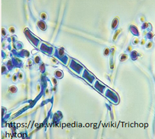
Trichophyton gallinae (Achorion gallinae). |
||
|
Susceptibility |
in the brooder stage in domestic birds (chicks, Quails, pheasants, Turkey, Pigeon and ducks) in the form of an acute out-break in young chicks adult birds occasionally may be affected as sporadic cases. infect chicks and turkey poult at or below 10 days of age but may affect birds up to 10 weeks. Turkeys highly susceptible than chickens. |
² Chickens, turkeys, pigeons, pheasants, quail and grouse are species most commonly get affected as well as other domestic animals and humans. ² Poultry of all ages are susceptible to the effects of this organism. |
all ages under poor manage-mental conditions |
||
|
Mode of transmit-ssion |
The bird comes in contact with the organisms through contaminated feed, litter or premises. Egg borne disease: the fungi penetrate the egg shell due to contamination of egg shell with fungi during incubation and may lead to embryonic mortality or hatched infected chicks and infection to recently healthy hatched chicks by inhalation of spores this is called Hatchery borne disease.
not contagious and does not spread from one bird to another. ( Most healthy birds can withstand repeated exposure to these organisms when there is count within the permissible limit). Inhalation of large amounts of the infectious form
of the mold or reduced resistance of the bird apparently results in
infection. |
by ingestion of the causative organism in infected feed, water or environment. Unsanitary and unclean water troughs are an excellent reservoir of the Candida organism. The disease does not however, spread directly from bird to bird (not contagious). The organism grows especially well on corn, so infection can be introduced by feeding moldy feed. |
Infection spreads from one bird to
another by direct contacts. |
||
|
Clinical signs |
The disease is observed in one of two
forms: B- chronic condition affecting adult
birds: |
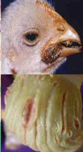
no specific symptoms Affected caged layer hens become obese and anemic. Some birds exhibit a vent inflammation that resembles a diarrhea induced condition having whitish in crustations of the feathers and skin around the area. Feed consumption may decrease by ten to twenty percent. |
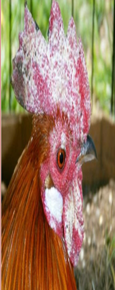
Lesions usually develop first on the comb, as the fungus spreads. white spots developed the surface of which scales off, and the comb may appear as through sprinkled with flour. The skin becomes thickened in the affected area and covered with scale and crusts especially around the feather follicles. |
||
|
|
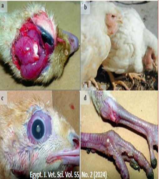
méningitis (a), dermatitis (b), uveitis (c) and arthritis (d) |
|
|
||
|
PM |
hard nodular areas in the lungs and an infection of the air sacs. Sometimes the air sac lesions are similar to those produced by infectious sinusitis or CRD. In some birds, colonies of mold growth can be seen on the air sac membranes which appear as yellowish nodules and cloudy air sacs. Necrotic foci may be found on liver, spleen, kidneys, proventriculus and other organs. Sometimes fungus may invade blood stream reaching the brain showing white to yellow necrotic foci. Ophthalmitis may occure and lead to blindness |
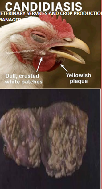
Are mostly confined to the crop, proventriculus and gizzard. The crop and proventriculus have whitish thickened areas that are often described as having a "turkish towel" appearance or pseudodiphtheritic membrane with necrotic batches with eroded mucosal surface. |
A moldy odor may be detected. |
||
|
Microscopic |
Aspergillosis |
Candidiasis |
Favus |
||
|
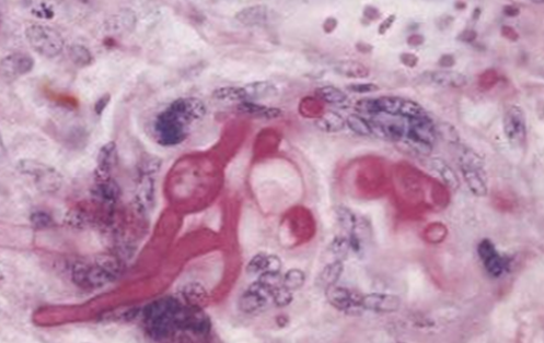
Direct microscopic detection by taking small portion of the nodules in kOH 10% on a microscopic slide covering with cover slide then apply genital heating mixed with ink dye lactophenol to show
blue stained separated branched hyphae |
round
cell and hyphae
|
branching fungal hyphae and Arthrospores.
|
|||
|
Lab. diagnosis |
Sabouraud’s agar at 35 c antibacterial drugs as chlormephenicol could added to suppress any bacterial growth. |
||||
|
The flat colonies are initially
white and turn blue green as conidia begin to mature. |
Colonies are whitish, creamy and highly
convex with yeast odor after 1-2 days at 37c. |
slow-growing, flat-to-powdery white-to-cream colonies that often produce a characteristic pink-to-red diffusible pigment into the agar. |
|||
|
Prevention and control |
avoiding
moldy litter, feed hoppers and watering places or premises are fertile area
for fungal growth of moulds unless frequent moving of utensils. |
||||
|
The disease can usually be prevented
by: |
An inexpensive, yet effective, water treatment is the continuous addition of household chlorine bleach to the drinking water at the rate of 5 parts per million (ppm). |
|
|||
|
Treatment |
Many broad-spectrum antibiotics will enhance this disease; therefore, they should not be used until after control of this condition is completed. |
||||
|
|
Tetracycline Sorbate at dose of 200
mg/L of drinking water for 5 days or Miconazole. |
Addition of Nystatin (100 g/Ton) or copper sulfate (2-3 lb/Ton) to the feed for seven to ten days should control moniliasis beside an adequate amount of Vitamin A.Methyl violet choline chloride and vitamin K prevent crop mycosis. |
Remove the crust and apply topical antifungal |
||
- Diagnosis for fungal diseases
-The diagnosis is typically dependent on a collection of data from the history, clinical presentation, postmortem findings, hematology, biochemistry, serology, radiological alterations, endoscopy, and fungus culture.
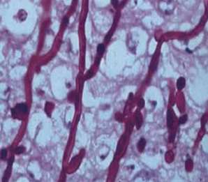
-Isolation and Identification of Causative Agent Mold can detected microscopically in tissues from infected. There are several ways to obtain tissue samples for testing, involves removing some of the affected tissue, swabbing the lesion, and pulling one of the plaques away. The diagnostic samples you collect must carefully handle using aseptic techniques. Simply immersing a tiny portion of the nodule in 10-20% potassium hydroxide (KOH), heating the slide over a flame for separating the material, and placing it on a microscope slide, these samples can studied under a microscope, Hyphae will appear
-Most commonly, samples placed on Sabouraud dextrose agar and incubating it for 24 hours- 7days hours at 37°C with a distinctive conidial head and colony, the pathogenic organism can be isolated.
- Histopathological examination using a special fungal stain reveals granulomas containing mycelium of the tissue.
-samples, such as the lungs, trachea, throat, and thoracic air sacs, treated before embedded in pieces of paraffin and stained using the haematoxylin and eosin (HE) procedure. Other specialized stains, such as Periodic acid-Schiff (PAS), Bauer and Gridley's, Groote's, and Gomori Methanamine Silver stains, make it simple to identify the hyphae and mycelia of fungus.
-Immunity and serologic tests Serological. Because the fungal antigens are generic, serologic testing have limited relevance. Enzyme linked immunosorbant assay (ELISA) and agar gel immunodiffusion measurements of the antibody response.
-Diagnostic imaging (Radiography and Endoscopy)
Radiographic examination is a quick and effective method for obtaining a preliminary assessment of the underlying disease in non-critical instances in valuable and research birds. When examining the bird's lungs and air sacs for indications of inflammation or granulomas and to determine the general health of the bird's lower respiratory tract, non-invasive imaging modalities such as lateral and dorso-ventral radiography images
Molecular diagnosis
PCR can be used to analyses fungal DNA from bodily fluids or serum, however although being extremely sensitive.
Differential diagnosis
The particular clinical signs of avian aspergillosis vary on the organ and systems affected. Early broiler chick mortality should screened out for Aspergillosis since mycotoxicosis, acute bacterial septicemia, or carbon monoxide poisoning may bring it on. Infectious laryngo-tracheitis, infectious bronchitis (coughing, gasping,and extension of the neck during inspiration), and Newcastle disease all cause dyspnea and watery, greenish diarrhea. mycobacteriosis, colibacillosis, dactylaria infection (nervous sign),
and nutritional encephalomalacia are among the differential diagnose. -The granulomatous lesions seen during necropsy typically used to distinguish pulmonary aspergillosis from other avian respiratory illnesses; however, Staphylococcus aureus pneumonia in chicks can resemble it.
- pneumonia and exudative fibrinous or fibrinoheterophilic air sacculitis are frequently observed in mycoplasmosis, colibacillosis, poultry cholera, and chlamydophilosis patients. -When granulomas predominate, mycobacteriosis and other mycoses must also take into consideration and Aspergillus ocular oedema resembles infectious coryza or vitamin A deficiency in chicks.
- Treatment of fungal diseases
-Use of one or more systemic antifungal medications is required for the treatment.
- Itraconazole, ketoconazole, clotrimazole, miconazole, and fluconazole are some of the medicines that are frequently utilised .
- Amphotericin B has traditionally been the medicine of choice for treating aspergillosis, although it has serious adverse effects and a high mortality rate. - Itraconazole and voriconazole after amphotericin B for better results.
- Prevention and control of fungal diseases
Major control measures include:
1) Minimizing exposure to the fungus and risk factors involve.
2) The removal of the birds from the polluted area.
3) Hatchery hygiene in young chickens through carefully cleaning and disinfection of the hatching equipment.
4) Contaminated material(s) removed to prevent additional exposure as removing the old litter and the mouldy feed.
5) To avoid an aspergillosis outbreak, resist using mouldy litter or feed.
6) Attempting to avoid disturbing the infected material(s) to reduce spore aerosolization further.
7) Higher air exchange rates or ventilation , lessens the severity of the epidemic.
8) Eggs that are seriously infected or fractured should not incubated because they stimulate fungal growth and since they may rupture and release spores into the hatching machine.
9) Related risk factors (bad ventilation, high temperature, humidity,…..)
- Dactylariosis ( Mycotic encephalitis)
Definition: Fungal disease characterized by nervous signs in turkey poults, quail chicks and chicks at 1-5 weeks old is the high susceptible age causing outbreaks.
Clinical signs:
Nervous signs: torticollis, incoordination, tremors, paralysis and death
Eye lesions and blindness also dyspnea may occur in rare cases.
P.M:
-Mycotic encephalitis
-Severe large, hardened, circumscribed cerebellar and cerebral lesion (necrosis is gray or yellow)
-Pulmonary granulomas are minimum
-Ocular lesion appear similar to aspergillosis
Diagnosis:
as signs, lesions, demonstration of brownish conidia and large number of gaint cells from brain.
Differential diagnoses include:
- Infectious Bronchitis Virus (IBV)
- Newcastle Disease (NCD)
- mycobacteriosis
- colibacillosis
- other mycoses (e.g., ochroconosis, zygomycosis)
- oncogenic tumors (e.g., Marek’s Disease, Avian Leukosis)
- nutritional encephalomalacia (Vit A ,E def.)
- post vaccinal reaction leads to gasping.
- MYCOTOXICOSIS
Mycotxin: is a complex chemical by –product produced from some species of fungi which elaborate them into surrounding ration.
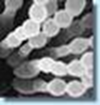
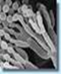
MYCOTOXICOSIS: is a lethal disease caused by toxins of pathogenic fungi. Characterized by hemorrhagic syndrome and aplastic anemia. Many fungi, such as Aspergillus flavus could produce toxin.
|
Major classes of mycotoxin- producing fungi |
Fungi species |
Mycotoxins |
|
-Aspergillus
-Pencillium
-Fusarium
|
A. flavus |
Aflatoxin |
|
A. ochraceus P. verrucosum |
Ochratoxin
|
|
|
Fusarium. graminearum
|
Type A
Trichothecenes
|
|
Mycotoxin |
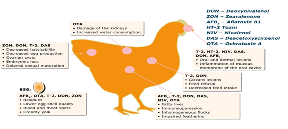
Possible effects |
|
Aflatoxins |
Liver diseases (hepatotoxic, hepatocarcinogenic); carcinogenic and teratogenic effects; hemorrhages (intestinal tract, kidneys); reduced growth rate; diminution of performance; immune suppression |
|
Ochratoxins |
Nephrotoxic; carcinogenic; mild liver damage; enteritis; teratogenic effects; poor feed conversion; reduced growth rate; immune suppression |
|
Trichothecenes |
Digestive disorders (vomiting, diarrhea, feed refusal); reduced weight gain; hemorrhages (stomach, heart, intestine, lung, bladder, kidney); edema; oral lesions; dermatitis; blood disorders; infertility; degeneration of bone marrow; slow growth; immune suppression |
|
Cyclopiazonic acid |
Necrotic effects (liver, gastrointestinal tissue, kidneys, skeletal muscles); carcinogenic; neurotoxic |
|
Citrinin |
Nephrotoxic; teratogenic; hepatotoxic |
- General transmission of Mycotoxins
Conditions to Encourage Fungal Growth?
- Relative humidity over 70%.
- Temperatures over 30 degrees Celsius for a period of a few days to a week.
- Stress to the affected plant, such as drought, flood, or insect infestation.
- High moisture content of crop (20% or higher).
- Must occur in conjunction, or fungal growth cycle will cease.
Mode of infection
- Mycotoxicosis is caused by ingestion of a diet containing more than the permissible limits (5ppm toxic substances for some mycotoxin) produced by molds growing on feed (prior or during harvesting or during storage), feed ingredients and possibly litter. Several types of fungi produce toxins that may cause problems in poultry, but of primary concern are substances produced by the Aspergillus flavus fungi and are thus called aflatoxins. Several other fungi also produce toxins that cause the disease.
- General clinical signs of Mycotoxins
- Lower feed conversion, Poor growth, Reduced carcass fat
- Altered immune mechanisms resulting in vaccine failures
- Increased susceptibility to infections
- Decreased ability to resist stress
- Lower egg production, Lower Hatchability, Smaller Eggs
- Fatty liver syndrome, Liver & kidney lesions, Decrease blood-clotting results in greater downgrading and condemnation of the birds, Increased susceptibility to bruising; كدمات
- Impaired kidney function, Kidney disorders
- Decreased semen volume and testes weight (Reduce hatchability)
- Abnormal behavior, alter feathering, Leg & bone problems
- Increase incidence of Coccidiosis
- Less carcass pigmentation, Egg yolks are paler
- Unthriftiness, pale shanks and combs or even paralysis.
- Breeder hens affected by aflatoxins are known to transfer measurable quantities to both the egg white and yolk.
- Differential diagnosis of mycotoxicosis
|
P.O.C. |
Aflatoxin |
Ochratoxins |
Tricothecenes |
||
|
Definition |
Liver diseases (hepatotoxic,
hepatocarcinogenic); carcinogenic and teratogenic effects |
Ochratoxin are Nephrotoxic;
carcinogenic; mild liver damage; enteritis; teratogenic effects; poor feed
conversion; reduced growth rate; immune suppression |
Digestive disorders (vomiting,
diarrhea, feed refusal) |
||
|
Host susceptible |
Ducklings are
10 times more sensitive
than young chicks for aflatoxicosis) and turkey poults are more susceptible than
chickens (Worldwide). |
||||
|
clinical signs |
(hepatotoxic, hepatocarcinogenic);
|
Nephrotoxic |
Digestive disorders |
||
|
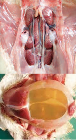
PM |
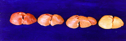
● In the day - old duckling, Aflatoxin
can produce hepatic tumors. |
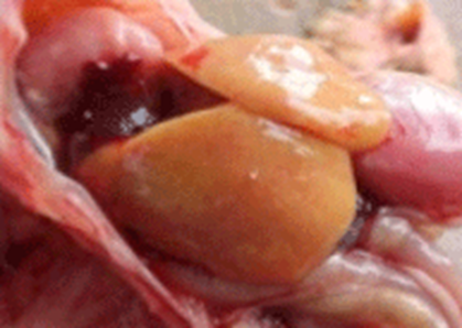
●Visceral urate, kidney may have
urate crystal which is white to tan in color |
●Sick birds may drop dead with few
signs of illness |
||
- General diagnosis of Mycotoxins
1. Based on flock history, symptoms, P.M lesions.
2. Confirmation by analysis of ration and storage places.
3. Culture for mold in the feed.
4.Analysis of crop and intestinal content.
4. Eliza competitive test for diagnosis (Rapid test).
5. Gas chromatography – Monoclonal Antibody technology, Spectrophotometer, HPLC,HPTLC
- General prevention and control of Mycotoxins
Molds are widespread in nature. The key is proper storage to control moisture and temperature to reduce growth of the molds. Although the mold is present, it cannot produce toxic products unless allowed to grow freely. Once the toxin is produced there is no known method for removing it from the feed or its harmful effects.
1. Providing a diet containing high fat and high protein levels and provide the ration with vitamin supplements may be of value.
2. Effective management of litter, feed and water.
3. Avoid using moldy feed, pelleting ration decrease the chance of fungal growth .
4. Antimycotic additives are helpful.
5. Adding Zeolites or bentionites or clinoptiolites, Sodium- aluminum silicate at 0.5%to ration act as toxin binder which actively searches "toxic mould" molecules - even in the chicken's gut. When it locates them, it completely binds and covers the "toxic mould" molecules so that they become harmless as the bird is unable to absorb the bound molecules through the gut wall. The inactivated molecules are then passed out in the faeces.
- Prevention and control of mycotoxicosis
- Mould-free feed and ingredients should used
- Good agricultural practices
- Management practices
- Prevention of mycotoxicoses in poultry should focus on using feed and ingredients free of mycotoxins and on management practices that prevent mould growth and mycotoxin production during feed transport and storage.
- Preharvest agricultural measures such as crop rotation, tilling, proper fertilization, and pest control can decrease some of the stressors that influence mycotoxin occurrence.
- Postharvest techniques such as regular inspection of feed storage and feeding systems can identify flow problems that allow residual feed and enhance fungal activity and mycotoxin formation.
- Mycotoxins can form in decayed, crusted feed in feeders, feed mills, and storage bins; thorough cleaning and correcting the problem can have immediate benefits. Temperature extremes cause moisture condensation and migration in bins and promote mycotoxin formation.
- Ventilation of poultry houses to avoid high relative humidity also decreases the moisture available for fungal growth and toxin formation in the feed.
- Antifungal agents added to feeds to prevent fungal growth have no effect on toxin already formed but may be cost-effective in conjunction with other feed management practices.
- Organic acids (0.5–1.5 g of propionic acid/kg of feed) are effective inhibitors; however, the effectiveness may be decreased by the particle size of feed ingredients and the buffering effect of certain ingredients.
- Sorbent compounds such as hydrated sodium calcium aluminosilicate (HSCAS) effectively bind and prevent absorption of aflatoxin.
- Esterified glucomannan, derived from the cell wall of the yeast Saccharomyces cerevisiae, is protective against aflatoxin B1 and ochratoxins. It decreases toxicity through the binding and decreased bioavailability of fumonisins, zearalenone, and T-2 toxin.
- Various other fermentation products, algae and plant extracts, and microbial feed additives have demonstrated ability to bind or degrade mycotoxins and may be applicable and appropriate for the situation.
- General treatment of Mycotoxins
- Remove contaminated material and replace
- Treat concurrent disease
- Improve management practices
- Provide supportive care
- Contaminated feed or bedding should be replaced. Concurrent diseases should be treated to alleviate disease interactions, and substandard management practices must be corrected.
- Some mycotoxins increase requirements for vitamins, electrolytes, trace minerals (especially selenium and zinc), protein, and lipids and can be compensated for by feed supplementation and water-based treatment.
- Nonspecific treatments using activated charcoal (digestive tract adsorption) in the feed have a sparing effect but are not practical for larger production units. In larger scale production, the use of clays and derivatives from yeast, bacteria, and plants has more practical applications.
- Certain enzymes have potential to biodegrade specific mycotoxins, such as fumonisins, into nontoxic metabolites (enzymatic biotransformation). However, availability of detoxifying enzymes is limited.
- Extra-label treatment with antifungal drugs labeled for use in other species (eg, nystatin and triazole antifungal agents) may not be permitted in certain jurisdictions.
- Is poly [1-(2-oxo-l-pyrroli-dinyl) ethylene 3%, used in the drinking water of poultry helps to control the poisonous substances produced by moulds and fungi including Aflatoxin, Cyclopiazonic Acid, Vomitoxin, Ochratoxin. Zearalenone, Penicillium and Fusarium. It also treats disorders caused by mycotoxins in poultry.
- References
Abou Elazm Textbook of poultry diseases Editor : Kamel Ibrahim Abou- Elazm.
American Association of Avian Pathologists (AAAP),Avian Disease Manual (8th Edition): https://www.aaap.info/avian-disease-manual
Andrew van Wijk. Aspergillosis in poultry (2016). Accessed at https://www.sava.co.za/vetnews/2016/2023%20February/VN%20CPD%20February%202023.pdf.
Arne, P., Thierry, S., Wang, D., et al. (2011). Aspergillus infections in birds: a review. Avian Diseases, 55(4), 527–546.https://pubmed.ncbi.nlm.nih.gov/21826144
Chaves Hernández AJ. Poultry and Avian Diseases. Encyclopedia of Agriculture and Food Systems. 2014:504–20. doi: 10.1016/B978-0-444-52512-3.00183-2. Epub 2014 Aug 21. PMCID: PMC7152037. https://pmc.ncbi.nlm.nih.gov/articles/PMC7152037/figure/f0020/
David E. Swayne et al.,Wiley-Blackwel (2019) Diseases of Poultry (14th Edition).
FDA (2001). Guidance for industry: fumonisin levels in human foods and animal feeds. United States Food and Drug Administration, Washington DC. pp. 56688-56689. Available at: https://www.fda.gov/regulatory-information/search-fda-guidancedocuments/guidance-industry-fumonisin-levels-human-foods-andanimal-feeds
FDA (2010). Guidance for industry and FDA: advisory levels for deoxynivalenol (DON) in finished wheat products for human consumption and grains and grain by-products used for animal feed.
Filazi,
A., Yurdakok-Dikmen, B., Kuzukiran, O., & Sireli, U. T. (2017).
Mycotoxins in Poultry. Poultry Science, InTech Open. DOI: 10.5772/66302.
https://www.kemin.com/na/en-us/markets/animal/poultry/feed-quality/resources/mold-and-mycotoxins/whats-your-risk
MSD Vet Manual. Aspergillosis in poultry. Kromm, M et al. 2020.
Murugesan, G. R., Ledoux, D. R., Naehrer, K., et al. (2015). Prevalence and effects of mycotoxins on poultry health and performance, and recent development in mycotoxin counteracting strategies. Poultry Science, 94(6), 1298–1315.
Shaapan, R., & Girh, Z. (2024). Overview of Aspergillosis in Poultry - A Review. Egyptian Journal of Veterinary Sciences, 55(2), 407-419. doi: 10.21608/ejvs.2023.234624.1602
Swayne, D et al. Diseases of Poultry. 14th Edition. 2019.
United States Food and Drug Administration: Silver Spring, MD, USA, 2010. Available at: https://www.fda.gov/regulatoryinformation/search-fda-guidance-documents/guidance-industryand-fda-advisory-levels-deoxynivalenol-don-finished-wheatproducts-human