Traumatic Acute Subdural Hematoma
| Site: | EHC | Egyptian Health Council |
| Course: | Neurosurgery Guidelines |
| Book: | Traumatic Acute Subdural Hematoma |
| Printed by: | Guest user |
| Date: | Thursday, 5 November 2026, 3:36 AM |
Description
"last update: 27 October 2026" Download Guideline
- Executive Summary
These guidelines are concerned with management of traumatic acute subdural hemorrhage.
Recommendations
Initial Management: (as a part of initial TBI management)
- Avoid hypoxia.
Strong recommendation.
- Secure the airway (endotracheal intubation) in patients with GCS ≤8 who are unable to maintain their airway or who remain hypoxic despite supplemental O2.
Strong recommendation.
- Avoid hypotension.
Strong recommendation.
- Brain imaging (CT) must be available and repeated if needed. we recommend also ICU admission, close neurosurgical observation (if any of these is not available, refer to a tertiary center).
Strong recommendation.
- The availability of equipped neurosurgery operating room is essential for management (if it is not available, refer to a tertiary center).
Strong recommendation.
- Anti-seizure medications (ASM):
Consider the use of ASMs (e.g., phenytoin, valproate, or carbamazepine) to decrease the incidence of early PTS (within 7 days of TBI).
Conditional recommendation
➡️Definitive treatment
n An acute subdural hematoma (SDH) with a thickness greater than 10 mm or a midline shift greater than 5 mm on computed tomographic (CT) scan should be surgically evacuated as soon as possible
Strong recommendation
- An acute SDH less than 10 mm thick and a midline shift less than 5 mm should undergo surgical evacuation as soon as possible, if the patient develops one or more of the following criteria:
- A sustained decrease of the GCS score by 2 or more points from that measured at the time of first assessment that is refractory to conservative management (after exclusion of general causes).
- Signs of lateralization.
- Increased intracranial pressure (ICP).
Strong recommendation
- If surgical evacuation of an acute SDH is indicated, it should be performed using a craniotomy/craniectomy ( preferably with duraplasty)
Strong recommendation.
- Recommendations
Initial management
|
Items: |
Strength of Recommendation |
|
|
- Avoid hypoxia |
Strong |
Moderate-Quality Evidence5 |
|
- Secure the airway (endotracheal intubation) in patients with GCS ≤8 who are unable to maintain their airway or who remain hypoxic despite supplemental O2 |
Strong |
Moderate-Quality Evidence5 |
|
- Avoid hypotension
|
Strong |
Moderate-Quality Evidence5 |
|
- Brain imaging (CT) must be available and repeated if needed. we recommend also ICU admission, close neurosurgical observation (if any of them not available, refer to a tertiary center). |
Strong
|
High-Quality Evidence6
|
|
· The availability of equipped neurosurgery operating room is essential for management (if it is not available, refer to a tertiary center). |
Strong
|
High-Quality Evidence6
|
|
· Antiseizure medications (ASM): Consider the use of ASMs (e.g., phenytoin, valproate, or carbamazepine) to decrease the incidence of early PTS (within 7 days of TBI). |
Conditional |
Moderate-Quality Evidence5 |
Definitive treatment
|
|
Level of recommendation |
Level of evidence |
|
- An acute subdural hematoma (SDH) with a thickness greater than 10 mm or a midline shift greater than 5 mm on computed tomographic (CT) scan should be surgically evacuated as soon as possible, guided by the patient’s Glasgow Coma Scale (GCS) score. |
Conditional
|
Intermediate certainty evidence.7
|
|
- A comatose patient (GCS score less than 9) with an SDH less than 10 mm thick and a midline shift less than 5 mm should undergo surgical evacuation as soon as possible, if the patient develops one or more of the following criteria: - A sustained decrease of the GCS score by 2 or more points from that measured at the time of first assessment that is refractory to conservative management (after exclusion of general causes). - Signs of lateralization. - Increased intracranial pressure (ICP). |
Conditional |
Intermediate certainty evidence7 |
|
- If surgical evacuation of an acute SDH is indicated, it should be performed using a traumatic flap craniotomy/craniectomy preferable with duraplasty. |
Good practice statement |
Intermediate certainty7 evidence |
- Committee:
Chair of the Committee:
Nasser M. F. El-Ghandour, MD
Members: (Alphabetically-arranged)
Abd-Elhafiz Shehab-Eldien,MD
Ahmed S. Kamel AbdelWahed, MD
Ahmed Zohdi, MD
Ayman Mohamed Ismail, MD
Diaa Eldin Galal Radwan, MD
Ihab El Refaee, MD
Mohamed Ahmed Hewedy, MD
Mohamed Khallaf, MD
Mohammed Salah Sedeek, MD
Omar Elwardany, MD
Ossama Abdelaziz, MD
Sameh Saleh, MD
Waleed Abbass, MD
Zeiad Yossry Fayed, MD
- Abbreviations
ASM: Anti-seizure medications
CT: computed tomography
GCS: Glasgow Coma score
ICP: intracranial pressure
PTS: post-traumatic seizure
SDH: subdural hematoma
TBI: traumatic brain injury
WHO: world health organization
- Introduction
Acute subdural hematoma (SDH) represents a major clinical entity in traumatic brain injury (TBI), diagnosed on computed tomography (CT) as extraaxial, hyperdense, crescent lesion between the dura, and brain parenchyma. Between 12% and 29% of patients admitted with a severe traumatic brain injury (TBI) have an acute SDH. When combining all patients with TBI, 11% present with an SDH. Acute traumatic SDH has been traditionally considered a lesion that should be treated surgically, although some might be approached conservatively. Because of its complex pathophysiology, mortality of acute SDH has been remaining high, in spite of advances in emergency medical systems, diagnostic modalities, and treatment techniques.1
Theoretically, intracranial hypertension due to acute SDH may lead to transtentorial cerebral herniation and secondary ischemic injury of the brain. The purpose of surgery is to release from herniation and to reduce secondary ischemic injury minimally. Thus, acute SDH causing significant mass effect, which may be effectively reduced by surgery, is supposed to be an indication for surgery. In general, initial surgical indication may be based on the patients GCS score, pupillary examination, and CT findings. Neurological deterioration and/or increase in ICP may also be an important factor in delayed decision.2
The choice of surgical procedures may depend on the surgeon’s expertise, training, neurological status of patients, duration from deterioration, pre-operative radiological findings, degree of intraoperative brain swelling, and availability of operating theater. In principle, the purpose of surgery is to release brain from intracranial hypertension to suppress secondary injury minimally. Thus, large craniotomy with hematoma evacuation may be principle. When intra- and/or post-operative brain swelling is strongly expected, decompressive craniectomy may be suitable. However, as it has been known that bony decompression result in apparent exacerbation of edema, the superiority of decompressive craniectomy to craniotomy is still controversial.3, 4
- Scope and Purpose
This Guideline is concerned with diagnosis and treatment decision of isolated acute traumatic subdural hematoma. The purpose of this multidisciplinary guideline is to identify improvement in the diagnostic tools and treatment strategies in managing patients with acute traumatic subdural hematoma and to create actionable recommendations to implement these strategies in clinical practice.
- The target audience
The guideline is intended for all clinicians who are likely to diagnose and manage patients with traumatic brain injuries, and it applies to any setting in which patients with acute subdural hematoma would be identified, monitored, or managed.
- Methods
A comprehensive online search for guidelines and articles was undertaken to identify the most relevant articles to be reviewed and guidelines to consider for adaptation.
Inclusion/exclusion criteria followed in the search were:
- Selecting only national and/or international guidelines.
- Specific range of dates for publication (using guidelines published or updated 2015 and later)
- A large series of case studies using surgical or nonsurgical management have been reviewed.
- Selecting peer reviewed publications only.
- Selecting guidelines written in English language.
- Selected articles were evaluated for design, prognostic significance, therapeutic efficacy, and overall outcome.
- Case series on cases less than 10 in the study were excluded.
- Excluding guidelines written by a single author, not on behalf of an organization to be valid and comprehensive, a guideline ideally requires multidisciplinary input
- Excluding guidelines published without references
- The following characteristics were summarized in a table:
- Developing organization/authors
- Date of publication, posting, and release
- Country/language of publication
- Date of posting and/or release
- Dates of the search used by the source guideline developers
All retrieved Guidelines were screened and appraised using the AGREE II instrument (www.agreetrust.org) by at least two members. The panel decided on a cut-off point or ranked the guidelines (any guideline scoring above 50% on the rigor dimension was retained). We choose for adaptation:
Guidelines for the Management of Severe Traumatic Brain Injury 4th Edition: Brain Trauma Foundation: September 2016.5
- Evidence assessment
According to WHO handbook for Guidelines we used the GRADE (Grading of Recommendations, Assessment, Development and Evaluation) approach to assess the quality of a body of evidence, develop and report recommendations. GRADE methods are used by WHO because these represent internationally agreed standards for making transparent recommendations.
The four levels of evidence are:
- High certainty evidence
- Moderate certainty evidence
- Low certainty evidence
- Very low certainty evidence
Table (1): Quality of evidence in GRADE
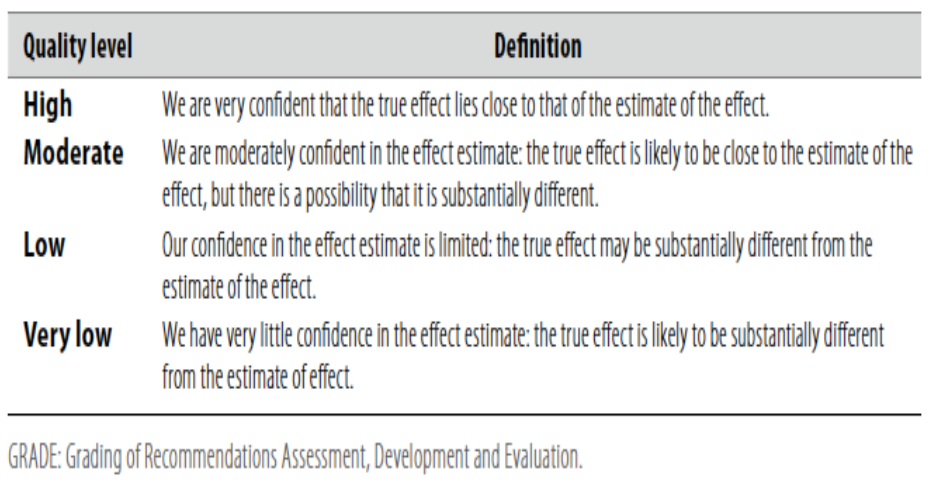
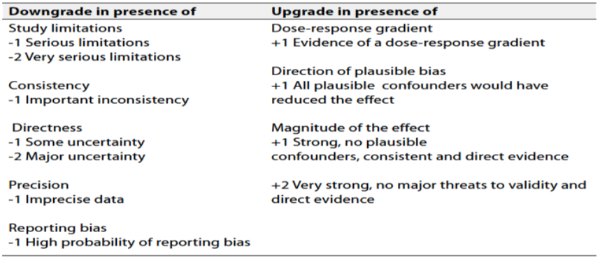
Table (2): Factors that determine How to upgrade or downgrade the quality of Evidence
The strength of the recommendation
The strength of a recommendation communicates the importance of adherence to the recommendation.
Strong recommendations
With strong recommendations, the guideline communicates the message that the desirable effects of adherence to the recommendation outweigh the undesirable effects. This means that in most situations the recommendation can be adopted as policy.
Conditional recommendations
These are made when there is greater uncertainty about the four factors above or if local adaptation has to account for a greater variety in values and preferences, or when resource use makes the intervention suitable for some, but not for other locations. This means that there is a need for substantial debate and involvement of stakeholders before this recommendation can be adopted as policy.
When not to make recommendations
When there is lack of evidence on the effectiveness of an intervention, it may be appropriate not to make a recommendation.
- Implementation Considerations
training for neurosurgeons on guidelines application.
- Clinical Indicators
1) Glasgow outcome score on admission and to be repeated every 2 hours.
2) Cranial CT scan on admission for monitoring
- Research Gaps
Role of neuroprotective agents in improving outcome after surgery for traumatic acute subdural hematoma.
- Updating the guideline
To keep these recommendations up to date and ensure its validity it will be periodically updated. This will be done whenever strong new evidence is available and necessitates updating.
- Annexes
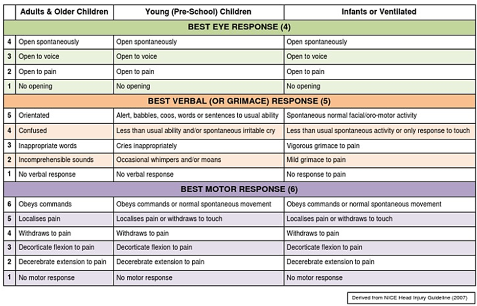
Glasgow Coma Scale
- References
1. Bajsarowicz P, Prakash I, Lamoureux J, Saluja RS, Feyz M, Maleki M, Marcoux J: Nonsurgical acute traumatic subdural hematoma: what is the risk? In J Neurosurg 2015, 123:1176–1183.
2. Karibe H, Hayashi T, Hirano T, Kameyama M, Nakagawa A, Tominaga T: Surgical Management of Traumatic Acute Subdural Hematoma in Adults: A Review Article. In Neurol Med Chir 2014, 54, 887–894.
3. Chen SH, Chen Y, Fang WK, Huang DW, Huang KC, Tseng SH: Comparison of craniotomy and decompressive craniectomy in severely head-injured patients with acute subdural hematoma. J Trauma 2011,71: 1632–1636.
4. Woertgen C, Rothoerl RD, schebesch KM, Albert R: Comparison of craniotomy and craniectomy in patients with acute subdural haematoma. J Clin Neurosci 2006,13: 718–721.
5. Guidelines for the Management of Severe Traumatic Brain Injury 4th Edition: Brain Trauma Foundation: September 2016.
6. Greenberg’s Handbook of Neurosurgery: Head Trauma – General Information, Grading, Initial Management July 2022; 53: e1000–e1018.
7. Bullock MR, Chesnut R, Ghajar J, Gordon D, Hartl R, Newell DW, et al: Surgical Management Of acute subdural hematomas. In Neurosurgery 2006, 58: S2-16-S2-24.