العمل الإجرائي للأمراض القلبية
| الموقع: | EHC | Egyptian Health Council |
| المقرر الدراسي: | دلائل الاجراءات التمريضية لأقسام الداخلي |
| كتاب: | العمل الإجرائي للأمراض القلبية |
| طبع بواسطة: | مستخدم ضيف |
| التاريخ: | السبت، 20 يونيو 2026، 10:59 PM |
الوصف
"last update: 25 March 2025" تحميل الدليل
- اعداد
تحت اشراف
- أ.د/ محمد لطيف الرئيس التنفيذي للمجلس الصحي المصري - د/ كوثر محمود نقيب عام التمريض المصري – عضو مجلس الشيوخ
اعداد
م | الاسم | الوظيفة |
1 | أ. د /امل احمد خليل مرسي | نائب رئيس الجامعة لشئون التعليم والطلاب – جامعة بورسعيد |
2 | أ.د / عفاف عبد العزيز عبد العزيز بصل | عميد كلية التمريض –استاذ تمريض باطنه وجراحي جامعة طنطا |
3 | أ.د/ زينب حسين على محمد سعد | وكيل الكلية لشئون البيئة وخدمة المجتمع – كلية التمريض – جامعه حلوان |
4 | أ.د /امل سعيد طه رفاعي | أستاذ ورئيس قسم التمريض الباطني الجراحي – جامعة بنها |
5 | أ.د /حنان احمد السباعي على | استاذ التمريض التمريض الباطني الجراحي- كلية التمريض – جامعة القاهرة |
6 | د /نيفين عبدربه النبي محمد عبد النبي | رئيس الإدارة المركزية ندباَ –وزاره الصحة |
7 | د /مايسه حسني احمد تمام | مدير عام للإدارة ندباَ – وزاره الصحة |
8 | د نانسي علاء الدين عبد الباسط على | المشرف على التعليم الفني- الهيئة العامة للرعاية الصحية |
9 | د شيرين محمد محمد سعدالدين | المشرف على تطوير الخدمات التمريضية –الهيئة العامة للرعاية الصحية |
10 | د/ مى محمود العسال | مدير عام الإدارة العامة لشئون المعاهد الفنية الصحية |
11 | أ.م.د/ هبة محمود محمد | أستاذ مساعد تمريض صحة الام وحديثي الولادة –كلية التمريض - جامعة عين شمس |
المشاركين | ||
12 | /أنهلة كامل مصطفي | مسئول التمريض بالهيئة العامة للرعاية الصحية فرع الإسماعيلية |
13 | /أمها سعد محمد النادي | عضو إدارة التمريض بالهيئة العامة للرعاية الصحية فرع الإسماعيلية |
14 | /أثروت عبد العال محمد | عضو إدارة التمريض بالهيئة العامة للرعاية الصحية فرع الإسماعيلية |
15 | أ /منى على عبد الرحمن الكتامى | أخصائي تمريض بالإدارة العامة للتمريض- وزارة الصحة |
16 | أ / شيرين عبد الحكيم عبد الحكيم خطاب | أخصائي تمريض بالإدارة العامة للتمريض- وزارة الصحة |
17 | أ/بهاء فؤاد برسوم | أخصائي تمريض بالإدارة العامة للتمريض- وزارة الصحة |
- قسم المهنة
أن أخلص فى عملى و أتقى الله فى مهنتى و أحترم قوانينها و أنظمتها و أؤدى مهامى بكل كفاءة و إخلاص
و أن أستند فى أدائى على المعرفة المستمدة من علوم التمريض
و أبذل قصارى جهدى لرعاية كل من وكل إلى رعايتهم و أحفظ كرامتهم و أكتم سرهم و أدافع عن حقوقهم و حمايتهم من أى أذى
و ألا أخشى فى قول الحق لومة لائم و أوفر بيئة أمنة للمريض و الأسرة و المجتمع
و أن أستمر فى تطوير نفسى و أوقر من علمنى و أحترمه
و أتعاون مع زملائى فى المهنة على البر و التقوى
والله على ما أقول شهيد"
- رؤية ورسالة الرعاية التمريضية
رؤية الرعاية التمريضية بوحدة رعاية مرضى الداخلى
يتطلع أفراد هيئة التمريض بوحدة رعاية مرضى الداخلى للإرتقاء بمهنة التمريض وتقديم الرعاية التمريضية لمرضى الوحدة بفاعلية وأمان وجودة طبقاً للمعايير المصرية والقومية والعالمية.
رسالة الرعاية التمريضية بوحدة رعاية مرضى الداخلى
يلتزم أفراد هيئة التمريض بوحدة رعاية مرضى الداخلى برفع المستوى الصحي للمرضى وتقديم أفضل رعاية تمريضية لهم بما يتماشى مع أهداف وإجراءات المستشفى وإدارة التمريض وكذلك تعمل على الإرتقاء بالمستوى العلمي والعملي لجميع أفراد هيئة التمريض بالوحدة وتغيير إتجاهاتهم نحو الإتجاهات الحديثة فى العلوم التمريضية والطبية .
مقدمة عن أمراض القلب والأوعية الدموية
أمراض القلب و الاوعية الدموية
Cardiovascular diseases (CVDs)
مقدمة عن أمراض القلب و الاوعية الدموية
- أمراض القلب والأوعية الدموية هي السبب الرئيسي للوفاة على المستوى العالميّ.
- توفي، بحسب التقديرات، نحو 17,9 مليون شخص بسبب أمراض القلب والأوعية الدموية في عام 2019، أي ما يمثل نسبة 32٪ من مجموع وفيات العالم، منها نسبة 85٪ كانت وفيات ناجمة عن النوبات القلبية والسكتات الدماغية.
- تحدث أكثر من ثلاثة أرباع الوفيات الناجمة عن أمراض القلب والأوعية الدموية في البلدان المنخفضة والمتوسّطة الدخل.
- تسبّبت أمراض القلب والأوعية الدموية في وفيات نسبتها 38٪ من أصل الوفيات المبكرة (دون سن 70 عاماً) البالغ عددها 17 مليون وفاة بسبب الأمراض غير السارية في عام 2019.
- يمكن الوقاية من معظم أمراض القلب والأوعية الدموية بمعالجة عوامل الخطر السلوكية والبيئية المسبّبة لها، مثل التدخين والنظام الغذائي غير الصحّي والسمنة والخمول البدني وتعاطي الكحول على نحو ضار وتلوّث الهواء.
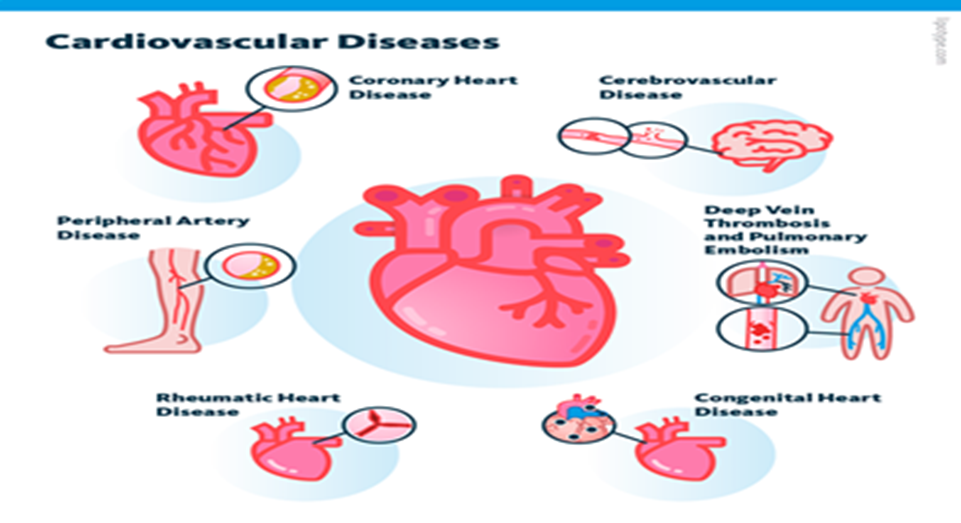
أهم امراض القلب و الاوعية الدموية
- أمراض الشرايين التاجية Coronary heart diseases (CHD)
- أمراض الأوعية الدموية الدماغية Cerebrovascular diseases
- أمراض الشرايين الطرفية Peripheral artery diseases
- أمراض القلب الروماتزمية Rheumatic heart diseases
- أمراض العيوب الخلقية congenital heart diseases
- الجلطات الوريدية العميقة و السدة الرئوية Deep venous thrombosis and pulmonary embolism
- الذبحة الصدرية
التعريف:
هي عبارة عن معاناة العضلة القلبية من نقصان حاد مفاجئ في الأوكسجين الضروري لعملها بسبب ترويه شريانية قلبية غير كافية و التى تسبب شعور بالعصر أو الضغط أو الثقل أو الضيق أو الألم في الصدر. وقد يؤدي ذلك إلى الشعور بثقل مُلقى على الصدر.
أنواع الذبحة الصدرية:
1- الذبحة الصدرية الثابتة المستقرة:
إن هذا النوع يمكن أن ينتقل وخلال مدة إلي مرحلة يتكيف فيها المريض مع مرضه ويكون عامل الوقت قد لعب دوره وسمح للدوران الجانبي المعواض بالنمو فإذا بنوبات الألم لا تحدث إلا نادرا وإذا ما حدثت فإنها تكون خفيفة وغالبا ما يقي العلاج الطبي ويساعد على راحة المريض تماما.
2- نوبات الذبحة الغير مستقرة (التناذر المنذر):
وفيها الألم ينتاب المريض فجأة أثناء الراحة او باقل مجهود وبشكل نوبات متعددة ومتزايدة و تكون الذبحة الصدرية غير المستقرة شديدة عادةً وتستمر فترة أطول من الذبحة الصدرية المستقرة، فقد تستمر لمدة 20 دقيقة أو أكثر. ولا يختفي الألم مع الراحة أو مع تناول أدوية الذبحة الصدرية المعتادة (النيتروجلسرين).
3- حالات الذبحة الصدرية المتكررة:
وفي هذا النوع ينتاب المريض أكثر من نوبة في اليوم الواحد ويزداد تناوله لأقراص النيتوجلسرين / الواحد بعد الآخر حتى لتصل أحيانا إلي عشرة أو عشرين حبة في اليوم وهذه الحالة ممكن أن تستمر أسابيع عدة فإنما يفترض في حصولها أحد أمرين اثنين فإما أن تكون تعبيرا عن إصابات متعددة ومهمة لجذوع الشرايين الثلاثة وإما أن تكون الإصابة عادية ولكن المريض يقدم لها بنفسية القلقة وحالته العصبية المميزة.
4- النوبات الملتوية أو "المموهة":
لهذه الحالات أهمية خاصة ونعني بها النوبات التي تحدث عند مرضى يشكون مشاكل وحالات مرضية أخرى غير القصور الإكليلي . الألم في مثل هذه الحالات غالبا ما يدل في امتداداته وأوقات حصوله وتغيراته على موضع وأسباب الإصابة أو المرض المعني كما هي الحالة مثلا في حالات الروماتيزم الفقري الرقبي (الديسك) أو اضطرابات الجهاز الهضمي كما في قرحة المعدة ويجب البحث إذا كانت تلك الحالات وحدها مسئولة عما يحدث من أعراض أم أنها تتوافق فعلا مع قصور إكليلي حقيقي تزيد من حدته وأهميته.
علامات وأعراض الذبحة الصدرية:
· تنتاب الذبحة الصدرية صاحبها بشكل نوبات مؤلمة والألم يظهر في مقدمة الصدر وخلف عظم القص ينمو ويتطور ولكن يبقى في الوسط بين الثديين أو ينتشر صعودا نحو أعلى القص والعنق أو نحو الفك الأسفل في جانبه ، والذراع الأيسر غالبا ما ينال حصته في جانبه الداخلي المقابل للجذع أو بشكل طوق حول الأصبعين الأخريين لليد اليسرى.
· وأكثر الأحيان يكون الألم عاصرا وأحيانا يكون ساحقا أو يشكل ضغط أو كماشة تطبق على الصدر بشدة أو ضيق بسط أو ثقل على الصدر أحيانا إلي حريق داخله.
· ويكون الشخص شاحب اللون – زيادة العرق وزيادة في عدد ضربات القلب واضطرابات فيها.
الطرق التشخيصية للذبحة الصدرية :
1- التاريخ المرضي:
وفيها يعطى المريض تاريخ كامل عن مرضه وفيها يوصف المريض النوبة وعما إذا كانت تصاحب مجهود / عصبية أو بعد تناول وجبة دسمة وفيها يوصف خصائص الألم ومكانه ومدته والمناطق التي يسمح فيها وإزالته.
2- أشعة على الصدر:
وفيها يظهر إذا كان هناك تضخم في القلب أو احتقان في الرئة.
3- تصوير الشرايين:
وفيها يفحص المريض لتشخيصه واختيار المرضى الذين يتم لهم عملية الوصل (الأبهري – التاجي) الـ By Pass .
4- رسم القلب العادي :
وفيها يحدث انخفاض في St Segment وذلك يحدث مع T wave inversion
5- رسم القلب الجهدي :
وفيها يقوم المريض بمجهود وذلك أثناء ركوب المريض دراجة معينة أو أثناء سيره في مكانه على بساط كهربائي متحرك وباتجاه معاكس لسيره أو صعود السلم ونتيجة لذلك يحدث زيادة في ضربات القلب وضغط الدم وعمل القلب وعندئذ يظهر الألم الذبحة الصدرية أو تغيرات في رسم القلب.
العلاج الطبي للذبحة الصدرية :
1- الأسبرين :
ويعمل على منع تجمع الصفائح الدموية وبالتالي يقلل من نسبة حدوث جلطة وتقليل حالات الوفاة في المرضى المصابين بالذبحة الصدرية.
2- الهيبارين :
ويعطي لمنع حدوث جلطة ويعطي 5000 وحدة وريد كل من 4 إلي 6 ساعات مع استمرار عمل رسم قلب لتحديد فاعلية عمل الهيبارين . ويتم عمل PTT أو PT بعد ساعتين من إعطاء الهيبارين في حالة إعطائه بالوريد كل 4 ساعات وبعد ذلك يوميا.
3- الأدوية المعالجة لألم الذبحة الصدرية (Nitro glycerin) نيتروجلسرين :
تزيد من توسع الشرايين وبالتالي من قدرتها على حمل كمية أكبر من الدم وهذه الأدوية وأدوية أخرى عديدة تعد من ناحية ثانية بتوسيعها لشرايين القلب الصغيرة مساعدة على نشوء ونمو شبكة الشرايين الجانبية التي تحمل الدم إلي الأماكن الناقصة الترويه وتسمى (الدوران الجانبي) وهي أيضا تقوم بتوسيع أوعية الجسم المحيطة مما يخفف الضغط داخلها وبالتالي العبء الملقى على القلب وتدخل هذه الأدوية في حيز عمل الفئة الثانية من العقاقير التي ينحصر عملها في تقليل عمل العضلة القلبية ويخفف استهلاكها للأوكسجين.
4- Beta adrenergic blockcing:
تعتبر هذه الأدوية ذات أهمية كبيرة إذ أنها لها القدرة على تقليل عدد ضربات القلب وضغط الدم والانقباضات (عضلة القلب) وهي أيضا تقلل من احتياجات عضلة القلب للأوكسجين وهم أيضا لهم القدرة على منع حدوث اضطرابات في ضربات القلب (arrhythmias) ويقللوا نسبة حدوث جلطة القلب والموت المفاجئ . مثال تينورمين.
5- Calcium channel blocking :
تعتبر هذه المجموعة من أهم الأدوية التي لها القدرة على تقليل انقباض عضلة القلب وارتخاء العضلات الرفيعة الموجودة في الأوعية الفرعية والقلبية Coronary and Peripheral vascular smooth muscle وهم أيضا لهم القدرة على التحكم في ألم الصدر في الذبحة الغير مستقرة. مثال التيازيم.
6- العلاج الجراحي (عملية " التجسير " أو " الوصل " الأبهري – التاجي) :
وقد سميت بهذا الأسم لأنها تقضي بنزع وريد من فخذ المريض نفسه "الوريد العافن" ويوضع بشكل جسر بين مكان في الشريان الأبهر وآخر في الشريان التاجي المصلب بعد موضع التضييق فيه.
وبعد عملية الزرع يجري الدم في هذا الجسر "الجوي" الوريدي من الأبهر حيث يندفع الدم بقوة باتجاه العضلة القلبية المعنية عبر بقية الشريان المصاب نفسه تاركا التضييق نفسه وشأنه ومعوضا عنه بزيادة تروية هذه العضلة وتزويدها بالأوكسجين.
- احتشاء عضلة القلب
ما هو احتشاء عضلة القلب؟
احتشاء عضلة القلب (Myocardial Infarction) يُستخدم كمصطلح مرادف لانسداد الشريان التاجي والنوبة القلبية، إلا أن مصطلح "احتشاء عضلة القلب" هو الأكثر تفضيلًا لأن نقص تروية عضلة القلب يؤدي إلى متلازمة الشريان التاجي الحادة (ACS) التي يمكن أن تؤدي إلى موت عضلة القلب.
في حالة احتشاء عضلة القلب، يتم تدمير منطقة من عضلة القلب بشكل دائم بسبب تمزق الترسبات الدهنية وتكوين الجلطة الدموية الذي ينتج عنه انسداد كامل في الشريان. تشمل نطاقات متلازمة الشريان التاجي الحادة الذبحة الصدرية غير المستقرة، واحتشاء عضلة القلب بدون ارتفاع في مقطع ST، واحتشاء عضلة القلب مع ارتفاع في مقطع ST.
الفسيولوجيا المرضية
في كل حالة من حالات احتشاء عضلة القلب، يوجد اختلال كبير بين تزويد الأكسجين لعضلة القلب واحتياجها.
الذبحة الصدرية غير المستقرة: يوجد تدفق دم منخفض في الشريان التاجي، وغالبًا ما يكون ذلك بسبب تمزق ترسبات تصلب الشرايين، ولكن الشريان لا يكون مسدودًا تمامًا.
تطور الاحتشاء: عندما تحرم الخلايا من الأكسجين، يحدث نقص التروية، وتتعرض الخلايا للإصابة، ويؤدي نقص الأكسجين إلى الاحتشاء أو تضرر الخلايا.
الأسباب
تعود أسباب احتشاء عضلة القلب بشكل رئيسي إلى الاوعية الدموية.
· تشنج الأوعية الدموية: هو انقباض أو تضيق مفاجئ للشريان التاجي.
· انخفاض إمدادات الأكسجين: يحدث انخفاض في إمدادات الأكسجين بسبب فقدان الدم الحاد، أو فقر الدم، أو انخفاض ضغط الدم.
· زيادة الطلب على الأكسجين: يسبب معدل ضربات القلب السريع، أو التسمم الدرقي، أو تعاطي الكوكايين .
الأعراض السريرية
يعاني بعض المرضى من أعراض سابقة أو تشخيص مسبق بمرض الشريان التاجي، ولكن حوالي نصفهم لا يبلغون عن أي أعراض سابقة.
· موقع ألم الصدر أثناء احتشاء عضلة القلب ألم الصدر: هو العرض الرئيسي لاحتشاء عضلة القلب. يتميز بألم مستمر وساحق في منطقة ما تحت القص وقد يمتد إلى الذراع الأيسر، الفك، الرقبة، أو لوحي الكتف. غالبًا ما يوصف الألم بأنه ثقيل، ضاغط، أو ساحق وقد يستمر لأكثر من 12 ساعة.
· ضيق التنفس: يحدث نتيجة زيادة الطلب على الأكسجين وانخفاض في إمداد الأكسجين.
· عسر الهضم: يظهر نتيجة تحفيز الجهاز العصبي.
· تسرع القلب وتسرع التنفس: لزيادة تعويض نقص إمدادات الأكسجين، يتسارع معدل ضربات القلب والتنفس.
· استجابة الكاتيكولامينات: قد يعاني المريض من برودة في الأطراف، عرق، قلق، وتوتر.
· الحمى: نادرًا ما تحدث في بداية احتشاء عضلة القلب، ولكن قد يظهر ارتفاع طفيف في درجة الحرارة خلال الأيام القليلة التالية.
التقييم و الطرق التشخيصية
· يعتمد تشخيص احتشاء عضلة القلب (MI) بشكل عام على الأعراض الظاهرة.
· التاريخ المرضي للمريض: يتضمن التاريخ المرضي وصف الأعراض الحالية، وسجل الأمراض القلبية السابقة وأي أمراض أخرى، بالإضافة إلى التاريخ العائلي لأمراض القلب.
· رسم القلب الكهربى (ECG):
· ارتفاع في مقطع ST يشير إلى نقص التروية.
· موجة T مرتفعة أو مقلوبة تدل على الإصابة.
· تطور موجات Q يشير إلى نقص التروية لفترة طويلة أو تلف بالخلايا.
· الإنزيمات القلبية والإنزيمات المتماثلة:
· CPK-MB (الإنزيم المتماثل في عضلة القلب): يرتفع خلال 4-8 ساعات، ويبلغ ذروته في 12-20 ساعة، ويعود إلى طبيعته في 48-72 ساعة.
· LDH: يرتفع خلال 8-24 ساعة، ويبلغ ذروته خلال 72-144 ساعة، وقد يستغرق ما يصل إلى 14 يومًا للعودة إلى الطبيعي. يُعد نسبة LDH1 أعلى من LDH2 (النسبة المقلوبة) عاملاً مساعدًا لتأكيد/تشخيص احتشاء عضلة القلب إذا لم يتم اكتشافه في المرحلة الحادة.
· التروبونين: تروبونين I (cTnI) وتروبونين T (cTnT): ترتفع المستويات خلال 4-6 ساعات، وتبلغ الذروة في 14-18 ساعة، وتعود إلى المستوى الطبيعي خلال 6-7 أيام. تتمتع هذه الإنزيمات بزيادة في التحديد بتلف بالخلايا وتكون مفيدة في تشخيص احتشاء عضلة القلب بعد العمليات الجراحية عندما قد يرتفع CPK-MB بسبب إصابة الهيكل العظمي.
· الميوجلوبين: بروتين الهيم ذو الوزن الجزيئي الصغير الذي يتم إفرازه بسرعة من الأنسجة العضلية التالفة مع ارتفاع خلال ساعتين بعد احتشاء عضلة القلب الحاد، وتحدث مستويات الذروة خلال 3-15 ساعة.
· الاملاح و المعادن: يمكن أن يؤدي عدم توازن الصوديوم والبوتاسيوم إلى تغيير التوصيل الكهربائي وإضعاف القدرة الانقباضية للقلب.
· كريات الدم البيضاء (WBC): يظهر ارتفاع الكريات البيض (10,000-20,000) عادة في اليوم الثاني بعد احتشاء عضلة القلب بسبب حدوث الالتهاب.
· معدل ترسيب كريات الدم الحمراء (ESR): يرتفع في اليوم الثاني أو الثالث بعد احتشاء عضلة القلب، مما يدل على استجابة للالتهاب.
· غازات الدم الشرياني (ABGs) / مقياس تأكسج النبض: قد تشير إلى نقص الأكسجة أو عمليات مرضية حادة/مزمنة في الرئة.
· الدهون (إجمالي الدهون، HDL، LDL، VLDL، إجمالي الكوليسترول، الدهون الثلاثية، الفوسفوليبيدات): قد تعكس الزيادات تصلب الشرايين كسبب لتضيق أو تشنج الشرايين التاجية.
· الأشعة السينية على الصدر: قد تكون طبيعية أو تظهر القلب متضخما مما يشير إلى فشل القلب أو تمدد البطين.
· ايكو القلب ثنائي الأبعاد: يمكن إجراؤه لتحديد أبعاد الغرف القلبية، وحركة جدار الحاجز/البطين، ونسبة التدفق EF(تدفق الدم)، وتكوين/وظيفة الصمامات.
· دراسات التصوير النووي: بيرسانتين أو ثاليوم: تُقيِّم تدفق الدم إلى عضلة القلب وحالة الخلايا العضلية القلبية، مثل موقع/مدى احتشاء عضلة القلب الحاد أو السابق.
· تصوير الدم القلبي/MUGA: يُقيِّم الأداء البطيني العام والمحدد، وحركة جدار المنطقة، ونسبة تدفق الدم (Ejection Friction
· تصوير الشرايين التاجية: يُظهر تضيق/انسداد الشرايين التاجية وعادة ما يتم إجراؤه بالتزامن مع قياس ضغط الغرف القلبية وتقييم وظيفة البطين الأيسر (EF). لا يتم إجراء هذا الإجراء عادةً في المرحلة الحادة من احتشاء عضلة القلب إلا إذا كانت عملية اصلاح الشرايين أو جراحة القلب الطارئة وشيكة.
· تصوير الشرايين الرقمي (DSA): تقنية تستخدم لتصور حالة التحويلات الجراحية للشريان التاجي وكشف أمراض الشرايين الطرفية.
· التصوير بالرنين المغناطيسي (MRI): يُتيح تصور تدفق الدم، والغرف القلبية أو الحاجز البطيني، والصمامات، الاوعية، وتكوينات الترسبات، ومناطق تلف الخلايا/الاحتشاء، والجلطات الدموية.
· اختبار الإجهاد البدني: يُحدد استجابة القلب والأوعية الدموية للنشاط (غالبًا ما يتم إجراؤه بالتزامن مع تصوير الثاليوم في مرحلة التعافي).
العلاج الطبى
يهدف
العلاج الطبى للذبحة
إلى تقليل تلف عضلة القلب، والحفاظ على وظيفة عضلة القلب، ومنع المضاعفات .
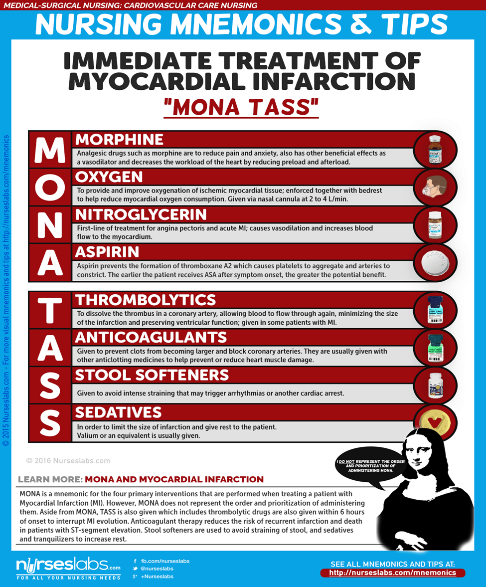
العلاج الدوائي
- المورفين: يُعطى المورفين في جرعات عبر الوريد لعلاج احتشاء عضلة القلب (MI) بهدف تقليل الألم والقلق.
- مثبطات الإنزيم المحول للأنجيوتنسين (ACE Inhibitors): تمنع هذه المثبطات تحويل الأنجيوتنسين I إلى الأنجيوتنسين II لتخفيض ضغط الدم وتشجيع الكلى على إفراز الصوديوم والسوائل، مما يقلل من الطلب على الأكسجين في القلب.
- الأدوية المذيبة للجلطات (Thrombolytics): تذيب هذه الأدوية الجلطة في الشريان التاجي، مما يسمح بتدفق الدم مرة أخرى عبر الشريان التاجي، ويقلل من حجم الاحتشاء ويحافظ على وظيفة البطين.
التدخل الطارئ للشريان التاجي عن طريق الجلد (Emergent Percutaneous Coronary Intervention)
- يُستخدم هذا الإجراء لفتح الشريان التاجي المسدود وتعزيز إعادة التروية إلى المنطقة التي حرمت من الأكسجين.
- قد يُوصى بإجراء PCI أيضًا للمرضى الذين يعانون من الذبحة الصدرية غير المستقرة واحتشاء عضلة القلب بدون ارتفاع في مقطع ST (NSTEMI)، خاصةً للمرضى المعرضين لخطر مرتفع بسبب نقص التروية المستمر.
الرعاية التمريضية
الرعاية التمريضية للمصابين باحتشاء عضلة القلب حاسمة ومنهجية، ويتطلب تقديم الرعاية بكفاءة للمريض.
التقييم التمريضي - يُعد التقييم من أهم جوانب رعاية المريض المصاب باحتشاء عضلة القلب.
- تقييم ألم الصدر الذي لا يزول بالراحة أو الأدوية.
- مراقبة العلامات الحيوية، وخاصةً ضغط الدم ومعدل النبض.
- تقييم وجود ضيق في التنفس، تسارع التنفس، وتواجد أصوات خشخشة في الرئتين.
- تقييم حالات الغثيان والقيء.
- تقييم انخفاض كمية البول.
- مراجعة التاريخ المرضي للمريض.
- إجراء تقييم جسدي دقيق وشامل للكشف عن المضاعفات والتغيرات في حالة المريض.
- تقييم مواقع الوريد باستمرار.
التشخيص التمريضى
استنادًا إلى الأعراض السريرية، والتاريخ المرضي، وبيانات التقييم التشخيصي، قد تشمل التشخيصات التمريضية الرئيسية:
- ضعف في تروية أنسجة القلب نتيجة لتقليل تدفق الدم التاجي.
- خطر ضعف تروية الأنسجة الطرفية نتيجة انخفاض النتاج القلبي بسبب خلل في وظيفة البطين الأيسر.
- نقص المعرفة فيما يتعلق بالرعاية الذاتية بعد احتشاء عضلة القلب.
التخطيط والأهداف
يهدف التخطيط في الرعاية التمريضية إلى تحديد الأهداف لتحقيق أفضل نتائج صحية للمريض، مثل تقليل الألم، تحسين وظيفة القلب، وزيادة معرفة المريض بالرعاية الذاتية بعد الاحتشاء.
لوضع خطة الرعاية، يجب التركيز على الأمور التالية:
- تخفيف الألم أو علامات وأعراض نقص التروية.
- منع تلف عضلة القلب.
- عدم وجود خلل في الجهاز التنفسي.
- الحفاظ على أو تحقيق تروية أنسجة كافية.
- تقليل القلق.
- عدم وجود مضاعفات أو الكشف المبكر عنها.
- غياب/السيطرة على آلام الصدر.
- معدل/نظم القلب كافٍ للحفاظ على النتاج القلبي الكافي وتروية الأنسجة.
- تحقيق مستوى من النشاط يكفي للرعاية الذاتية الأساسية.
- تقليل/إدارة القلق.
- فهم عملية المرض، وخطة العلاج، والتنبؤات.
- وضع خطة لتلبية الاحتياجات بعد الخروج من المستشفى.
الأولويات التمريضية:
- تخفيف الألم والقلق.
- تقليل عبء العمل على القلب.
- منع/الكشف المبكر والمساعدة في علاج الاضطرابات النظمية أو المضاعفات المهددة للحياة.
- تعزيز صحة القلب والرعاية الذاتية.
التدخلات التمريضية:
يجب أن تستند التدخلات التمريضية إلى الأهداف الموضحة في خطة الرعاية التمريضية.
- إعطاء الأكسجين مع العلاج الدوائي للمساعدة في تخفيف الأعراض.
- تشجيع الراحة في الفراش مع رفع مسند الظهر للمساعدة في تقليل انزعاج الصدر وضيق التنفس.
- تشجيع تغيير الوضعيات بشكل متكرر لمنع تجمع السوائل في قواعد الرئتين.
- فحص درجة حرارة الجلد والنبضات الطرفية بشكل متكرر لمراقبة تروية الأنسجة.
- تقديم المعلومات بطريقة صادقة وداعمة.
- مراقبة المريض عن كثب لأي تغيرات في معدل ونظم القلب، أصوات القلب، ضغط الدم، ألم الصدر، الحالة التنفسية، كمية البول، تغيرات في لون الجلد، والقيم المخبرية.
التقييم:
بعد تنفيذ التدخلات في الوقت المحدد، يجب على الممرضة التحقق مما إذا كان:
- عدم وجود ألم أو علامات وأعراض نقص التروية.
- تم منع تلف عضلة القلب.
- عدم وجود خلل في الجهاز التنفسي.
- الحفاظ على تروية أنسجة كافية.
- تم تقليل القلق.
إرشادات الخروج والرعاية المنزلية:
الطريقة الأكثر فعالية لزيادة احتمال أن يلتزم المريض بخطة الرعاية الذاتية بعد الخروج هي تحديد أولويات المريض.
- التثقيف: يُعتبر التثقيف حول نمط الحياة الصحي للقلب أحد الأولويات التي يجب على الممرضة تعليمها للمريض.
- الرعاية المنزلية: تساعد الممرضة المريض في جدولة مواعيد المتابعة والالتزام بها، وكذلك في الالتزام بإدارة إعادة التأهيل القلبي الموصوفة.
- المراقبة والمتابعة: قد يحتاج المريض إلى تذكيرات حول متابعة المراقبة بما في ذلك الفحوصات المخبرية الدورية وتخطيط كهربية القلب، وكذلك الفحص الصحي العام.
- الالتزام: يجب أن تراقب الممرضة أيضًا التزام المريض بالقيود الغذائية والأدوية الموصوفة.
إرشادات التوثيق:لضمان أن كل إجراء موثق هو إجراء تم تنفيذه، يجب تأمين التوثيق. ويجب توثيق ما يلي:
- النتائج الفردية.
- العلامات الحيوية، نظم القلب، وجود اضطرابات النظم.
- خطة الرعاية والأشخاص المشاركين في التخطيط.
- خطة التعليم.
- الاستجابة للتدخلات، التعليم، والإجراءات المنفذة.
- العناية التمريضية لمريض موت جزء من عضلة القلب
العالم كله يعاني من هذا المرض بل إن نسبة الوفيات ترتفع نسبة حدوثها بسبب هذا المرض في العالم كله، وتكثر الوفيات بين الرجال من سن (40 – 70 سنة).
وتندر الوفيات بين السيدات قبل سن اليأس وبالطبع حين نتحدث عن أسباب موت عضلة القلب، فإن ذلك نرده إلي نفس أسباب تصلب الشرايين.
ومعنى موت عضلة القلب أنها تفقد وظيفتها وعملها وتصبح عديمة الجدوى بسبب شئ بسيط (جلطة) يغلق شريانا يغذي عضلة القلب ، فيمنع وصول الدم فتموت العضلة في هذا الجزء، ويتليف هذا الجزء من القلب تتبدل خلاياه ، وتتبدل معها حياة هذا الجزء وهو غالبا في البطين الأيسر والحاجز بين البطينين لسمك جدارها وارتفاع ضغط الدم فيها وبالطبع فإن مكان هذه الإصابة ومدى امتدادها يتوقف على الشريان المصاب، وعلى كفاءة الشرايين الجانبية لدورة الدم في القلب (غالبا ما يكون الشريان التاجي الأيسر).
الأعراض :
· يحدث الألم الحاد على حين غرة غير مسبوق بإجهاد أو توتر (أو ما يسبق الذبحة الصدرية) ويشتد الألم وتكون قمة اشتداده خلف عظمة القص وتنتشر لتصل إلي منتصف الصدر والرقبة والفكين ومنطقة ما يسمى (فم المعدة) وكلا الكتفين، خاصة الكتف الأيسر، والذراع الأيسر.
· الألم مشابه لألم الذبحة في سماته وموقعه وانتشاراته غير أنه أشد حدة وأطول وقتا.
· والألم يتجاوز الساعات وأحيانا يمتد ليوم أو يومين، ولا يتحسن أو يختفي بالراحة ولا 3بتلك الحبة تحت اللسان.
· قد يسبق الألم ذبحة صدرية أو أن الألم يأتي شديدا منذ اللحظة الأولى.
· يصحب الألم عرق غزير وأحيانا يصاحب ذلك غثيان أو قيئ.
· الدليل النهائي لدينا على حدوث تليف جزء من عضلة القلب (موتها) هو رسم القلب المميز لهذه الحالة.
المضاعفات :
· يصاب المريض ببعض المضاعفات أهمها اعتلال القلب، واضطراب نبضاته وزيادتها عن المعدل (70 – 80% دقيقة).
· هبوط القلب، وخلل في الصمامات القلبية (الصمام الميترالي) بصفة خاصة، وتكون تجلطات صغيرة نرى أثرها في من تطول فترة راحتهم أثناء المرض فتظهر الجلطات في الساقين والفخذين.
· احيانا يحدث انتفاخ في بعض أجزاء القلب مما يؤدي إلي فشل القلب، وتكون التجلطات الصغيرة واضطرابات في النبض.
· قد يحدث نزف تحت غشاء القلب يؤدي إلي الوفاة فجأة.
· ويتوزع مصير مرضى (التليف في عضلة القلب أو موتها) بين وفاة (خمس الحالات موتا فجائيا) وقبل أن يذهب إلي الطبيب أو يذهب إليه الطبيب.
· وهناك خمس آخر يحدث خلال شهر بعد الأزمة (نتيجة مضاعفات الحالة).
· وآخرون حوالي (60%) لا تظهر عليهم أعراض، أو قد تهاجمهم أزمات الذبحة الصدرية.
العلاج :
· للتعامل مع المريض يجب أن نعرف أنه إذا أعطى المريض راحة تامة بدنيا ونفسيا فإنه يحدث له تحسن تلقائي ومعروف أن الغرض من إعطاء العقاقير تخفيف الأعراض ومنع أو التعامل مع المضاعفات.
· يجب أن يتعامل مع الألم ولابد أن يتعامل معه في الحال ويكون التعامل بجدية واهتمام (والمورفين ساعتها هو الحل) أو بدائله بمعرفة الطبيب (10 ملليجرام عضل أووريد)، ويكرر إذا لزم الأمر. ويجب أن يلاحظ الطبيب أنه لا تتعدى الجرعات المعطاة في الاثنى عشر ساعة الأولى (60 ملليجرام) حتى لا يحدث هبوط للجهاز التنفسي للمريض أو حدوث نوبات من القيئ
· الراحة ضرورية جدا حتى يحدث التحسن وقد تطول هذه الراحة، لذا يجب ان يراعى الأعراض الجانبية للنوم الطويل في الفراش مثل: (قرحات الفراش– تجلطات في الساقين– التهاب رئوي– احتباس بول– إمساك .. الخ).
· وتكون مدة الراحة في الحالات التي لا تصاحبها مضاعفات (راحة من العمل بعد هذه الفترة) حوالي 3 أسابيع وفي حدوث مضاعفات يكون 6 أسابيع ويعطى المريض مهدئا خفيفا لتخفيف حدة القلق عنده أو منوم.
· يراعى طعام المريض خاصة في الأسابيع الأولى، يحتوي على سعرات أقل– ملح أقل– خفيف سهل الهضم– كميات صغيرة– (وجبات متكررة ).
· الامتناع عن الشاي والقهوة والتدخين امتناعا تاما خاصة في المراحل الأولى.
· ينصح بوضع المريض في غرفة العناية المركزة بالمستشفى للتعامل مع الحالة في وقتها، ومتابعة المضاعفات والتعامل الطبي معها
- العناية بمرضي الشرايين التاجية
العناية بمرضي الشرايين التاجية :
تعتبر الرعاية التمريضية المستمرة في وحدة الرعاية المركزة لها دور حيوي حتى تستقر حالة المريض فعلى الممرضة ملاحظة الآتي :
1. ملاحظة العلامات الحيوية مثل النبض– التنفس– الحرارة وضغط الدم.
2. الراحة التامة في الفراش.
3. تحريك الساقين لتجنب حدوث جلطة الساق.
4. تنفيذ العلاج كما أمر الطبيب بدقة ويلاحظ الأعراض الجانبية للدواء.
5. تقييم وملاحظة السوائل الداخلة والخارجة.
6. ملاحظة جيدة– صعوبة في التنفس– عدم الراحة– ألم بالصدر– الطعام– الأدوية المعطاة.
يجب على الممرضة ابداء بعض الملاحظات على المريض حتى تتمكن من تقييم حالته والاكتشاف المبكر للمضاعفات وتكون الملاحظة إما مباشرة مثل النظر والسمع ولمس المريض أو غير مباشرة مثل قياس ضغط الدم واستعمال المونيتور وتكون ملاحظة المريض كاملة من رأسه حتى قدمه كما يلي :
العين | زرقان في مقلة العين– أو تورم الجفون نتيجة أوديما |
الفم | ملاحظة زرقان حول الشفة أوغشاء الفم أوسماع تزييق أثناء التنفس– كحة في وجود أوعدم وجود بلغم/ شكوى المريض من صعوبة التنفس– ألم بالفك وقيئ. |
الرقبة | احتقان الوريد بالرقبة أو ألم من وإلي الصدر |
الصدر | ملاحظة التنفس وسرعته وعمقه وقياس النبض من القلب |
الذراع | ورم باليد والرسغ نتيجة أوديما زرقان الأظافر. |
البطن | غثيان– قيئ– سوء هضم |
الأرجل | أوديما بالفخذ أوالساق والكعب والقدم وزرقة بأظافر القدم |
الجلد | زرقة أوإصفرار– برودة الجلد– قوى– رطب– علامة نزيف مثل كدمة– نقط حمراء |
تغيرات السلوك | قلق– خوف من موت محتم– الشعور بالكآبة– ألم بالصدر نتيجة مجهود أو صعود السلم |
تغيرات في العلامات الحيوية | زيادة في درجة الحرارة يعني وجود عدوى أوالتهاب- أي تغيير في معدل أوانتظام النبض يدل على وظيفة القلب مثلا مريض القصور بالدورة الدموية يعاني من عدم انتظام النبض ويكون النبض سريع أو بطئ غير طبيعي– زيادة معدل التنفس يعني أن المريض يحتاج إلي المساعدة بالأوكسجين نظرا لقلة الأوكسجين الواصل للأنسجة– الزيادة في ضغط الدم تعتبر من العوامل الخطرة المؤدية إلي أمراض الشرايين التاجية وعلى الممرضة تبليغ الطبيب بعد أن تقيس العلامات الحيوية. |
واجبات أفراد هيئة التمريض :
1. يجب على الممرضة توجيه مجهودها تجاه الاكتشاف المبكر للمضاعفات ومنع إصابات اخرى لعضلة القلب وإعطاء المريض احساسه بالراحة.
2. الملاحظة المستمرة للمونيتور أثناء وجود المريض في وحدة الرعاية المركزة وذلك في الرعاية المتوسطة والقدرة على تفسير وقراءة الـ ECG وذلك لبحث المضاعفات واضطرابات في ضربات القلب.
3. بالنسبة لحدوث إعاقة في عملية تبادل الغازات : يجب على الممرضة أن تكون على دراية بعلامات قلة الأوكسجين بالمخ hypoxia وهي تعتبر في : ضغط الدم– اضطرابات في ضربات القلب– صعوبة في التنفس– دوخه– صداع– عدم اتزان– غثيان– قلق– احساس بعدم الراحة ولذلك يجب عليها ابلاغ الطبيب.
4. إعطاء المريض اكسجين حسب حالته: ويجب على الممرضة القيام بالعناية بالفم والأسنان والشفاه التي قد يحدث تشققات بها نتيجة استعمال الاوكسجين (كريم).
· يجب على الممرضة : أن تقوم بسماع صوت التنفس– وحساب عدد مرات التنفس والعمق والنظام كل 1 ساعة.
· اعطاء المريض مدرات البول : مع ملاحظة أملاح الجسم.
· لإزالة ألم الصدر : يجب على الممرضة أن تقوم بتقييم وتسجيل وصف كامل للألم والنشاط الذي يقوم به لتحديد سبب الألم.
· الراحة الكاملة للمريض لتقليل استهلاك الأكسجين.
· عمل رسم قلب أثناء الألم.
· إعطاء أدوية مزيلة للألم وموسعة للشرايين.
· يجب على الممرضة أن تشجع المريض على ايقاف التدخين لأنه سبب رئيسي في حدوث المرض.
· إعطاء الرعاية التمريضية للمريض الذي يعاني من قيئ وغثيان وتشمل:
· وضع المريض في وضع مريح (نصف جالس).وضع حوض كلوي بجانب المريض.
· التسجيل والتبليغ بمحتويات ولون وكمية ورائحة القيئ.
· إعطاء وجبات صغيرة ومتكررة وسوائل.
· إعطاء أدوية مضادة للقيئ حسب أوامر الطبيب.
· الرعاية التمريضية للفم.
5. بالنسبة للغذاء: قد يوضع المريض على نظام غذائي خاص حسب حالته مثل تقليل الملح بدرجات مختلفة مثل في ارتفاع ضغط الدم والأوديما كذلك التقليل من الدهون والكولسترول كما في حالة المرضى ذو الكولسترول المرتفع كذلك تقليل السعرات الحرارية وأن يكون الغذاء من 5 إلي 6 وجبات صغيرة خالية من الدهون وكذلك تجنب الأطعمة التي تؤدي إلي تكوين غازات وانتفاخ البطن كذلك يجب تجنب الطعام ساخن جدا أو بارد جدا.
6. يجب على الممرضة أن تساعد المريض أن يقلل من نسبة القلق والاضطراب بواسطة طمأنته واحساسه بالراحة وتشجيعه على أن يعبر عما بداخله بالنسبة لمرضه من خوف وتساعده على أن يتأقلم مع حالته المرضية. يجب على الممرضة أن تشجع المريض على القيام ببعض الأنشطة حتى يقلل من نسبة خوفه وذلك مثال قراءة الجرائد أو الكتب.
7. بالنسبة للنشاط الجنسي : هناك بعض النصائح يجب على الممرضة إعطائها للمريض حتى تساعده هو وزوجته على الشعور بالمتعة وإشباع العلاقة الجنسية مع تقليل المجهود على القلب:
· الراحة الكافية قبل العملية الجنسية.
· إيجاد الوضع المريح له ولزوجته.
· تناول بعض العقاقير مثل النيتروجلسرين قبل العملية الجنسية كوقاية لعدم حدوث ألم بالصدر.
· تأجيل العملية الجنسية من 1 –½ 1 ساعة بعد تناول وجبة دسمة.
· تبليغ الطبيب ببعض الأعراض إذا حدثت مصاحبة للعملية الجنسية مثل:
- زيادة عدد ضربات القلب واستمرارها أكثر من 15 دقيقة.
- ألم بالصدر لم يستجيب للنيتروجلسرين.
8. يجب على الممرضة أن تنصح المريض بتجنب التمرينات العضلية العنيفة والذهنية وأن يقوم ببعض التمرينات المتوسطة التي لا تسبب ألم بالصدر مثل المشي الذي يبدأ في مسافة قصيرة خلال الحجرة وبعد ذلك يبدأ في الزيادة بالتدريج خلال فترة أسبوع مع ملاحظة مستمرة من الطبيب.
9. مساعدة المريض في الإخراج: فالغالبية العظمى من المرضى قد يعانون من إمساك:
◀️ فيجب على الممرضة إعطائهم ألياف في الطعام المقدم لهم وسوائل كافية.
◀️ منع الأطعمة الحريفة وإعطاء ملينات.
◀️ إعطاء القصرية للمرضى الغير مسموح لهم بالحركة ويراعى الفردية والسرية.
◀️ عمل خطة للنشاط حسب الحالة والمسموح به.
10. بالنسبة لنقص المعلومات عن المريض عن طبيعة الدواء والوقت والجرعة وعدد مرات أخذه والأعراض الجانبية وتأثيره الدوائي المتوقع يجب على الممرضة أن توضح للمريض عمل والجرعة والوقت والأعراض الجانبية للأدوية التي يتناولها وتحث المريض على أن يكرر هذه المعلومات:
◀️ تشجيع المريض على أن يأخذ قسط من الراحة إذا شعر بالدوخه بعد تناول الأدوية.
◀️ يجب على المريض أن يسجل عدد مرات ألم الصدر والأدوية التي أخذها الجرعة والعدد (Isordil).
◀️ تجنب مشتقات الكحول أثناء تناول الدواء.
◀️ ملاحظة الأعراض الجانبية للدواء وإبلاغ الطبيب.
11. يجب على الممرضة أن تضع أو تخطط برنامج تعليمي لكل مريض على حدة عند خروجه إلي المنزل (للمريض وعائلته) وذلك يشمل:
◀️ التحكم في العوامل الخطرة المختلفة ومدى الاستفادة من ذلك.
◀️ النشاط الجنسي .
◀️ الأدوية المعطاة (الاسم – الجرعة – فاعلية الدواء – الأعراض الجانبية).
◀️ وضع برنامج للتمرينات الرياضية التي يقوم بها بالمنزل.
◀️ أهمية المتابعة والفحص الطبي الدوري. العلامات والأعراض الجانبية التي تتطلب اللجوء إلي الطبيب (ألم بالصدر لم يزول مع استعمال النيتروجلسرين – خفقان – اضطراب في انتظام ضربات القلب – دوخه).
◀️ تعليم أفراد الأسرة خطوات انعاش القلب والصدر.
◀️ أهمية تناول النيتروجلسرين قبل الدخول في أي نشاط أومجهود.
◀️ طرق عد النبض.
◀️ تناول الأطعمة قليلة الملح– الكوليسترول– الدهون والتحكم في السعرات الحرارية .
◀️ التثقيف الصحي لمريض Pacemaker .
◀️ إرشاد عائلة المريض على أهمية توفير جو لطيف وهادئ ومشوب بالحب والاحترام. احترام الإنسان فيه وعدم استعمال السلبية معه وإنما وسائل التعقل والتفهم على أن يكون ذلك في نطاق توصيات الطبيب وتعليماته.
◀️ بالنسبة للعمل يجب أن يتجنب عمل أى مجهود له سواء كان ذلك جسديا أو نفسيا وأن تحاول تبديل الأعمال المجهدة بأخرى أقل مشقة منها إذا أمكن ذلك.
◀️ بالنسبة لحياته اليومية يجب أن تكون بعيدة كل البعد عن الصخب والإزعاج والإجهاد وهذا لا يعني العزلة والابتعاد عن الناس.
◀️ على المريض أن يأخذ القدر الكافي من الراحة اليومية.
◀️ يجب أن يكون السكن جامعا للخصائص من حيث هدوء أجوائه وجيد التهوية يدخله الهواء والشمس.
◀️ يجب أن يكون الماء عند الاستحمام فاترا لا باردا ولا ساخنا ولمدة بسيطة مع وجوب تجنب الوقوف الطويل والأوضاع المتعبة أثناءه.
◀️ بالنسبة للزيارة يجب أن تكون مبعث راحة واطمئنان وفرح للمريض أثناء نقاهته المنزلية ويكون عدد الزائرين قليلا وتكون مدة الزيارة قصيرة ما أمكن ولا تكون عرضه لنقاشات وأحاديث مملة أو مثيرة للحساسية والحماس.
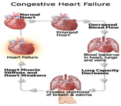
- العناية التمريضية لمريض هبوط القلب
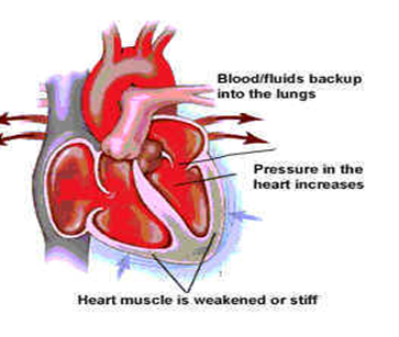
العناية التمريضية لمريض هبوط القلب
Congestive heart Failure
التعريف :
هو فشل عضلة القلب لضخ كمية دم مناسبة لتقابل حاجة الجسم هذا الهبوط إما أن يكون هبوط حاد يؤدى إلى توقف مفاجئ لعضلة القلب لضخ الدم مما ينتج عنه الدم إما أن يكون هبوط مزمن يحدث بالتدريج وتكون أعراضه خفيفة ويتميز هبوط القلب بحدوث عوامل معوضة لكي تعوض هبوط القلب مثل :
• سرعة دقات القلب
• تضخم القلب خاصة البطين ventricular hypertrophy
• توسع حجم البطين ventricular dilatation
• تجمع كميات غير طبيعى من الدم فى الجهاز التنفسىpulmonary congestion
أسبابه :
يحدث هبوط القلب إذا تجمع سبب أو اكثر من الأسباب آلاتية :
1 - كمية الدم الوارد إلى القلب قليلة نتيجة حدوث نزيف اوجفاف .
2 - كمية الدم الوارد إلى القلب تزيد عن الضروري نتيجة لكثرة المحاليل .
3 - كمية الدم الوارد ألي القلب قليلة نتيجة ضيق فى إحدى الصمامات اوالشرايين .
4 - إصابة من أي نوع لعضلة القلب نفسها .
5 - زيادة الاحتياجات الغذائية للجسم نتيجة للحمى الشديدة اوالحمل .
الهدف من العناية بمرض هبوط القلب :
الهدف الاساسى هو التخلص اوالتقليل من الأسباب المؤدية للمرض
1 -راحة تامة جسمية وعضلية .
2 - ديجتاليز DIGITALIS ديجوكسين .
3 - وجبة دسمة مناسب - صوديوم بوتاسيوم
4 - موسع للشرايين والأوردة .
تقليل تجمع الماء والصوديوم داخل الجسم وذلك بأتباع آلاتي :
• تقليل صوديوم فى الوجبات .
• مدر للبول
• تقليل كمية السوائل الداخلة للجسم
• الحد من التوتر للمريض
• إعطاء المريض اكسجين
• إزالة الارتشاح البلوري والارتشاح البريتونى
• إخبار المريض عن كل شئ عن مرضه لكيفية التعامل معه
دور أفراد هيئة التمريض نحو المريض :
1 - وضع المريض فى حجرة هادئة باردة لمساعدته على النوم .
2 - وضع كل الأشياء فى متناول يد المريض .
3 - جعل المريض فى وضع راحة باستمرار .
4 - تحاول أن تطمئن المريض عن صحته .
5 - تكلم المريض من اهمية الراحة له عند عودته للمنزل .
6 - تعريف المريض أعراض التسمم digitalis :
- غثيان - قئ - إسهال - صداع - اكتئاب - توتر - دوخة - تشنج - هلوثة - فقد للذاكرة - زيادة دقات القلب أو قلة دقات القلب - ارتكاريا
- إذا حدث أي من الاعراض السابقة يجب التوقف عن العلاج .
- إعطاء المريض كلوريد البوتاسيوم .
يجب قبل إعطاء digitalis :
1- اخذ نبض المريض لدقيقة كاملة بالسماعة من على صدر المريض
2- نلاحظ بعناية معدل النبضات إذا كانت منتظمة اوغير منتظمة وتسجيلها
3- إذا كان نبض المريض سريع اواقل من 60 نبضة فى الدقيقة يجب إيقاف الجرعة وأبلاغ اطبيب
4- يجب ملاحظة المريض بعناية لأي عرض من أعراض التسمم بالديجتاليز
5- يجب إعطاء المريض وجبات غنية بالبوتاسيوم
6- تجنب إعطاء المريض أي طعام يحتوى على أملاح الصوديوم مثل الفول السودانى – الشيبسى والبعد عن الملح نهائى
7- وزن المريض كل يوم فى نفس الوقت ونفس الميزان وعادة قبل الإفطار
8- العناية بجلد المريض لان الجلد المتورم يكون اكثر عرضه للتشقق
9- العلاج بالأكسجين فى حالة صعوبة التنفس
10- يجب عمل خريطة .
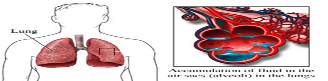
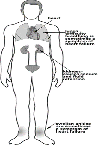
- العناية التمريضية لمريض الارتشاح الرئوي
التعريف :
الارتشاح الرئوى هو احتقان الرئة نتيجة زيادة كمية الدم فى الأوعية الدموية الموجودة في الجهاز التنفسى مما يؤدى إلى صعوبة كبيرة فى التنفس .
هذا الارتشاح يحدث غالبا نتيجة فشل الجزء الأيسر من القلب مما ينتج عنه صعوبة فى عودة الدم من الجهاز التنفسى الى القلب فيؤدى الى تراكم الدم فى الرئة ويؤدى إلى الارتشاح الرئوى هذا الارتشاح يؤدى إلى نهاية حياة المريض من الاختناق
الأعراض :
1- صعوبة شديدة فى التنفس خاصة فى الوضع الأفقي
2- شحوب فى الوجه
3- زيادة عدد دقات القلب
4- افراز كميات كبيرة من البلغم المخلوط بالدم 5- ازرقاق
الهدف الأساسي لرعايتنا لهذا المريض هو مساعدة المريض لكي يأخذ كمية الأكسجين المحتاج لها
دور أفراد هيئة التمريض نحو المريض :
1 - نوم المريض بزاوية 90 أو 45 درجة أو جلوسه على كرسى لان هذا الوضع يسهل عملية التنفس للمريض
2- إمداد المريض ب8 لتر أكسجين رطب
3- إعطاء المريض ديجوكسين
4- إعطاء المريض مدر للبول مثل لازكس 40 جم إلى 120 حجم وريد ببطء
5- إعطاء امينوفللين 250 إلى 500 حجم لتوسيع الشعب الهوائية
6- أحيانا يحتاج المريض لجهاز تنفس صناعى لإنقاذ حياته.
- المراجع
- National Institutes of Health (NIH): https://www.nih.gov/
- Centers for Disease Control and Prevention (CDC): https://www.cdc.gov/
- American Heart Association (AHA): https://www.heart.org/
- Braunwald's Heart Disease: A Textbook of Cardiovascular Medicine by Eugene Braunwald
- Harrison's Principles of Internal Medicine (various editions)