تخطى إلى المحتوى الرئيسي

Endovascular Intervention

الموقع: EHC | Egyptian Health Council
المقرر الدراسي: طب المخ والأعصاب
كتاب: Endovascular Intervention
طبع بواسطة: Guest user
التاريخ: الأربعاء، 6 مايو 2026، 3:45 AM

الوصف

"last update: 27 July  2025"                                                                                                        Download Guideline

- Executive Summary

These guidelines are concerned with clinical practice standards of endovascular intervention neurology procedures. It will discuss practicing physician qualifications, specific requirement for treating centers and specific pre & postoperative care and indications of different endovascular intervention neurology procedures.


I)  PHYSICIAN QUALIFICATIONS

1-    Practicing physician must have a valid license to practice medicine within their respective countries.

2-    Practicing physician specialization criteria should be defined at a national level according to national medical regulations. They must have accomplished training in one of the following medical specialties: Neurology, Neurosurgery, Intervention radiology.

3-    Practicing physician must have completed an accredited post graduate dedicated training in intervention neurology subspecialty. This program should have not less than 24 months mandatory dedicated training in intervention neurology.

Good practice statement.

II)   REQUIREMENTS FOR PRACTICING INSTITUTIONS/DEPARTMENTS

1- Intervention neurology practicing must take place in institutions/departments operating in accordance with the national standards of medical service providence.

2- All patients would be treated at a center offering a full spectrum of neuroendovascular care.

3- Treating centers should have the following requirements at least to provide safe and efficient intervention neurology services:

a. Offers full spectrum of neuroendovascular therapy (including aneurysm treatment, surgical and endovascular, arteriovenous malformations, arteriovenous fistulas, etc.)

b. At least 250 case per year of stroke patients’ management in a dedicated neuroscience department.

c. Dedicated intensive care unit/stroke unit to manage pre- and post-operative patients.

d.Standardized care pathways should be implemented with clinical practice guidelines, order sets, and other tools to ensure consistent care delivery and minimize practice variability. This should apply to providers, and nursing and ancillary staff.

Strong recommendation


III) PREPROCEDURE PATIENT CARE

1-    Preprocedural documentation for elective diagnostic cervicocerebral/spinal catheter angiography, must contain the following:

a.  Clinically significant history, including indications for the procedure.

b.  Clinically significant physical examination and diagnostic imaging findings, including neurological and vascular examinations appropriate to the procedure performed, and a general examination of relevant organ systems.

c.  Laboratory evaluation as appropriate, including but not limited to measurement of hemoglobin, hematocrit, creatinine, electrolytes, and coagulation parameters.

d.  Informed consent must be in compliance with all local laws and policies.

Strong recommendation

IV)PATIENT SELECTION, INDICATIONS AND OUTCOMES

A. Diagnostic Angiography

1- Diagnostic cervicocerebral/spinal catheter angiography is a proven, safe, and effective procedure for evaluating many intracranial and extracranial disorders, especially vascular abnormalities of the head, neck, and brain. Strong recommendation

2- Diagnostic cervicocerebral/spinal catheter angiography has been considered the gold standard for judging the accuracy of other intracranial or extracranial vascular imaging modalities. Strong recommendation

3-The following list of indications helps to focus on the primary indications for diagnostic cervicocerebral/spinal catheter angiography and therefore helps to avoid unnecessary testing:

a. Definition of the presence and extent of atherosclerotic occlusive disease and thromboembolic phenomena and as an aid in planning intervention.

b. Definition of the etiology of cervicocerebral/spinal hemorrhage.

c.  Definition of the presence, location, and anatomy of extra- or intracranial and spinal aneurysms and vascular malformations.

d. Evaluation of vasospasm related to subarachnoid hemorrhage or drug-induced vasculopathy.

e. Definition of the vascular supply to cervicocerebral/spinal tumors.

f.   Diagnosis and definition of the nature and extent of cervicocerebral/spinal congenital or acquired vascular abnormalities.

g. Definition of the presence of venous occlusive disease.

h. Definition of the relevant vascular anatomy for planning or evaluating a therapeutic intervention.

Strong recommendation

4-    The threshold for these indications is 99%. When fewer than 99% of the procedures are for these indications, the institution should review the process of patient selection. Strong recommendation

5-    There are no absolute contraindications to diagnostic cervicocerebral catheter angiography. Relative contraindications include hypotension, severe hypertension, and coagulopathy, clinically significant sensitivity to iodinated contrast material, renal insufficiency, and congestive heart failure. Strong recommendation

6-    Patient management should address these relative contraindications prior to the procedure. When possible, every effort should be made to correct or control these clinical situations before the procedure. Strong recommendation

7-    Diagnostic cervicocerebral/spinal catheter angiographic examinations must be performed by or under the supervision of and interpreted by a physician who has the appropriate qualification and training in the field of cervicocerebral/spinal catheter angiography. Strong recommendation


B. Acute Ischemic Stroke (Mechanical Thrombectomy)
From 0 to 6 hours From Onset:

1-    Patients should receive mechanical thrombectomy with a stent retriever if they meet all the following criteria: (1) pre stroke mRS score of 0 to 1; (2) causative occlusion of the internal carotid artery or MCA segment 1 (M1); (3) age ≥18 years; (4) NIHSS score of ≥6; (5) ASPECTS of ≥6; and (6), treatment can be initiated (groin puncture) within 6 hours of symptom onset. Strong recommendation

2-  Although the benefits are uncertain, the use of mechanical thrombectomy with stent retrievers may be reasonable for carefully selected patients with AIS in whom treatment can be initiated (groin puncture) within 6 hours of symptom onset and who have causative occlusion of the MCA segment 2 (M2) or MCA segment 3 (M3) portion of the MCAs. Conditional recommendation

3-  Although its benefits are uncertain, the use of mechanical thrombectomy with stent retrievers may be reasonable for patients with AIS in whom treatment can be initiated (groin puncture) within 6 hours of symptom onset and who have pre stroke mRS score >1, ASPECTS <6, or NIHSS score<6, and causative occlusion of the internal carotid artery (ICA) or proximal MCA (M1). Conditional recommendation

4-  Although the benefits are uncertain, the use of mechanical thrombectomy with stent retrievers may be reasonable for carefully selected patients with AIS in whom treatment can be initiated (groin puncture) within 6 hours of symptom onset and who have causative occlusion of the anterior cerebral arteries, vertebral arteries, basilar artery, or posterior cerebral arteries. Conditional recommendation

From 6 to 24 hours From Onset:

1- In selected patients with AIS within 6 to 16 hours of last known normal who have LVO in the anterior circulation and meet other DAWN or DEFUSE 3 eligibility criteria, mechanical thrombectomy is recommended. Strong recommendation

2- In selected patients with AIS within 16 to 24 hours of last known normal who have LVO in the anterior circulation and meet other DAWN eligibility criteria, mechanical thrombectomy is reasonable. Conditional recommendation

Thrombectomy Technique:

1- The use of stent retrievers is indicated in preference to the Mechanical Embolus Removal in Cerebral Ischemia (MERCI) device. Strong recommendation

2- The use of mechanical thrombectomy devices other than stent retrievers as first-line devices for mechanical thrombectomy may be reasonable in some circumstances, but stent retrievers remain the first choice. Conditional recommendation

3- The use of a proximal balloon guide catheter or a large-bore distal-access catheter, rather than a cervical guide catheter alone, in conjunction with stent retrievers may be beneficial. Conditional recommendation

4- The technical goal of the thrombectomy procedure should be reperfusion to a modified Thrombolysis in Cerebral Infarction (mTICI) grade 2b/3 angiographic result to maximize the probability of a good functional clinical outcome. Strong recommendation

5- To ensure benefit, reperfusion to mTICI grade 2b/3 should be achieved as early as possible within the therapeutic window. Strong recommendation

6- Use of salvage technical adjuncts including intra-arterial thrombolysis may be reasonable to achieve mTICI 2b/3 angiographic results. Conditional recommendation

7- In the 6- to 24-hour thrombectomy window evaluation and treatment should proceed as rapidly as possible to ensure access to treatment for the greatest proportion of patients. Strong recommendation

8- Direct aspiration thrombectomy as first-pass mechanical thrombectomy is recommended as noninferior to stent retriever for patients who meet all the following criteria: (1) pre-stroke mRS score of 0 to 1; (2) causative occlusion of the internal carotid artery or M1; (3) age ≥18 years; (4) NIHSS score of ≥6; (5) ASPECTS ≥6; and (6) treatment initiation (groin puncture) within 6 hours of symptom onset. Strong recommendation

9- It is reasonable to select an anesthetic technique during EVT for AIS on the basis of individualized assessment of patient risk factors, technical performance of the procedure, and other clinical characteristics. Conditional recommendation

10- Treatment of tandem occlusions (both extracranial and intracranial occlusions) when performing mechanical thrombectomy may be reasonable. Conditional recommendation

11- The safety and efficacy of IV glycoprotein IIb/IIIa inhibitors administered during endovascular stroke treatment are uncertain. Conditional recommendation

Other Endovascular Therapies:

1-    Mechanical thrombectomy with stent retrievers is recommended over intra-arterial fibrinolysis as first-line therapy. Strong recommendation

2-    Intra-arterial fibrinolysis initiated within 6 hours of stroke onset in carefully selected patients who have contraindications to the use of IV alteplase might be considered, but the consequences are unknown. Conditional recommendation

C. Endovascular Management For Secondary Prevention Of Ischemic Stroke

Intracranial Large Artery Atherosclerosis

1-  In patients with severe stenosis (70%-99%) of a major intracranial artery and actively progressing symptoms or so-called medical failures; recurrent TIA or stroke after institution of aspirin and clopidogrel therapy, achievement of SBP< 140 mmHg, and high-intensity statin therapy, the usefulness of angioplasty alone or stent placement to prevent ischemic stroke in the territory of the stenotic artery is recommended. Strong recommendation

2-  In patients with stroke or TIA attributable to severe stenosis (70%–99%) of a major intracranial artery, angioplasty, and stenting should not be performed as an initial treatment, even for patients who were taking an antithrombotic agent at the time of the stroke or TIA. Conditional recommendation

3- In patients with a stroke or TIA attributable to moderate stenosis (50%–69%) of a major intracranial artery, angioplasty or stenting is associated with excess morbidity and mortality compared with medical management alone. Conditional recommendation

4- In patients with stroke or TIA attributable to 50% to 99% stenosis or occlusion of a major intracranial artery, extracranial-intracranial bypass surgery may be considered when indicated. Conditional recommendation

Extracranial Carotid stenosis

1- In patients with a TIA or non-disabling ischemic stroke within the past 6 months and ipsilateral severe (70%–99%) carotid artery stenosis, carotid endarterectomy (CEA) is recommended to reduce the risk of future stroke, provided that perioperative morbidity and mortality risk is estimated to be<6%. Strong recommendation

2- In patients with ischemic stroke or TIA and symptomatic extracranial carotid stenosis who are scheduled for carotid artery stenting (CAS) or CEA, procedures should be performed by operators with established periprocedural stroke and mortality rates of<6%. Strong recommendation

3- In patients with carotid artery stenosis and a TIA or stroke, intensive medical therapy, with antiplatelet therapy, lipid lowering therapy, and treatment of hypertension, is recommended to reduce stroke risk. Strong recommendation

4- In patients with recent TIA or ischemic stroke and ipsilateral moderate (50%–69%) carotid stenosis as documented by catheter-based imaging or noninvasive imaging, CEA is recommended to reduce the risk of future stroke, depending on patient-specific factors such as age, sex, and comorbidities, if the perioperative morbidity and mortality risk is estimated to be<6%. Strong recommendation

5- In patients ≥70 years of age with stroke or TIA in whom carotid revascularization is being considered, it is reasonable to select CEA over CAS to reduce the periprocedural stroke rate. Conditional recommendation

6- In patients in whom revascularization is planned within 1 week of the index stroke, it is reasonable to choose CEA over CAS to reduce the periprocedural stroke rate. Conditional recommendation

7- In patients with TIA or nondisabling stroke, when revascularization is indicated, it is reasonable to perform the procedure within 2 weeks of the index event rather than delay surgery to increase the likelihood of stroke-free outcome. Conditional recommendation

8- In patients with symptomatic severe stenosis (≥70%) in whom anatomic or medical conditions are present that increase the risk for surgery (such as radiation-induced stenosis or restenosis after CEA) it is reasonable to choose CAS to reduce the periprocedural complication rate. Conditional recommendation

9- In symptomatic patients at average or low risk of complications associated with endovascular intervention, when the ICA stenosis is ≥70% by noninvasive imaging or >50% by catheter-based imaging and the anticipated rate of periprocedural stroke or death is<6%. CAS may be considered as an alternative to CEA for stroke prevention, particularly in patients with significant cardiovascular comorbidities predisposing to cardiovascular complications with endarterectomy. Conditional recommendation

10- In patients with a recent stroke or TIA (past 6 months), the usefulness of trans carotid artery revascularization (TCAR) for the prevention of recurrent stroke and TIA is uncertain. Conditional recommendation

11- In patients with recent TIA or ischemic stroke and when the degree of stenosis is< 50%, revascularization with CEA or CAS to reduce the risk of future stroke is not recommended. Conditional recommendation

12- In patients with a recent (within 120 days) TIA or ischemic stroke ipsilateral to atherosclerotic stenosis or occlusion of the middle cerebral or carotid artery, extracranial intracranial bypass surgery is not recommended Conditional recommendation

Extracranial Vertebral artery stenosis

1-  In patients with recently symptomatic extracranial vertebral artery stenosis, intensive medical therapy (antiplatelet therapy, lipid-lowering, BP control) is recommended to reduce stroke risk. Strong recommendation

2- In patients with ischemic stroke or TIA and extracranial vertebral artery stenosis who are having symptoms despite optimal medical treatment, the usefulness of stenting is not well established. Conditional recommendation

3- In patients with ischemic stroke or TIA and extracranial vertebral artery stenosis who are having symptoms despite optimal medical treatment, the usefulness of open surgical procedures, including vertebral endarterectomy and vertebral artery transposition, is not well-established. Conditional recommendation

Moyamoya Disease

1- In patients with Moyamoya disease and a history of ischemic stroke or TIA, surgical revascularization with direct or indirect extracranial-intracranial bypass can be beneficial for the prevention of ischemic stroke or TIA. Conditional recommendation

2- Inpatients with moyamoya disease and a history of ischemic stroke or TIA, the use of antiplatelet therapy, typically aspirin monotherapy, for the prevention of ischemic stroke or TIA may be reasonable. Conditional recommendation

Carotid Web

1- In patients with carotid web in the distribution of ischemic stroke and TIA, without other attributable causes of stroke, antiplatelet therapy is recommended to prevent recurrent ischemic stroke or TIA. Strong recommendation

2- In patients with carotid web in the distribution of ischemic stroke refractory to medical management, with no other attributable cause of stroke despite comprehensive workup, carotid stenting or CEA may be considered to prevent recurrent ischemic stroke. Conditional recommendation

Fibromuscular Dysplasia

1- In patients with fibromuscular dysplasia (FMD) and a history of ischemic stroke or TIA without other attributable causes, antiplatelet therapy, BP control, and lifestyle modification are recommended for the prevention of future ischemic events. Strong recommendation

2- In patients with a history of ischemic stroke or TIA attributable to dissection, with FMD, and no evidence of intraluminal thrombus, it is reasonable to administer antiplatelet therapy for the prevention of future ischemic events. Conditional recommendation

3- In patients with cervical carotid artery FMD and recurrent ischemic stroke without other attributable causes despite optimal medical management, carotid angioplasty with or without stenting may be reasonable to prevent ischemic stroke. Conditional recommendation

Dolichoectasia

1- In patients with vertebrobasilar dolichoectasia and a history of ischemic stroke or TIA without other attributable causes, the use of antiplatelet or anticoagulant therapy is reasonable for the prevention of recurrent ischemic events according to proposed pathogenesis of ischemic event. Conditional recommendation

D. Endovascular Management Of Aneurysmal Subarachnoid Hemorrhage

1-    In patients with spontaneous SAH with high level of concern for aneurysmal source and a negative or CT angiography, digital subtraction angiography is indicated to diagnose/ exclude cerebral aneurysm(s). Strong recommendation

2- In patients with SAH from confirmed cerebral aneurysm(s), DSA can be useful to determine optimal strategy for aneurysm intervention. Conditioned recommendation

3- For patients with aSAH, timely transfer from hospitals with low case volume to higher-volume centers with multidisciplinary neurointensive care services, comprehensive stroke center capabilities, and experienced cerebrovascular surgeons/ neuroendovascular interventionalists is recommended to improve outcomes. Strong recommendation

4- For patients with aSAH, surgical or endovascular treatment of the ruptured aneurysm should be performed as early as feasible after presentation, preferably within 24 hours of onset, to improve outcome. Strong recommendation

5- For patients with aSAH, the ruptured aneurysm should be evaluated by specialist(s) with endovascular and surgical expertise to determine the relative risks and benefits of surgical or endovascular treatment according to patient and aneurysm characteristics. Strong recommendation

6- For patients with aSAH, complete obliteration of the ruptured aneurysm is indicated whenever to reduce the risk of rebleeding and retreatment. Strong recommendation

7- For patients with aSAH in whom complete obliteration of the ruptured aneurysm by either clipping or primary coiling treatment is not feasible in the acute phase, partial obliteration to secure the rupture site and retreatment in a delayed fashion in those with functional recovery are reasonable to prevent rebleeding. Conditioned recommendation

8- For patients with aSAH from ruptured aneurysms of the posterior circulation that are amenable to coiling, coiling is indicated in preference to clipping to improve outcome. Strong recommendation

9- For patients >70 years of age with aSAH, the superiority of coiling or clipping to improve outcome is not well established. Conditioned recommendation

10- For patients <40 years of age with aSAH, clipping of the ruptured aneurysm might be considered the preferred mode of treatment to improve durability of the treatment and outcome. Conditioned recommendation 

11- For patients with aSAH from ruptured wide-neck aneurysms not amenable to surgical clipping or primary coiling, endovascular treatment with stent-assisted coiling or flow diverters is reasonable to reduce the risk of rebleed. Conditioned recommendation

12- For patients with aSAH from ruptured fusiform/blister aneurysms, the use of flow diverters is reasonable to reduce mortality. Conditioned recommendation

13- In patients with aSAH and severe vasospasm, use of intra-arterial vasodilator therapy may be reasonable to reverse cerebral vasospasm and reduce the progression and severity of DCI. Conditioned recommendation

14- In patients with aSAH and severe vasospasm, cerebral angioplasty may be reasonable to reverse cerebral vasospasm and reduce the progression and severity of DCI. Conditioned recommendation

E. Brain Arteriovenous Fistula And Malformation Embolization

1.     Digital subtraction catheter cerebral angiography (DSA)—including 2D, 3D, and reformatted cross-sectional views when appropriate—is recommended in the pre-treatment assessment of cerebral AVMs. Strong recommendation It is recommended that endovascular embolization of cerebral AVMs be performed in the context of a complete multidisciplinary treatment plan aiming for obliteration of the AVM and cure. Strong recommendation

2.     Embolization of brain AVMs before surgical resection can be useful to reduce intraoperative blood loss, morbidity, and surgical complexity. Conditioned recommendation

3.     The role of primary curative embolization of cerebral AVMs is uncertain, particularly as compared to microsurgery and radiosurgery with or without adjunctive embolization. Further research is needed, particularly with regard to risk for AVM recurrence. Conditioned recommendation

4.     Targeted embolization of high-risk features of ruptured brain AVMs may be considered to reduce the risk for recurrent hemorrhage. Conditioned recommendation

5.     Palliative embolization may be useful to treat symptomatic AVMs in which curative therapy is otherwise not possible. Conditioned recommendation

6.     Imaging follow-up after apparent cure of brain AVMs is recommended to assess for recurrence. Although non-invasive imaging may be used for longitudinal follow-up, DSA remains the gold standard for residual or recurrent AVM detection in patients with concerning imaging and/or clinical findings. Strong recommendation

7.     Improved national and international reporting of patients of all ages with brain AVMs, their treatments, side effects from treatment, and their long-term outcomes would enhance the ability to perform clinical trials and improve the rigor of research into this rare condition. Strong recommendation

F.  Spinal Arteriovenous Fistula And Malformation Embolization

1- Digital subtraction angiography (DSA), given its higher spatial and temporal resolution, remains superior to non-invasive modalities in identifying relevant dural AVF or AVM angioarchitecture features as compared with non-invasive modalities. Strong recommendation

2- Angioarchitecture features, including feeding artery aneurysms, nidus aneurysms, large-caliber arteriovenous fistulous connections, and venous outflow stenoses, can be visualized to lesser or greater degrees by non-invasive imaging such as MR angiography (MRA) and CT angiography (CTA). Conditioned recommendation

3- The presence of spinal dural AVF is an indication for treatment in all patients; embolization maybe contraindicated in those patients in whom the anterior spinal artery originates from the same pedicle as the spinal dural AVF. Conditioned recommendation

4- The indications for embolization of spinal cord AVMs include all symptomatic patients with lesions that can be cured; adjuvant therapy before surgery/radiosurgery; and palliative therapy when total obliteration is not practical and the patient suffers from progressive neurologic deficit or high risk of hemorrhage (associated aneurysm or pseudoaneurysm, previous hemorrhage) or when partial embolization is thought to be of benefit (presence of AVF, outflow restriction with venous ectasia). Strong recommendation

G.  Head, Neck And Brain Tumor Embolization

1-    Endovascular embolization of highly vascular head, neck and brain tumors is undertaken to devascularise the lesion with the goal of minimizing blood loss and decreasing operating time. Conditioned recommendation

2-    In certain instances, embolization may be used as the sole treatment for palliation by decreasing the size of the tumor and reducing pain in patients who are deemed non-operable candidates. Conditioned recommendation

3-    The following list summarizes vascular tumors that are commonly treated with adjunct embolization prior to operative resection:

a. Juvenile nasopharyngeal angiofibroma (JNA)

b. Hemangiopericytoma

c. Glomus jugulare and other paragangliomas

d. Meningiomas

e. Hemangioblastoma

Conditioned recommendation

4- Digital subtraction angiography may provide additional information to supplement the clinical examination and findings on CT or MRI imaging. Angiography allows identification of displaced feeders to the tumor, facilitating their localization and ligation during surgery. In addition, the extent of tumor growth around the internal carotid, as well as the presence of collateral flow distal to the involved carotid, are important pieces of information. Conditioned recommendation

5-    Combined with a balloon occlusion test, catheter angiography can help determine the feasibility of carotid sacrifice during surgery if needed. Conditioned recommendation

6-    The goal of embolization should be to reduce the amount of tumor blush by approximately 80% or more. Conditioned recommendation

7-    Despite added resources used for embolization procedures compared with resection alone, the benefits of embolization may still be cost-effective. Conditioned recommendation

8-    Surgical resection should be carried out 1-8 days after embolization in order to maximize the benefits of the embolization procedure. Conditioned recommendation

9-    Major complications are rare with extracranial tumor embolization. However, stroke and intracerebral hemorrhage have been reported in up to 3-6% during intracranial embolization. Conditioned recommendation

H.  Venous Sinus Stenting Procedure

1-    Idiopathic intracranial hypertension may be caused by significant bilateral venous sinus stenoses shown by MRV studies. Conditioned recommendation

2-    Endovascular venous stenting may improve visual deficits. There is insufficient evidence to determine role of endovascular approach in restoration of venous outflow in cerebral venous stenosis. Conditioned recommendation

3-    Endovascular therapy may be considered in patients with clinical deterioration or persistent symptoms despite adequate medical therapy. Conditioned recommendation

V) POSTPROCEDURE CARE:

1-    A procedure note should be completed for all patients. It should summarize the findings of the study, its major technical aspects, and any immediate complications. The report should be available for review by the referring physician in a timely manner. Strong recommendation

2-    All patients should be at bed rest and observed for indicators of procedural complications in the initial postprocedural period. Strong recommendation

3-    During the initial postprocedural period an experienced licensed provider should periodically monitor the puncture site and the status of the distal vascular distribution. Strong recommendation


- Recommendations

I) PHYSICIAN QUALIFICATIONS

1-    Practicing physician must have a valid license to practice medicine within their respective countries.

2-    Practicing physician specialization criteria should be defined at a national level according to national medical regulations. They must have accomplished training in one of the following medical specialties: Neurology, Neurosurgery, Intervention radiology.

3-    Practicing physician must have completed an accredited post graduate dedicated training in intervention neurology subspecialty. This program should have not less than 24 months mandatory dedicated training in intervention neurology.

Good practice statement

II)  REQUIREMENTS FOR PRACTICING INSTITUTIONS/DEPARTMENTS

1- Intervention neurology practicing must take place in institutions/departments operating in accordance with the national standards of medical service providence.

2- All patients would be treated at a center offering a full spectrum of neuroendovascular care.

3- Ideally, treating centers should have the following requirements at least to provide safe and efficient intervention neurology services:

a. Offers full spectrum of neuroendovascular therapy (including aneurysm treatment, surgical and endovascular, arteriovenous malformations, arteriovenous fistulas, etc.)

b. At least 250 case per year of stroke patients’ management in a dedicated neuroscience department.

c. Dedicated intensive care unit/stroke unit to manage pre- and post-operative patients.

d. Standardized care pathways should be implemented with clinical practice guidelines, order sets, and other tools to ensure consistent care delivery and minimize practice variability. This should apply to providers, and nursing and ancillary staff.

Strong recommendation & High level of evidence (Pierot et al., 2018)

III) PREROCEDURE PATIENT CARE

1- Preprocedural documentation for elective diagnostic cervicocerebral/spinal catheter angiography, must contain the following:

a. Clinically significant history, including indications for the procedure.

b. Clinically significant physical examination and diagnostic imaging findings, including neurological and vascular examinations appropriate to the procedure performed, and a general examination of relevant organ systems.

c. Laboratory evaluation as appropriate, including but not limited to measurement of hemoglobin, hematocrit, creatinine, electrolytes, and coagulation parameters.

d. Informed consent must be in compliance with all local laws and policies.

Strong recommendation & High level of evidence (ACR-ASNR-SIR-SNIS Practice Parameter, 2021)

IV)  PATIENT SELECTION, INDICATIONS AND OUTCOMES
A. Diagnostic Angiography

1- Diagnostic cervicocerebral/spinal catheter angiography is a proven, safe, and effective procedure for evaluating many intracranial and extracranial disorders, especially vascular abnormalities of the head, neck, and brain. Strong recommendation & Moderate level of evidence (Wojak, J.C. et al., 2015)

2- Diagnostic cervicocerebral/spinal catheter angiography has been considered the gold standard for judging the accuracy of other intracranial or extracranial vascular imaging modalities. Strong recommendation & High level of evidence (Wojak, J.C. et al., 2015)

3- The following list of indications helps to focus on the primary indications for diagnostic cervicocerebral/spinal catheter angiography and therefore helps to avoid unnecessary testing:

a. Definition of the presence and extent of atherosclerotic occlusive disease and thromboembolic phenomena and as an aid in planning intervention.

b. Definition of the etiology of cervicocerebral/spinal hemorrhage.

c.  Definition of the presence, location, and anatomy of extra- or intracranial and spinal aneurysms and vascular malformations.

d. Evaluation of vasospasm related to subarachnoid hemorrhage or drug-induced vasculopathy.

e. Definition of the vascular supply to cervicocerebral/spinal tumors.

f. Diagnosis and definition of the nature and extent of cervicocerebral/spinal congenital or acquired vascular abnormalities.

g. Definition of the presence of venous occlusive disease.

h. Definition of the relevant vascular anatomy for planning or evaluating a therapeutic intervention.

Strong recommendation & High level of evidence (ACR-ASNR-SIR-SNIS Practice Parameter, 2021)

4-  The threshold for these indications is 99%. When fewer than 99% of the procedures are for these indications, the institution should review the process of patient selection. Strong recommendation & High level of evidence (ACR-ASNR-SIR-SNIS Practice Parameter, 2021)

5- There are no absolute contraindications to diagnostic cervicocerebral catheter angiography. Relative contraindications include hypotension, severe hypertension, and coagulopathy, clinically significant sensitivity to iodinated contrast material, renal insufficiency, and congestive heart failure. Strong recommendation & Moderate level of evidence (ACR-ASNR-SIR-SNIS Practice Parameter, 2021)

6- Patient management should address these relative contraindications prior to the procedure. When possible, every effort should be made to correct or control these clinical situations before the procedure. Strong recommendation & Moderate level of evidence (ACR-ASNR-SIR-SNIS Practice Parameter, 2021)

7-  Diagnostic cervicocerebral/spinal catheter angiographic examinations must be performed by or under the supervision of and interpreted by a physician who has the appropriate qualification and training in the field of cervicocerebral/spinal catheter angiography. Strong recommendation & High level of evidence (ACR-ASNR-SIR-SNIS Practice Parameter, 2021)

B. Acute Ischemic Stroke (Mechanical Thrombectomy)
From 0 to 6 hours From Onset:

1-    Patients should receive mechanical thrombectomy with a stent retriever if they meet all the following criteria: (1) pre stroke mRS score of 0 to 1; (2) causative occlusion of the internal carotid artery or MCA segment 1 (M1); (3) age ≥18 years; (4) NIHSS score of ≥6; (5) ASPECTS of ≥6; and (6), treatment can be initiated (groin puncture) within 6 hours of symptom onset. Strong recommendation & high level of evidence (Bush CK et al., 2016)

2-    Although the benefits are uncertain, the use of mechanical thrombectomy with stent retrievers may be reasonable for carefully selected patients with AIS in whom treatment can be initiated (groin puncture) within 6 hours of symptom onset and who have causative occlusion of the MCA segment 2 (M2) or MCA segment 3 (M3) portion of the MCAs. Conditional recommendation & moderate level of evidence (Lemmens R et al., 2016)

3-    Although its benefits are uncertain, the use of mechanical thrombectomy with stent retrievers may be reasonable for patients with AIS in whom treatment can be initiated (groin puncture) within 6 hours of symptom onset and who have pre stroke mRS score >1, ASPECTS <6, or NIHSS score<6, and causative occlusion of the internal carotid artery (ICA) or proximal MCA (M1). Conditional recommendation & moderate level of evidence (Lemmens R et al., 2016)

4-    Although the benefits are uncertain, the use of mechanical thrombectomy with stent retrievers may be reasonable for carefully selected patients with AIS in whom treatment can be initiated (groin puncture) within 6 hours of symptom onset and who have causative occlusion of the anterior cerebral arteries, vertebral arteries, basilar artery, or posterior cerebral arteries. Conditional recommendation & moderate level of evidence (Lemmens R et al., 2016)

From 6 to 24 hours From Onset:

1- In selected patients with AIS within 6 to 16 hours of last known normal who have LVO in the anterior circulation and meet other DAWN or DEFUSE 3 eligibility criteria, mechanical thrombectomy is recommended. Strong recommendation & high level of evidence (Nogueira RG, et al., 2017 and Albers GW et al., 2017)

2- In selected patients with AIS within 16 to 24 hours of last known normal who have LVO in the anterior circulation and meet other DAWN eligibility criteria, mechanical thrombectomy is reasonable. Conditional recommendation & moderate level of evidence (Nogueira RG, et al., 2017 and Albers GW et al., 2017)

Thrombectomy Technique:

1-     The use of stent retrievers is indicated in preference to the Mechanical Embolus Removal in Cerebral Ischemia (MERCI) device. Strong recommendation & high level of evidence (Lapergue B, et al., 2017)

2-     The use of mechanical thrombectomy devices other than stent retrievers as first-line devices for mechanical thrombectomy may be reasonable in some circumstances, but stent retrievers remain the first choice. Conditional recommendation & moderate level of evidence (Lapergue B, et al., 2017)

3-  The use of a proximal balloon guide catheter or a large-bore distal-access catheter, rather than a cervical guide catheter alone, in conjunction with stent retrievers may be beneficial. Conditional recommendation & high level of evidence (Dippel DW et al., 2016)

4- The technical goal of the thrombectomy procedure should be reperfusion to a modified Thrombolysis in Cerebral Infarction (mTICI) grade 2b/3 angiographic result to maximize the probability of a good functional clinical outcome. Strong recommendation & high level of evidence (Marks MP et al., 2013)

5- To ensure benefit, reperfusion to mTICI grade 2b/3 should be achieved as early as possible within the therapeutic window. Strong recommendation & high level of evidence (Marks MP et al., 2013)

6-  Use of salvage technical adjuncts including intra-arterial thrombolysis may be reasonable to achieve mTICI 2b/3 angiographic results. Conditional recommendation & low level of evidence (Dippel DW et al., 2016)

7- In the 6- to 24-hour thrombectomy window evaluation and treatment should proceed as rapidly as possible to ensure access to treatment for the greatest proportion of patients. Strong recommendation & moderate level of evidence (Dippel DW et al., 2016)

8-  Direct aspiration thrombectomy as first-pass mechanical thrombectomy is recommended as noninferior to stent retriever for patients who meet all the following criteria: (1) pre stroke mRS score of 0 to 1; (2) causative occlusion of the internal carotid artery or M1; (3) age ≥18 years; (4) NIHSS score of ≥6; (5) ASPECTS ≥6; and (6) treatment initiation (groin puncture) within 6 hours of symptom onset. Strong recommendation & moderate level of evidence (Dippel DW et al., 2016)

9-     It is reasonable to select an anesthetic technique during EVT for AIS on the basis of individualized assessment of patient risk factors, technical performance of the procedure, and other clinical characteristics. Conditional recommendation & moderate level of evidence (Lowhagen P et al., 2017)

10-  Treatment of tandem occlusions (both extracranial and intracranial occlusions) when performing mechanical thrombectomy may be reasonable. Conditional recommendation & moderate level of evidence (Lowhagen P et al., 2017)

11-  The safety and efficacy of IV glycoprotein IIb/IIIa inhibitors administered during endovascular stroke treatment are uncertain. Conditional recommendation & low level of evidence (Lowhagen P et al., 2017)

Other Endovascular Therapies:

1-    Mechanical thrombectomy with stent retrievers is recommended over intra-arterial fibrinolysis as first-line therapy. Strong recommendation & high level of evidence (Lapergue B, et al., 2017)

2-    Intra-arterial fibrinolysis initiated within 6 hours of stroke onset in carefully selected patients who have contraindications to the use of IV alteplase might be considered, but the consequences are unknown. Conditional recommendation & low level of evidence (Lapergue B, et al., 2017)

C. Endovascular Management For Secondary Prevention Of Ischemic Stroke

Intracranial Large Artery Atherosclerosis

1-    In patients with severe stenosis (70%-99%) of a major intracranial artery and actively progressing symptoms or recurrent TIA or stroke after institution of aspirin and clopidogrel therapy, achievement of SBP< 140 mmHg, and high-intensity statin therapy (so-called medical failures), the usefulness of angioplasty alone or stent placement to prevent ischemic stroke in the territory of the stenotic artery is unknown. Strong recommendation, high [AA1] level of evidence (Chimowitz MI et al., 2005)

2-    In patients with stroke or TIA attributable to severe stenosis (70%–99%) of a major intracranial artery, angioplasty, and stenting should not be performed as an initial treatment, even for patients who were taking an antithrombotic agent at the time of the stroke or TIA. Conditional recommendation & high level of evidence (Chimowitz MI et al., 2011)

3-    In patients with a stroke or TIA attributable to moderate stenosis (50%–69%) of a major intracranial artery, angioplasty or stenting is associated with excess morbidity and mortality compared with medical management alone. Conditional recommendation & moderate level of evidence (Chimowitz MI et al., 2005 and Amarenco P et al., 2020)[AA2] 

4-    In patients with stroke or TIA attributable to 50% to 99% stenosis or occlusion of a major intracranial artery, extracranial-intracranial bypass surgery is not recommended. Conditional recommendation & moderate level of evidence (kwon SU et al., 2005)

Extracranial Carotid stenosis

1-    In patients with a TIA or non-disabling ischemic stroke within the past 6 months and ipsilateral severe (70%–99%) carotid artery stenosis, carotid endarterectomy (CEA) is recommended to reduce the risk of future stroke, provided that perioperative morbidity and mortality risk is estimated to be<6%. Strong recommendation & high level of evidence (Rothwell PM et al., 2003)

2- In patients with ischemic stroke or TIA and symptomatic extracranial carotid stenosis who are scheduled for carotid artery stenting (CAS) or CEA, procedures should be performed by operators with established periprocedural stroke and mortality rates of<6%. Strong recommendation & high level of evidence ( Barnett HJM et al., 1991)

3- In patients with carotid artery stenosis and a TIA or stroke, intensive medical therapy, with antiplatelet therapy, lipid lowering therapy, and treatment of hypertension, is recommended to reduce stroke risk. Strong recommendation & high level of evidence (Barnett HJM et al., 1991)

4- In patients with recent TIA or ischemic stroke and ipsilateral moderate (50%–69%) carotid stenosis as documented by catheter-based imaging or noninvasive imaging, CEA is recommended to reduce the risk of future stroke, depending on patient-specific factors such as age, sex, and comorbidities, if the perioperative morbidity and mortality risk is estimated to be<6%. Strong recommendation & moderate level of evidence( Barnett HJM et al., 1991)

5- In patients ≥70 years of age with stroke or TIA in whom carotid revascularization is being considered, it is reasonable to select CEA over CAS to reduce the periprocedural stroke rate. Conditional recommendation & high level of evidence (Howard G, et al., 2016)

6-    In patients in whom revascularization is planned within 1 week of the index stroke, it is reasonable to choose CEA over CAS to reduce the periprocedural stroke rate. Conditional recommendation & high level of evidence (Rantner B, et al., 2017)

7- In patients with TIA or nondisabling stroke, when revascularization is indicated, it is reasonable to perform the procedure within 2 weeks of the index event rather than delay surgery to increase the likelihood of stroke-free outcome. Conditional recommendation & high level of evidence ( Rothwell PM, et al., 2004)

8-    In patients with symptomatic severe stenosis (≥70%) in whom anatomic or medical conditions are present that increase the risk for surgery (such as radiation-induced stenosis or restenosis after CEA) it is reasonable to choose CAS to reduce the periprocedural complication rate. Conditional recommendation & high level of evidence (Yadav JS, et al., 2004)

9- In symptomatic patients at average or low risk of complications associated with endovascular intervention, when the ICA stenosis is ≥70% by noninvasive imaging or >50% by catheter-based imaging and the anticipated rate of periprocedural stroke or death is<6%. CAS may be considered as an alternative to CEA for stroke prevention, particularly in patients with significant cardiovascular comorbidities predisposing to cardiovascular complications with endarterectomy. Conditional recommendation & moderate level of evidence (Bratt TG, et al., 2010)

10- In patients with a recent stroke or TIA (past 6 months), the usefulness of trans carotid artery revascularization (TCAR) for the prevention of recurrent stroke and TIA is uncertain. Conditional recommendation & moderate level of evidence (Schermerhon ML et al., 2019)

11- In patients with recent TIA or ischemic stroke and when the degree of stenosis is< 50%, revascularization with CEA or CAS to reduce the risk of future stroke is not recommended. Conditional recommendation & high level of evidence (Rothwell PM et al., 2003)

12- In patients with a recent (within 120 days) TIA or ischemic stroke ipsilateral to atherosclerotic stenosis or occlusion of the middle cerebral or carotid artery, extracranial intracranial bypass surgery is not recommended Conditional recommendation & high level of evidence (Powers WJ et al., 2011)

Extracranial Vertebral artery stenosis

1- In patients with recently symptomatic extracranial vertebral artery stenosis, intensive medical therapy (antiplatelet therapy, lipid-lowering, BP control) is recommended to reduce stroke risk. Strong recommendation & high level of evidence ( Markus HS et al., 2019)

2- In patients with ischemic stroke or TIA and extracranial vertebral artery stenosis who are having symptoms despite optimal medical treatment, the usefulness of stenting is not well established. Conditional recommendation & weak level of evidence( Markus HS et al., 2019)

3- In patients with ischemic stroke or TIA and extracranial vertebral artery stenosis who are having symptoms despite optimal medical treatment, the usefulness of open surgical procedures, including vertebral endarterectomy and vertebral artery transposition, is not well-established. Conditional recommendation & weak level of evidence( Markus HS et al., 2019)

Moyamoya Disease

1-  In patients with Moyamoya disease and a history of ischemic stroke or TIA, surgical revascularization with direct or indirect extracranial-intracranial bypass can be beneficial for the prevention of ischemic stroke or TIA. Conditional recommendation & moderate level of evidence (Deng X et al., 2018)

2-  Inpatients with moyamoya disease and a history of ischemic stroke or TIA, the use of antiplatelet therapy, typically aspirin monotherapy, for the prevention of ischemic stroke or TIA may be reasonable. Conditional recommendation & moderate level of evidence ( Jeon JP et al., 2018)

Carotid Web

1- In patients with carotid web in the distribution of ischemic stroke and TIA, without other attributable causes of stroke, antiplatelet therapy is recommended to prevent recurrent ischemic stroke or TIA. Strong recommendation & high level of evidence (Haussen DC et al., 2017)

2- In patients with carotid web in the distribution of ischemic stroke refractory to medical management, with no other attributable cause of stroke despite comprehensive workup, carotid stenting or CEA may be considered to prevent recurrent ischemic stroke. Conditional recommendation & weak level of evidence (Haussen DC et al., 2017)

Fibromuscular Dysplasia

1- In patients with fibromuscular dysplasia (FMD) and a history of ischemic stroke or TIA without other attributable causes, antiplatelet therapy, BP control, and lifestyle modification are recommended for the prevention of future ischemic events. Strong recommendation & high level of evidence( Gornik HL et al., 2019)

2- In patients with a history of ischemic stroke or TIA attributable to dissection, with FMD, and no evidence of intraluminal thrombus, it is reasonable to administer antiplatelet therapy for the prevention of future ischemic events. Conditional recommendation & moderate level of evidence( Gornik HL et al., 2019)

3- In patients with cervical carotid artery FMD and recurrent ischemic stroke without other attributable causes despite optimal medical management, carotid angioplasty with or without stenting may be reasonable to prevent ischemic stroke. Conditional recommendation & moderate level of evidence( Smith LL et al., 1987)

Dolichoectasia

1- In patients with vertebrobasilar dolichoectasia and a history of ischemic stroke or TIA without other attributable causes, the use of antiplatelet or anticoagulant therapy is reasonable for the prevention of recurrent ischemic events. Conditional recommendation & moderate level of evidence (Passero SG et al., 2008)

D. Endovascular Management Of Aneurysmal Subarachnoid Hemorrhage

1-  In patients with spontaneous SAH with high level of concern for aneurysmal source and a negative or CT angiography ,digital subtraction angiography is indicated to diagnose/ exclude cerebral aneurysm(s). Strong recommendation & low level of evidence (Catapano JS et al., 2019 & Howard BM et al., 2019)

2- In patients with SAH from confirmed cerebral aneurysm(s), DSA can be useful to determine optimal strategy for aneurysm intervention. Conditioned recommendation & low level of evidence (Nagai M & Watanabe E, 2010)

3-  For patients with aSAH, timely transfer from hospitals with low case volume to higher-volume centers with multidisciplinary neurointensive care services, comprehensive stroke center capabilities, and experienced cerebrovascular surgeons/ neuroendovascular interventionalists is recommended to improve outcomes. Strong recommendation & moderate level of evidence (Buscot MJ et al., 2022 & Phuong NT et al., 2021)

4- For patients with aSAH, surgical or endovascular treatment of the ruptured aneurysm should be performed as early as feasible after presentation, preferably within 24 hours of onset, to improve outcome. Strong recommendation & low level of evidence  (Oudshoorn SC, et al., 2014)

5- For patients with aSAH, the ruptured aneurysm should be evaluated by specialist(s) with endovascular and surgical expertise to determine the relative risks and benefits of surgical or endovascular treatment according to patient and aneurysm characteristics. Strong recommendation & low level of evidence (Hoh, BL et al., 2023)

6- For patients with aSAH, complete obliteration of the ruptured aneurysm is indicated whenever to reduce the risk of rebleeding and retreatment. Strong recommendation & low level of evidence (Pierot L et al., 2020)

7- For patients with aSAH in whom complete obliteration of the ruptured aneurysm by either clipping or primary coiling treatment is not feasible in the acute phase, partial obliteration to secure the rupture site and retreatment in a delayed fashion in those with functional recovery are reasonable to prevent rebleeding. Conditioned recommendation & low level of evidence (Hoh, BL et al., 2023)

8- For patients with aSAH from ruptured aneurysms of the posterior circulation that are amenable to coiling, coiling is indicated in preference to clipping to improve outcome. Strong recommendation & high level of evidence (Lindgren A et al., 2018)

9- For patients >70 years of age with aSAH, the superiority of coiling or clipping to improve outcome is not well established. Conditioned recommendation & high level of evidence (Ryttlefors M et al., 2008)

10- For patients <40 years of age with aSAH, clipping of the ruptured aneurysm might be considered the preferred mode of treatment to improve durability of the treatment and outcome. Conditioned recommendation  with very low level of evidence (Ryttlefors M et al., 2008)

11- For patients with aSAH from ruptured wide-neck aneurysms not amenable to surgical clipping or primary coiling, endovascular treatment with stent-assisted coiling or flow diverters is reasonable to reduce the risk of rebleed. Conditioned recommendation & very low level of evidence (Ten Brinck MFM et al., 2022)

12- For patients with aSAH from ruptured fusiform/blister aneurysms, the use of flow diverters is reasonable to reduce mortality. Conditioned recommendation & very low level of evidence (Zhu D et al., 2018)

13- In patients with aSAH and severe vasospasm, use of intra-arterial vasodilator therapy may be reasonable to reverse cerebral vasospasm and reduce the progression and severity of DCI. Conditioned recommendation, low level of evidence (Ido K et al., 2020 & Giorgianni A et al., 2022)

14- In patients with aSAH and severe vasospasm, cerebral angioplasty may be reasonable to reverse cerebral vasospasm and reduce the progression and severity of DCI. Conditioned recommendation low level of evidence (Schacht H et al., 2020)

E. Brain Arteriovenous Fistula And Malformation Embolization

1-    Digital subtraction catheter cerebral angiography (DSA)—including 2D, 3D, and reformatted cross-sectional views when appropriate—is recommended in the pre-treatment assessment of cerebral AVMs. Strong recommendation & low level of evidence (De Leacy, R. et al., 2022)

2-    It is recommended that endovascular embolization of cerebral AVMs be performed in the context of a complete multidisciplinary treatment plan aiming for obliteration of the AVM and cure. Strong recommendation & low level of evidence (De Leacy, R. et al., 2022)

3-    Embolization of brain AVMs before surgical resection can be useful to reduce intraoperative blood loss, morbidity, and surgical complexity. Conditioned recommendation & low level of evidence (De Leacy, R. et al., 2022)The role of primary curative embolization of cerebral AVMs is uncertain, particularly as compared to microsurgery and radiosurgery with or without adjunctive embolization. Further research is needed, particularly with regard to risk for AVM recurrence. Conditioned recommendation & very low level of evidence (De Leacy, R. et al., 2022)

4-    Targeted embolization of high-risk features of ruptured brain AVMs may be considered to reduce the risk for recurrent hemorrhage. Conditioned recommendation & very low level of evidence (De Leacy, R. et al., 2022)

5-    Palliative embolization may be useful to treat symptomatic AVMs in which curative therapy is otherwise not possible. Conditioned recommendation & low level of evidence (De Leacy, R. et al., 2022)

6-    Imaging follow-up after apparent cure of brain AVMs is recommended to assess for recurrence. Although non-invasive imaging may be used for longitudinal follow-up, DSA remains the gold standard for residual or recurrent AVM detection in patients with concerning imaging and/or clinical findings. Strong recommendation & very low level of evidence (De Leacy, R. et al., 2022)

7-    Improved national and international reporting of patients of all ages with brain AVMs, their treatments, side effects from treatment, and their long-term outcomes would enhance the ability to perform clinical trials and improve the rigor of research into this rare condition. Strong recommendation & very low level of evidence (De Leacy, R. et al., 2022)

F. Spinal Arteriovenous Fistula And Malformation Embolization

1- Digital subtraction angiography (DSA), given its higher spatial and temporal resolution, remains superior to non-invasive modalities in identifying relevant dural AVF or AVM angioarchitecture features as compared with non-invasive modalities. Strong recommendation & High level of evidence (De Leacy, R. et al., 2022)

2- Angioarchitecture features, including feeding artery aneurysms, nidus aneurysms, large-caliber arteriovenous fistulous connections, and venous outflow stenoses, can be visualized to lesser or greater degrees by non-invasive imaging such as MR angiography (MRA) and CT angiography (CTA). Conditioned recommendation & Moderate level of evidence (Settecase F and Rayz VL, 2021)

3- The presence of spinal dural AVF is an indication for treatment in all patients; embolization maybe contraindicated in those patients in whom the anterior spinal artery originates from the same pedicle as the spinal dural AVF. Conditioned recommendation & moderate level of evidence (Narayanan, S. et al., 2012)

4- The indications for embolization of spinal cord AVMs include all symptomatic patients with lesions that can be cured; adjuvant therapy before surgery/radiosurgery; and palliative therapy when total obliteration is not practical and the patient suffers from progressive neurologic deficit or high risk of hemorrhage (associated aneurysm or pseudoaneurysm, previous hemorrhage) or when partial embolization is thought to be of benefit (presence of AVF, outflow restriction with venous ectasia). Strong recommendation & Moderate level of evidence (Narayanan, S. et al., 2012)

G. Head, Neck And Brain Tumor Embolization

1-  Endovascular embolization of highly vascular head, neck and brain tumors is undertaken to devascularise the lesion with the goal of minimizing blood loss and decreasing operating time. Conditioned recommendation & Moderate level of evidence (Duffis, E.J. et al., 2012)

2- In certain instances, embolization may be used as the sole treatment for palliation by decreasing the size of the tumor and reducing pain in patients who are deemed non-operable candidates. Conditioned recommendation & Moderate level of evidence (Tasar M & Yetiser S, 2004)

3- The following list summarizes vascular tumors that are commonly treated with adjunct embolization prior to operative resection:

a. Juvenile nasopharyngeal angiofibroma (JNA)

b. Hemangiopericytoma

c. Glomus jugulare and other paragangliomas

d. Meningiomas

e. Hemangioblastoma

Conditioned recommendation & Moderate level of evidence (Duffis, E.J. et al., 2012)

4- Digital subtraction angiography may provide additional information to supplement the clinical examination and findings on CT or MRI imaging. Angiography allows identification of displaced feeders to the tumor, facilitating their localization and ligation during surgery. In addition, the extent of tumor growth around the internal carotid, as well as the presence of collateral flow distal to the involved carotid, are important pieces of information. Conditioned recommendation & Moderate level of evidence (Persky et al., 2002)

5- Combined with a balloon occlusion test, catheter angiography can help determine the feasibility of carotid sacrifice during surgery if needed. Conditioned recommendation & Moderate level of evidence (Duffis, E.J. et al., 2012)

6-The goal of embolization should be to reduce the amount of tumor blush by approximately 80% or more. Conditioned recommendation & Moderate level of evidence (Tasar M & Yetiser S, 2004 and White JB et al., 2008)

7- Despite added resources used for embolization procedures compared with resection alone, the benefits of embolization may still be cost-effective. Conditioned recommendation & Moderate level of evidence (Dean BL et al., 1994)

8- Surgical resection should be carried out 1-8 days after embolization in order to maximize the benefits of the embolization procedure. Conditioned recommendation & Moderate level of evidence (Duffis, E.J. et al., 2012)

H. VENOUS SINUS STENTING PROCEDURE

1-  Idiopathic intracranial hypertension may be caused by significant bilateral venous sinus stenoses shown by MRV studies. Conditioned recommendation & Low level of evidence (Farb RI et al., 2003)

2- Endovascular venous stenting may improve visual deficits. There is insufficient evidence to determine role of endovascular approach in restoration of venous outflow in cerebral venous stenosis. Conditioned recommendation & very low level of evidence (Lee, S.-K. et al., 2018)

3- Endovascular therapy may be considered in patients with clinical deterioration or persistent symptoms despite adequate medical therapy. Conditioned recommendation & very low level of evidence (Lee, S.-K. et al., 2018)

V) POSTPROCEDURE CARE:

1- A procedure note should be completed for all patients. It should summarize the findings of the study, its major technical aspects, and any immediate complications. The report should be available for review by the referring physician in a timely manner. Strong recommendation & High level of evidence (Wojak, J.C. et al., 2015)

2- All patients should be at bed rest and observed for indicators of procedural complications in the initial postprocedural period. Strong recommendation & High level of evidence (ACR-ASNR-SIR-SNIS Practice Parameter, 2021)

3- During the initial postprocedural period an experienced licensed provider should periodically monitor the puncture site and the status of the distal vascular distribution. Strong recommendation & High level of evidence (ACR-ASNR-SIR-SNIS Practice Parameter, 2021)


- Acknowledgements

“We would like to acknowledge the Neurology Committee of National Egyptian Guidelines for adapting & reviewing these guidelines.

Scientific group members: Ahmed Elbassiouny, Wael Osman, Mohammed Elazzazy, Wessam Fathy, Hassan Hosni, Azza Abdlnasser, Mona Ahmed Nada, Magdy Khalaf, Tarek Rageh, Mohamed Foad, Ahmed Fawzi Amin, Khaled Mohamed Ossama, Romany Adly, Amro Said, Ahmed Elsaid, Ahmed Sarhan, Ayman Elsudany, Rady Yousef, Sherif Salah, Ahmed Ateia.

- Abbreviations

aSAH aneurysmal subarachnoid hemorrhage

BP blood pressure

CBF cerebral blood flow

CSF cerebrospinal fluid

CT computed tomography

CTA computed tomography angiography

CTP computed tomography perfusion

DCI delayed cerebral ischemia

DSA digital subtraction angiography

EEG electroencephalography

EVD external ventricular drain

GCS Glasgow Coma Scale

ICH intracerebral hemorrhage

ICP intracranial pressure

ICU intensive care unit

LOE Level of Evidence

LOS length of stay

LP lumbar puncture

MCA middle cerebral artery

MMSE Mini-Mental Status Examination

mRS modified Rankin Scale

NIHSS National Institutes of Health Stroke Scale

QOL quality of life

RR relative risk

SAH subarachnoid hemorrhage

TCD transcranial Doppler

TTM therapeutic temperature management

AF - Atrial fibrillation

ASC - Acute stroke center

ASPECTS - Alberta Stroke Program Early Computed Tomography Score

BP - Blood pressure

CT - Computed tomography

CTA - Computed tomography angiography

DOAC - Direct oral anticoagulant

DWI - Diffusion-weighted imaging

FLAIR - Fluid-attenuated inversion recovery

HDL - High density lipoprotein

ICH - Intracerebral haemorrhage

INR - International normalized ratio (for blood clotting time)

LDL - Low density lipoprotein

MCA - Middle cerebral artery

MR - Magnetic resonance

MRA - Magnetic resonance angiography

MRI - Magnetic resonance imaging

mRS - Modified Rankin Scale score

NHS - National Health Service

NICE - National Institute for Health and Care Excellence

NIHSS - National Institute of Health Stroke Scale

PAF - Paroxysmal atrial fibrillation

PFO - Patent foramen ovale

TIA - Transient ischemic attack

TOE - Transesophageal echocardiogram

VA - Vertebral artery

VKA - Vitamin K antagonist

WHO - World Health Organization

- Glossary

Acute stroke service - Consists of: a) a comprehensive stroke center (CSC) providing hyperacute, acute and inpatient rehabilitation including thrombectomy (thrombectomy center) and neurosurgery; or b) an acute stroke center (ASC) providing hyperacute, acute and inpatient rehabilitation.  All components of a specialist acute stroke service should be based in a hospital that can investigate and manage people with acute stroke and their medical and neurological complications.

Alteplase - A medicine used for thrombolysis.

Anticoagulants - A group of medicines used to reduce the risk of clots by thinning the blood.

Antiplatelets - A group of medicines used to prevent the formation of clots by stopping platelets in the blood sticking together.

Antithrombotics - The generic name for all medicines that prevent the formation of blood clots. This includes antiplatelets and anticoagulants.

Atherosclerosis - Fatty deposits that harden on the inner wall of the arteries (atheroma) and roughen its surface; this makes the artery susceptible to blockage either by narrowing or by formation of a blood clot.

Atrial fibrillation - A heart condition that causes an irregular heartbeat, often faster than the normal heart rate.

Cardiovascular disease - Disease of the heart and/or blood vessels. 

Carotid angioplasty - surgical procedure that widens the internal diameter of the carotid artery, after it has been narrowed by atherosclerosis.

Carotid arteries - Main blood vessels in the neck, which supply oxygenated blood to the brain.

Carotid stenosis - The narrowing of the carotid arteries in the neck. 

Clipping - A metal device used surgical to close the neck of aneurysm sticking

Coiling - A metal which deploy inside aneurysm to secured

Computed tomography (CT) - An X ray technique used to examine the brain. 

Computerized angiography – Anon invasive technique that allow for details of brain blood vessels

Cost-effectiveness - The extent to which the benefits of a treatment outweigh the costs. 

Diagnostic cervicocerebral catheter angiography – a complete patient encounter involving percutaneous passage of a catheter into the carotid or the vertebral arteries followed by injection of contrast material and imaging and diagnostic evaluation of the intracranial and extracranial circulation using film or digital imaging systems.

Doppler ultrasound -An imaging technique that measures blood flow and velocity through blood vessels. 

EVD – External ventricular drain which used to drain haemorrhage outside ventricle

Hyperlipidemia - Raised levels of lipids (cholesterol, triglycerides or both) in the blood serum. 

Hyperlipidemia - Raised levels of lipids (cholesterol, triglycerides or both) in the blood serum. 

Hypertension - Raised blood pressure.

Hypertension - Raised blood pressure. 

Indicator - a specific, quantifiable, and objective measure of quality.

Ischemic stroke - A stroke that happens when a blood clot blocks an artery that is carrying blood to the brain. 

Magnetic resonance imaging (MRI) - A non-invasive imaging technique that allows for detailed examination of the brain. 

Major complication –a stroke or other event that results in admission to the hospital for therapy (for outpatient procedures), requires an unplanned increase in the level of care resulting in prolonged hospitalization, or results in permanent adverse sequelae or death.

Mechanical ventilation - Advice used to secured respiration when patient had DCL. 

Minor complication – a transient ischemic event or other occurrence that results in no sequelae; however, such an event may require minimal therapy or a short hospital stay for observation (generally overnight).

MRI with diffusion-weighted imaging - This type of scan shows areas of recent ischemic brain damage.  

Seizure - abnormal brain discharge cause body seize

Stroke - A clinical syndrome, of presumed vascular origin, typified by rapidly developing signs of focal or global disturbance of cerebral functions lasting more than 24 hours or leading to death.

Stroke – a focal neurological deficit lasting longer than 24 hours, typically documented by imaging findings clinically relevant to the deficit.

Subarachnoid haemorrhage: a haemorrhage in subarachnoid space

Successful examination – a technically successful procedure and set of images resulting in identification or exclusion of the suspected pathology or other pathology capable of being identified with arteriography.

Threshold – a specific level of an indicator that should prompt the performance of a review.

Thrombectomy - The excision of a blood clot from a blood vessel.

Thrombolysis - The use of medicines to break up a blood clot. An example of thrombolysis medicine is alteplase, also sometimes called tPA. 

Transient ischemic attack (TIA) – a brief episode of neurological dysfunction caused by focal brain or retinal ischemia, with clinical symptoms typically lasting less than one hour, usually without imaging evidence of infarction (some TIAs are associated with diffusion restriction detected on MRI indicating ischemia or infarction with complete resolution of symptoms within 24 hours.)

- Introduction, purpose, scope and audience

INTRODUCTION

The field of interventional neurology has expanded dramatically in recent years. Beyond acute ischemic stroke, endovascular therapy may also be used for the treatment of intracranial aneurysms, embolization of arteriovenous malformations and fistulae, preoperative embolization of intracranial and cervical vascular tumors, and stenting of stenotic extracranial and intracranial vessels. The advent of neurointerventional therapy now even includes the possibility of treatment of conditions as diverse as intracranial hypertension, by stenting venous sinuses.

SCOPE AND PURPOSE

In light of the growth of the field and the increase in the potential to expand the performance of interventional endovascular treatments of stroke and other neurological conditions, this guideline define appropriate clinical indications and specific therapeutic approaches for these procedures in adults (i.e., people aged over 18 years).

This guideline is intended to help practicing physicians ensure that patients undergo cervicocerebral/spinal intervention catheter angiography for appropriate reasons, that the methods used and the periprocedural care provided are adequate to minimize complications, and that the quality of the studies obtained is sufficient to answer the clinical questions that prompted them. It will also outline required qualifications needed for either physician and neurointervention centers that will ensure safe & efficient practicing of neurointervention procedures.

This guideline is not intended to overrule regulations or standards concerning the provision of services and should be considered in conjunction with them. In considering and implementing this guideline, users are advised to also consult and follow all appropriate legislation, standards and good practice.


- TARGET AUDIENCE

The guideline is intended for:

·  those providing endovascular intervention neurology – neurologists, neurosurgeons and intervention radiologists physicians.

·  those commissioning, providing endovascular neuro-intervention services;

·  anyone seeking to improve the care of people undergoing endovascular neuro-intervention procedures.


- Methods

We adopted WHO proposed seven distinct steps for development of clinical guidelines to ensure a thorough and rigorous process.

The final research questions and consensus questions are structured using the ‘Population, Intervention, Control, Outcome’ (PICO) format. Each question is assigned to an appropriate topic group according to the scope.

A literature search is undertaken for each individual question to identify studies that help to answer the question and provide evidence that is robust enough to allow recommendations to be made. Literature searching is coordinated by the stroke guideline team. These initial searches look for guidelines, systematic reviews, and meta-analyses only and cover the following databases:

a. Cochrane Database of Systematic Reviews (CDSR)

b. MEDLINE.

A comprehensive search for guidelines was undertaken to identify the most relevant guidelines to consider for adaptation. inclusion/exclusion criteria followed in the search and retrieval of guidelines to be adapted:


·  Selecting only evidence-based guidelines (guideline must include a report on systematic literature searches and explicit links between individual recommendations and their supporting evidence)

·  Selecting only national and/or international guidelines Specific range of dates for publication (using Guidelines published or updated 2015 and later)

·  Selecting peer reviewed publications only

·  Selecting guidelines written in English language

·  Excluding guidelines written by a single author not on behalf of an organization in order to be valid and comprehensive, a guideline ideally requires multidisciplinary input.

·  Excluding guidelines published without references as the panel needs to know whether a thorough literature review was conducted and whether current evidence was used in the preparation of the recommendations

The following characteristics of the retrieved guidelines were summarized in a table:

·  Developing organization/authors

·  Date of publication, posting, and release

·  Country/language of publication

·  Date of posting and/or release

·  Dates of the search used by the source guideline developers

All Guidelines were screened and appraised using AGREE II instrument (www.agreetrust.org) by at least two members. The panel decided a cut-off point or rank the guidelines (any guideline scoring above 50% on the rigor dimension was retained). The Guideline Development Group has decided to adapt the current guidelines guided by most recent guidelines and standard of practice reported from ACR, ASNR, SIR & SNIS, AHA/ASA[AA1] . (Wojak, J.C. et al., 2015 and Pierot et al., 2018 and ACR-ASNR-SIR-SNIS Practice Parameter, 2021)

EVIDENCE ASSESSMENT

According to WHO handbook for Guidelines we used the GRADE (Grading of Recommendations, Assessment, Development and Evaluation) approach to assess the quality of a body of evidence, develop and report recommendations. GRADE methods are used by WHO because these represent internationally agreed standards for making transparent recommendations. Detailed information on GRADE is available through the GRC secretariat and on the following sites:

■ GRADE working group: https://www.gradeworkinggroup.org/

■ GRADE online training modules: http://cebgrade.mcmaster.ca/

■ GRADE profile software: http://ims.cochrane.org/revman/gradepro

THE STRENGTH OF THE RECOMMENDATION

The strength of a recommendation communicates the importance of adherence to the recommendation.

§  Strong recommendations

With strong recommendations, the guideline communicates the message that the desirable effects of adherence to the recommendation outweigh the undesirable effects. This means that in most situations the recommendation can be adopted as policy.

§  Conditional recommendations

These are made when there is greater uncertainty about the four factors above or if local adaptation has to account for a greater variety in values and preferences, or when resource use makes the intervention suitable for some, but not for other locations. This means that there is a need for substantial debate and involvement of stakeholders before this recommendation can be adopted as policy.

When not to make recommendations

When there is lack of evidence on the effectiveness of an intervention, it may be appropriate not to make a recommendation.


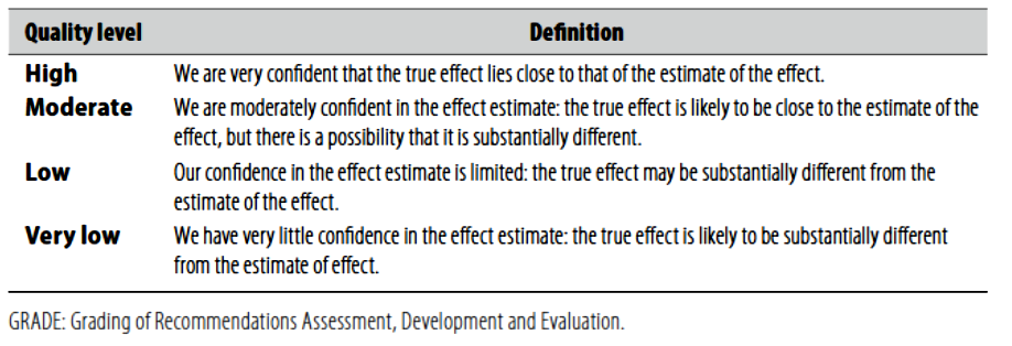
(Table-1) Quality of evidence in GRADE


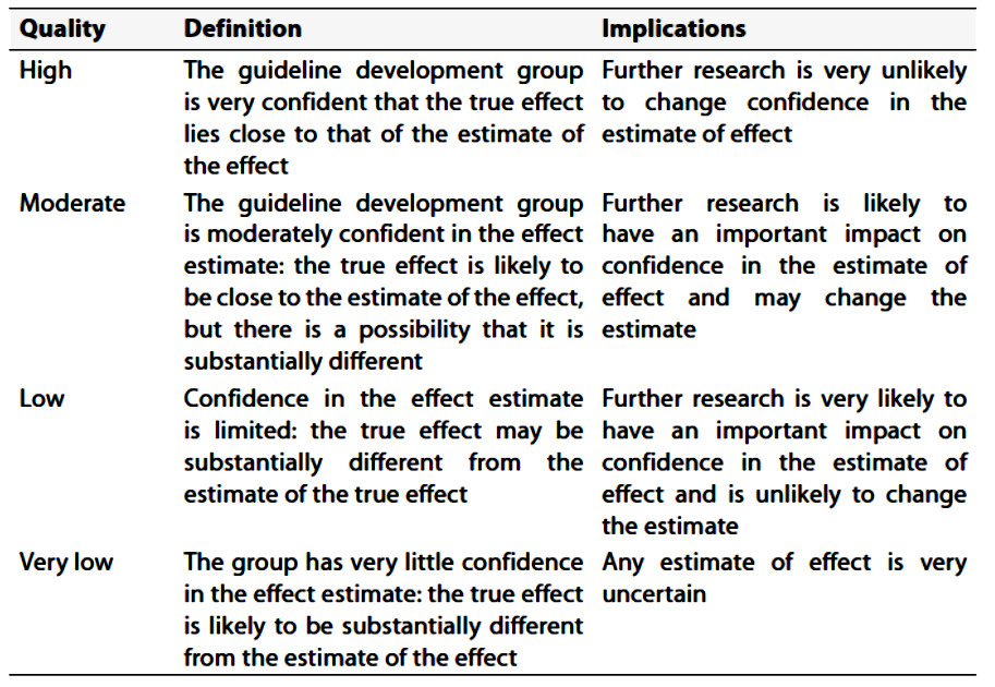
(Table-2) Significance of the four levels of evidence


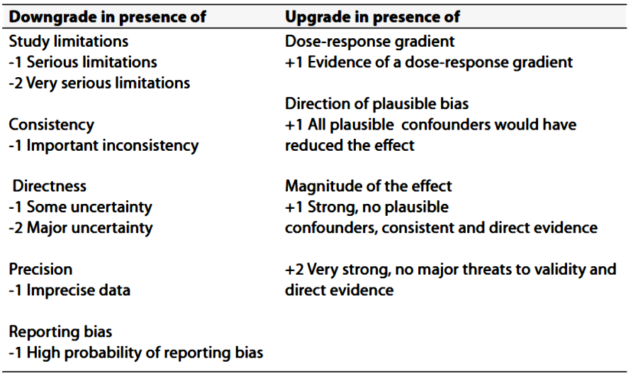
(Table-3) Factors that determine How to upgrade or downgrade the quality of Evidence


- Implementation considerations

In this section key recommendations are provided to guide the funding, planning and delivery of services along the entire pathway of stroke care. These recommendations will provide the best proposed outline for delivering endovascular intervention neurology services for stroke management.

Recommendations:

1.     Comprehensive stroke services should include the whole stroke pathway from prevention (including neurovascular services) through pre-hospital and acute care, early rehabilitation, secondary prevention, early supported discharge, community rehabilitation, systematic follow-up, palliative care and long-term support.

2.     A public education and professional training strategy should be developed and implemented to ensure that the public and emergency personnel (e.g. staff in emergency call centers) can recognize when a person has a suspected stroke or TIA and respond appropriately. This should be implemented in such a way that it can be formally evaluated.

3.     Along the pathway of stroke care, there should be protocols between healthcare providers and social services that enable seamless and safe transfers of care without delay.

4.     The provision of comprehensive acute stroke services may require the development of hub-and-spoke models of care (where a few hospitals in a region are designated to provide the hyperacute care for all patients), or telemedicine networks and other forms of cross-site working.

5.     The optimal disposition of acute stroke services will depend on the geography of the area served, with the objective of delivering the maximum number of time-critical treatments to the greatest number of people with stroke.

6.     Healthcare providers should enact all the secondary stroke prevention measures recommended in this guideline. Effective secondary prevention should be assured through a process of regular audit and monitoring.

7.     Healthcare authorities should play an active role in promoting secondary vascular prevention, which is a public health issue as well as being relevant to the individual person with stroke.


- Clinical indicators for monitoring

Values outside of the suggested thresholds should trigger a review of policies and/or practices within the department to determine the causes and to implement changes to manage the incidence of related indicators.

1.     For quality Improvement Issues related to cervicocerebral/spinal angiography; the following indicators and related thresholds may be used:

a.     Reported Success Rates of Diagnostic cervicocerebral/spinal catheter angiography is 98% (suggested threshold <98%).

b.     Reported Overall Procedure complication, either neurologic or systemic occurring within 24 hours of the angiography, is 2% (suggested threshold <2%)

2.     For quality improvement issues related to spinal dural AVF endovascular embolization; the following indicators and related thresholds may be used:

a.     The reported threshold indicators of the efficacy are technical success (81%) with occlusion of the targeted vessels and clinical success (60%) with improvement or stabilization of symptoms.

b.     The reported overall complication of dural AVF embolization is 10-15% (suggested threshold <5%).

3-    For quality improvement issues related to spinal AVM endovascular embolization; the following indicators and related thresholds may be used:

a.     The reported threshold indicators of the efficacy are technical success (90%) with occlusion of the targeted vessels and clinical success (50%) with improvement or stabilization of symptoms.

b.     The reported overall complication of dural AVF embolization is 10-15% (suggested threshold <10%).

4-    For quality improvement issues related to extracranial tumor endovascular embolization, major complications are rare. However, stroke and intracerebral hemorrhage have been reported in up to 3-6% during intracranial embolization (suggested threshold <5%).


- Research needs

1-  Cost effective studies for implementation of endovascular intervention procedures Vs best medical treatment Vs surgical intervention in certain situations in Egyptian population.

2-  Needs assessment studies for maximizing numbers of comprehensive stroke centers providing endovascular intervention stroke services.


- Updating the guideline

To keep these recommendations valid, all guidelines need to be periodically updated. This will be done whenever strong evidence is available and necessitates recommendation updates.


- References

1.      ACR-ASNR-SIR-SNIS Practice Parameter for the Performance of Diagnostic Cervicocerebral Catheter Angiography in Adults (amended 2021). Available at https://gravitas.acr.org/PPTS/GetDocumentView?docId=167+&releaseId=2.

2.      Albers GW, Marks MP, Kemp S, Christensen S, Tsai JP, Ortega-Gutierrez S, McTaggart R, Torbey MT, Kim-Tenser M, Leslie-Mazwi T, Sarraj A, Kasner SE, Ansari SA, Yeatts SD, Hamilton S, Mylnash M, Heit JJ, Zaharchuk G, Kim S, Carrozzella J, Palesch YY, Demchuk AM, Bammer R, Lavori PW, Broderick JP, Lansberg MG; on behalf of the DEFUSE 3Investigators. Thrombectomy for stroke with perfusion imaging selection at 6–16 hours. N Engl J Med. In press.

3.      Amarenco P, Denison H, Evans SR, Himmelmann A, James S, Knutsson M, Ladenvall P, Molina CA, Wang Y, Johnston SC; THALES Steering Committee and Investigators. Ticagrelor added to aspirin in acute non severe ischemic stroke or transient ischemic attack of atherosclerotic origin. Stroke. 2020;51:3504–3513. Doi: 10.1161/STROKEAHA.120.032239

4.      Barnett HJM, Taylor DW, Haynes RB, Sackett DL, Peerless SJ, Ferguson GG, Fox AJ, Rankin RN, Hachinski VC, Wiebers DO, et al. Beneficial effect of carotid endarterectomy in symptomatic patients with high-grade carotid stenosis. N Engl J Med. 1991;325:445–453.

5.      Brott TG, Hobson RW 2nd, Howard G, Roubin GS, Clark WM, Brooks W, Mackey A, Hill MD, Leimgruber PP, Sheffet AJ, et al; CREST Investigators. Stenting versus endarterectomy for treatment of carotid-artery stenosis. N Engl J Med. 2010;363:11–23. Doi: 10.1056/NEJMoa0912321

6.      Buscot MJ, Chandra RV, Maingard J, Nichols L, Blizzard L, Stirling C, Smith K, Lai L, Asadi H, Froelich J, et al. Association of onset-to-treatment time with discharge destination, mortality, and complications among patients with aneurysmal subarachnoid hemorrhage. JAMA Netw Open. 2022;5:e2144039. doi:10.1001/jamanetworkopen.2021.44039

7.      Bush CK, Kurimella D, Cross LJ, Conner KR, Martin-Schild S, He J, Li C, Chen J, Kelly T. Endovascular treatment with stent-retriever devices for acute ischemic stroke: a meta-analysis of randomized controlled trials. PLoS One. 2016;11:e0147287. Doi: 10.1371/journal.pone.0147287.

8.      Catapano JS, Lang MJ, Koester SW, Wang DJ, DiDomenico JD, Fredrickson VL, Cole TS, Lee J, Lawton MT, Ducruet AF, et al. Digital subtraction cerebral angiography after negative computed tomography angiography findings in non-traumatic subarachnoid hemorrhage. J Neurointerv Surg. 2020;12:526–530. Doi: 10.1136/neurintsurg-2019-015375

9.      Chimowitz MI, Lynn MJ, Derdeyn CP, Turan TN, Fiorella D, Lane BF, Janis LS, Lutsep HL, Barnwell SL, Waters MF, et al; SAMMPRIS Trial Investigators. Stenting versus aggressive medical therapy for intracranial arterial steno sis. N Engl J Med. 2011;365:993–1003. Doi: 10.1056/NEJMoa1105335

10.   Chimowitz MI, Lynn MJ, Howlett-Smith H, Stern BJ, Hertzberg VS, Frankel MR, Levine SR, Chaturvedi S, Kasner SE, Benesch CG, et al; Warfarin Aspirin Symptomatic Intracranial Disease Trial Investigators. Comparison of warfarin and aspirin for symptomatic intracranial arterial stenosis. N Engl J Med. 2005;352:1305–1316. Doi: 10.1056/NEJMoa043033

11.   De Leacy, R. et al. (2022) ‘Endovascular treatment in the multimodality management of brain arteriovenous malformations: Report of the society of neurointerventional surgery standards and Guidelines Committee’, Journal of Neurointerventional Surgery, 14(11), pp. 1118–1124. doi:10.1136/neurintsurg-2021-018632.

12.   Dean BL, Flom RA, Wallace RC, et al. Efficacy of endovascular treatment of meningiomas: evaluation with matched samples. AJNR Am J Neuroradiol 1994;15:1675e80.

13.   Deng X, Gao F, Zhang D, Zhang Y, Wang R, Wang S, Cao Y, Zhao Y, Pan Y, Liu X, et al. Direct versus indirect bypasses for adult ischemic-type moyamoya disease: a propensity score-matched analysis. J Neurosurg. 2018;128:1785–1791. Doi: 10.3171/2017.2.JNS162405

14.   Dippel DW, Majoie CB, Roos YB, van der Lugt A, van Oostenbrugge RJ, van Zwam WH, Lingsma HF, Koudstaal PJ, Treurniet KM, van den Berg LA, Beumer D, Fransen PS, Berkhemer OA; for the MR CLEAN Investigators. Influence of device choice on the effect of intra-arterial treatment for acute ischemic stroke in MR CLEAN (Multicenter Randomized Clinical Trial of Endovascular Treatment for Acute Ischemic Stroke in the Netherlands). Stroke. 2016;47:2574–2581. Doi: 10.1161/STROKEAHA.116.013929.

15.   Duffis, E.J. et al. (2012) ‘Head, neck, and Brain tumor embolization guidelines’, Journal of Neurointerventional Surgery, 4(4), pp. 251–255. doi:10.1136/neurintsurg-2012-010350.

16.   Farb RI, Vanek I, Scott JN, et al. Idiopathic intracranial hypertension: the prevalence and morphology of sinovenous stenosis. Neurology. 2003;60:1418–24.

17.   Giorgianni A, Agosti E, Molinaro S, Terrana AV, Vizzari FA, Nativo L, Garg K, Craparo G, Conti V, Locatelli D, et al. Flow diversion for acutely ruptured intracranial aneurysms treatment: a retrospective study and literature review. J Stroke Cerebrovasc Dis. 2022;31:106284. doi:10.1016/j.jstrokecerebrovasdis.2021.106284

18.   Gornik HL, Persu A, Adlam D, Aparicio LS, Azizi M, Boulanger M, Bruno RM, de Leeuw P, Fendrikova-Mahlay N, Froehlich J, et al. First international consensus on the diagnosis and management of fibromuscular dysplasia. Vasc Med. 2019;24:164–189. Doi: 10.1177/1358863X18821816

19.   Haussen DC, Grossberg JA, Bouslama M, Pradilla G, Belagaje S, Bianchi N, Allen JW, Frankel M, Nogueira RG. Carotid web (intimal fibromuscular dysplasia) has high stroke recurrence risk and is amenable to stenting. Stroke. 2017;48:3134–3137. Doi: 10.1161/STROKEAHA.117.019020

20.   Hoh, B.L. et al. (2023b) ‘2023 guideline for the management of patients with aneurysmal subarachnoid hemorrhage: A guideline from the American Heart Association/American Stroke Association’, Stroke, 54(7). doi:10.1161/str.0000000000000436.

21.   Howard BM, Hu R, Barrow JW, Barrow DL. Comprehensive review of imaging of intracranial aneurysms and angiographically negative subarachnoid hemorrhage. Neurosurg Focus. 2019;47:E20. Doi: 10.3171/2019.9.FOCUS19653

22.   Howard G, Roubin GS, Jansen O, Hendrikse J, Halliday A, Fraedrich G, Eckstein HH, Calvet D, Bulbulia R, Bonati LH, et al; Carotid Stenting Trialists’ Collaboration. Association between age and risk of stroke or death from carotid endarterectomy and carotid stenting: a meta-analysis of pooled patient data from four randomized trials. Lancet. 2016;387:1305 1311. Doi: 10.1016/S0140-6736(15)01309-4

23.   Ido K, Kurogi R, Kurogi A, Nishimura K, Arimura K, Nishimura A, Ren N, Kada A, Matsuo R, Onozuka D, et al; J-ASPECT Study Collaborators. Effect of treatment modality and cerebral vasospasm agent on patient outcomes after aneurysmal subarachnoid hemorrhage in the elderly aged 75 years and older. PLoS One. 2020;15:e0230953. doi:10.1371/journal.pone.0230953

24.   Jeon JP, Kim JE, Cho WS, Bang JS, Son YJ, Oh CW. Meta-analysis of the surgical outcomes of symptomatic moyamoya disease in adults. J Neurosurg. 2018;128:793–799. Doi: 10.3171/2016.11.JNS161688

25.   Jovin TG, Saver JL, Ribo M, Pereira V, Furlan A, Bonafe A, Baxter B, Gupta R, Lopes D, Jansen O, Smith W, Gress D, Hetts S, Lewis RJ, Shields R, Berry SM, Graves TL, Malisch T, Rai A, Sheth KN, Liebeskind DS, Nogueira RG. Diffusion-weighted imaging or computerized tomography perfusion assessment with clinical mismatch in the tri age of wake up and late presenting strokes undergoing neurointervention with Trevo (DAWN) trial methods. Int J Stroke. 2017;12:641–652. Doi: 10.1177/1747493017710341.

26.   Kwon SU, Cho YJ, Koo JS, Bae HJ, Lee YS, Hong KS, Lee JH, Kim JS. Cilostazol prevents the progression of the symptomatic intracranial arterial stenosis: the multicenter double-blind placebo-controlled trial of cilostazol in symptomatic intracranial arterial stenosis. Stroke. 2005;36:782–786. Doi: 10.1161/01.STR.0000157667.06542.b7

27.   Lapergue B, Blanc R, Gory B, Labreuche J, Duhamel A, Marnat G, Saleme S, Costalat V, Bracard S, Desal H, Mazighi M, Consoli A, Piotin M; ASTER Trial Investigators. Effect of endovascular contact aspiration vs stent retriever on revascularization in patients with acute ischemic stroke and large vessel occlusion: the ASTER randomized clinical trial. JAMA. 2017;318:443–452. Doi: 10.1001/jama.2017.9644.

28.   Leacy RD, Ansari SA, Schirmer CM, Cooke DL, Prestigiacomo CJ, Bulsara KR, et al. Endovascular treatment in the multimodality management of brain arteriovenous malformations: report of the Society of NeuroInterventional Surgery Standards and Guidelines Committee. Journal of NeuroInterventional Surgery. 2022 Nov 1;14(11):1118–24.

29.   Lee, S.-K. et al. (2018) ‘Current endovascular strategies for cerebral venous thrombosis: Report of the SNIS standards and Guidelines Committee’, Journal of NeuroInterventional Surgery, 10(8), pp. 803–810. doi:10.1136/neurintsurg-2018-013973.

30.   Lemmens R, Hamilton SA, Liebeskind DS, Tomsick TA, Demchuk AM, Nogueira RG, Marks MP, Jahan R, Gralla J, Yoo AJ, Yeatts SD, Palesch YY, Saver JL, Pereira VM, Broderick JP, Albers GW, Lansberg MG; DEFUSE 2, IMS III, STAR, and SWIFT Trialists; DEFUSE 2 IMS III STAR and SWIFT Trialists. Effect of endovascular reperfusion in relation to site of arterial occlusion. Neurology. 2016;86:762–770. Doi: 10.1212/WNL.0000000000002399.

31.   Lindgren A, Vergouwen MD, van der Schaaf I, Algra A, Wermer M, Clarke MJ, Rinkel GJ. Endovascular coiling versus neurosurgical clipping for people with aneurysmal subarachnoid haemorrhage. Cochrane Database Syst Rev. 2018;8:CD003085. Doi: 10.1002/14651858.CD003085.pub3

32.   Löwhagen Hendén P, Rentzos A, Karlsson JE, Rosengren L, Leiram B, Sundeman H, Dunker D, Schnabel K, Wikholm G, Hellström M, Ricksten SE. General anesthesia versus conscious sedation for endovascular treatment of acute ischemic stroke: the AnStroke trial (Anesthesia During Stroke). Stroke. 2017;48:1601–1607. Doi: 10.1161/ STROKEAHA.117.016554.

33.   Marks MP, Lansberg MG, Mlynash M, Kemp S, McTaggart R, Zaharchuk G, Bammer R, Albers GW; DEFUSE 2 Investigators. Correlation of AOL recanalization, TIMI reperfusion and TICI reperfusion with infarct growth and clinical outcome. J Neurointerv Surg. 2014;6:724–728. Doi: 10.1136/neurintsurg-2013-010973.

34.   Markus HS, Harshfield EL, Compter A, Kuker W, Kappelle LJ, Clifton A, van der Worp HB, Rothwell P, Algra A; Vertebral Stenosis Trialists’ Collaboration. Stenting for symptomatic vertebral artery stenosis: a preplanned pooled individual patient data analysis. Lancet Neurol. 2019;18:666–673. Doi: 10.1016/S1474-4422(19)30149-8

35.   Nagai M, Watanabe E. Benefits of clipping surgery based on three-dimensional computed tomography angiography. Neurol Med Chir (Tokyo).2010;50:630–637. Doi: 10.2176/nmc.50.630

36.   Narayanan, S. et al. (2012) ‘Standard of practice: Embolization of spinal arteriovenous fistulae, spinal arteriovenous malformations, and tumors of the spinal axis’, Journal of NeuroInterventional Surgery, 5(1), pp. 3–5. doi:10.1136/neurintsurg-2012-010551.

37.   Nogueira RG, Jadhav AP, Haussen DC, Bonafe A, Budzik RF, Bhuva P, Yavagal DR, Ribo M, Cognard C, Hanel RA, Sila CA, Hassan AE, Millan M, Levy EI, Mitchell P, Chen M, English JD, Shah QA, Silver FL, Pereira VM, Mehta BP, Baxter BW, Abraham MG, Cardona P, Veznedaroglu E, Hellinger FR, Feng L, Kirmani JF, Lopes DK, Jankowitz BT, Frankel MR, Costalat V, Vora NA, Yoo AJ, Malik AM, Furlan AJ, Rubiera M, Aghaebrahim A, Olivot JM, Tekle WG, Shields R, Graves T, Lewis RJ, Smith WS, Liebeskind DS, Saver JL, Jovin TG DAWN Trial Investigators. Thrombectomy 6 to 24 hours after stroke with a mismatch between deficit and infarct [published online ahead of print November 11, 2017]. N Engl J Med. Doi: 10.1056/NEJMoa1706442. http://www. nejm.org/Doi/full/10.1056/NEJMoa1706442.

38.   Ogilvy CS, Stieg PE, Awad I, Brown RD, Kondziolka D, Rosenwasser R, et al. Recommendations for the management of intracranial arteriovenous malformations. Stroke [Internet]. 2001 Jun 1;32(6):1458–71. Available from: https://doi.org/10.1161/01.str.32.6.1458

39.   Oudshoorn SC, Rinkel GJ, Molyneux AJ, Kerr RS, Dorhout Mees SM, Backes D, Algra A, Vergouwen MD. Aneurysm treatment <24 versus 24-72 h after subarachnoid hemorrhage. Neurocrit Care. 2014;21:4–13. doi:10.1007/s12028-014-9969-8

40.   Passero SG, Rossi S. Natural history of vertebrobasilar dolichoectasia. Neurology. 2008;70:66–72. Doi: 10.1212/01.wnl.0000286947.89193.f3

41.   Persky MS, Setton A, Niimi Y, et al. Combined endovascular and surgical treatment of head and neck paraganglioma team approach. Head Neck 2002;24:423e31.

42.   Phuong Nguyen T, Rehman S, Stirling C, Chandra R, Gall S. Time and predictors of time to treatment for aneurysmal subarachnoid haemorrhage (aSAH): a systematic review. Int J Qual Health Care. 2021;33:mzab019. doi:10.1093/intqhc/mzab019

43.   Pierot L, Barbe C, Herbreteau D, Gauvrit JY, Januel AC, Bala F, Ricolfi F, Desal H, Velasco S, Aggour M, et al. Rebleeding and bleeding in the year following intracranial aneurysm coiling: analysis of a large prospective multicenter cohort of 1140 patients: Analysis of Recanalization after Endovascular Treatment of Intracranial Aneurysm (ARETA) Study. J Neurointerv Surg. 2020;12:1219–1225. Doi: 10.1136/neurintsurg-2020-015971

44.   Pierot L, Jayaraman MV, Szikora I, AAFITN, ANZSNR, ASNR, CSNR, ESMINT, ESNR, ESO, JSNET, SFNR, SILAN, SNIS, SVIN, WSO, WFITN, et al Standards of practice in acute ischemic stroke intervention: international recommendations Journal of NeuroInterventional Surgery 2018;10:1121-1126.

45.   Powers WJ, Clarke WR, Grubb RL Jr, Videen TO, Adams HP Jr, Derdeyn CP; COSS Investigators. Extracranial-intracranial bypass surgery for stroke prevention in hemodynamic cerebral ischemia: the Carotid Occlusion Surgery Study randomized trial. JAMA. 2011;306:1983–1992. Doi: 10.1001/jama.2011.1610

46.   Rantner B, Kollerits B, Roubin GS, Ringleb PA, Jansen O, Howard G, Hendrikse J, Halliday A, Gregson J, Eckstein HH, et al; Carotid Stenosis Trialists’ Collaboration. Early endarterectomy carries a lower procedural risk than early stenting in patients with symptomatic stenosis of the in ternal carotid artery: results from 4 randomized controlled trials. Stroke. 2017;48:1580–1587. Doi: 10.1161/STROKEAHA.116.016233 Downloaded from http://ahajournals.org by on January 24, 2025

47.   Rothwell PM, Eliasziw M, Gutnikov SA, Fox AJ, Taylor DW, Mayberg MR, Warlow CP, Barnett HJ; Carotid Endarterectomy Trialists’ Collaboration. Analysis of pooled data from the randomized controlled trials of endarterectomy for symptomatic carotid stenosis. Lancet. 2003;361:107–116. Doi: 10.1016/s0140-6736(03)12228-3

48.   Rothwell PM, Eliasziw M, Gutnikov SA, Warlow CP, Barnett HJ; Carotid Endarterectomy Trialists Collaboration. Endarterectomy for symptomatic carotid stenosis in relation to clinical subgroups and timing of surgery. Lancet. 2004;363:915–924. Doi: 10.1016/S0140-6736(04)15785-1

49.   Ryttlefors M, Enblad P, Kerr RS, Molyneux AJ. International Subarachnoid Aneurysm Trial of neurosurgical clipping versus endovascular coiling: subgroup analysis of 278 elderly patients. Stroke. 2008;39:2720–2726. doi:10.1161/STROKEAHA.107.506030

50.   Saver JL, Goyal M, van der Lugt A, Menon BK, Majoie CB, Dippel DW, Campbell BC, Nogueira RG, Demchuk AM, Tomasello A, Cardona P, Devlin TG, Frei DF, du Mesnil de Rochemont R, Berkhemer OA, Jovin TG, Siddiqui AH, van Zwam WH, Davis SM, Castaño C, Sapkota BL, Fransen PS, Molina C, van Oostenbrugge RJ, Chamorro Á, Lingsma H, Silver FL, Donnan GA, Shuaib A, Brown S, Stouch B, Mitchell PJ, Davalos A, Roos YB, Hill MD; HERMES Collaborators. Time to treat ment with endovascular thrombectomy and outcomes from ischemic stroke: a meta-analysis. JAMA. 2016;316:1279–1288. Doi: 10.1001/ jama.2016.13647.

51.   Schacht H, Kuchler J, Boppel T, Leppert J, Ditz C, Schramm P, Neumann A. Transluminal balloon angioplasty for cerebral vasospasm after spontaneous subarachnoid hemorrhage: a single-center experience. Clin Neurol Neurosurg. 2020;188:105590. Doi: 10.1016/j.clineuro.2019.105590

52.   Schermerhorn ML, Liang P, Eldrup-Jorgensen J, Cronenwett JL, Nolan BW, Kashyap VS, Wang GJ, Motaganahalli RL, Malas MB. Association of trans-carotid artery revascularization vs transfemoral carotid artery stenting with stroke or death among patients with carotid artery stenosis. JAMA. 2019;322:2313–2322. Doi: 10.1001/jama.2019.18441

53.   Settecase F, Rayz VL. Advanced vascular imaging techniques. Handb Clin Neurol 2021;176:81–105.

54.   Smith LL, Smith DC, Killeen JD, Hasso AN. Operative balloon angioplasty in the treatment of internal carotid artery fibromuscular dysplasia. J Vasc Surg. 1987;6:482–487. Doi: 10.1067/mva.1987.avs0060482

55.   Tasar M, Yetiser S. Glomus tumors: therapeutic role of selective embolization. J Craniofac Surg 2004;15:497e505.

56.   Ten Brinck MFM, Shimanskaya VE, Aquarius R, Bartels R, Meijer FJA, Koopmans PC, de Jong G, Wakhloo AK, de Vries J, Boogaarts HD. Outcomes after flow diverter treatment in subarachnoid hemorrhage: a meta-analysis and development of a clinical prediction model (OUTFLOW). Brain Sci. 2022;12:394. doi:10.3390/brain sci12030394

57.   White JB, Link MJ, Cloft HJ. Endovascular embolization of paragangliomas: a safe adjuvant to treatment. J Vasc Interv Neurol 2008;1:37e41.

58.   Wojak, J.C. et al. (2015) ‘Quality Improvement Guidelines for adult diagnostic CERVICOCEREBRAL angiography: Update Cooperative study between the Society of Interventional Radiology (sir), American Society of Neuroradiology (ASNR), and Society of Neurointerventional Surgery (SNIS)’, Journal of Vascular and Interventional Radiology, 26(11), pp. 1596–1608. doi:10.1016/j.jvir.2015.07.002.

59.   Yadav JS, Wholey MH, Kuntz RE, Fayad P, Katzen BT, Mishkel GJ, Bajwa TK, Whitlow P, Strickman NE, Jaff MR, et al; Stenting and Angioplasty with Protection in Patients at High Risk for Endarterectomy Investigators. Protected carotid-artery stenting versus endarterectomy in high-risk patients. N Engl J Med. 2004;351:1493–1501. Doi: 10.1056/NEJMoa040127

60.   Zhu D, Yan Y, Zhao P, Duan G, Zhao R, Liu J, Huang Q. Safety and efficacy of flow diverter treatment for blood blister-like aneurysm: a systematic review and meta-analysis. World Neurosurg. 2018;118:e79–e86. doi:10.1016/j.wneu.2018.06.123