
Book
Procedural Work For Electrocardiography
- Cardiac Electrophysiology and How the Heart Beats
Definition of Cardiac Electrophysiology: The contraction of any muscle is accompanied by electrical currents known as depolarization, and these currents can be recorded using electrodes placed on the surface of the body. This allows the recording of muscle contractions throughout the body. However, to clearly record the contraction of only the heart muscles, the person should be at rest, and all other body muscles should be relaxed.
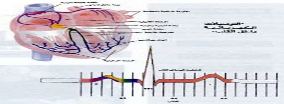
Although the heart contains four chambers, only two chambers are visible in the ECG recording because the atria contract together, as well as the ventricles.
Normal Pulse:
The regular rhythmic heartbeats originate from within the heart muscle tissue itself, as it is self-regulating. The normal pulse begins with an electrical signal generated by a specialized, tiny electrical and neuro-muscular pacemaker located in the wall of the right atrium, known as the sinoatrial node (SA node), which generates electrical impulses at a rate of 60-100 beats per minute. This electrical signal rapidly spreads to both atria, causing them to contract and push blood from the atria into the ventricles. The signal then moves to the atrioventricular node (AV node), which is a small backup pacemaker located between the atria and ventricles. The AV node allows the electrical signal to pass through electrical pathways branching from it into the ventricles within a fraction of a second, causing the ventricles to contract and push blood out of the heart. The right ventricle pumps deoxygenated blood to the lungs for oxygenation, while the left ventricle pumps oxygenated blood to all parts of the body, providing tissues with the oxygen-rich blood. After oxygen is extracted from the blood, it returns to the right side of the heart, completing one full cycle of blood circulation.
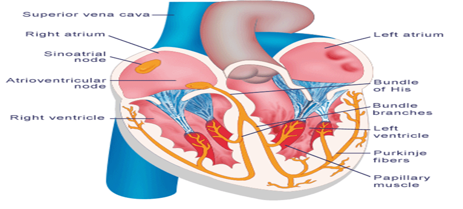
Thus, in a healthy
human, electrical signals originate from the sinoatrial
node (SA node), and this is known as the sinus rhythm. However, in some pathological cases,
electrical impulses can arise from other sources, such as the atrioventricular node (AV node), which then results
in a nodal rhythm. In other cases,
the electrical impulses might come from other areas of the heart, such as the
ventricular muscles themselves. This could be due to the failure of the
sinoatrial node to generate impulses, or because an external source elsewhere
in the heart beats at a higher rate than the primary pacemaker, thus taking
over the rhythm of the heart.
