
Book
Guidelines for Obstetrics and Gynecology Nursing Procedures
- Anatomy of the Reproductive System
Introduction
The reproductive system differs from other body systems because it remains inactive until puberty. The primary reproductive glands, or gonads, in females are the ovaries. These glands produce gametes (reproductive cells) as well as sex hormones.
The sex hormones—estrogen and progesterone—play a crucial role in both the development and function of the reproductive organs, as well as in sexual behavior. These hormones also influence the growth and development of various other organs and tissues in the body.
The Female Reproductive System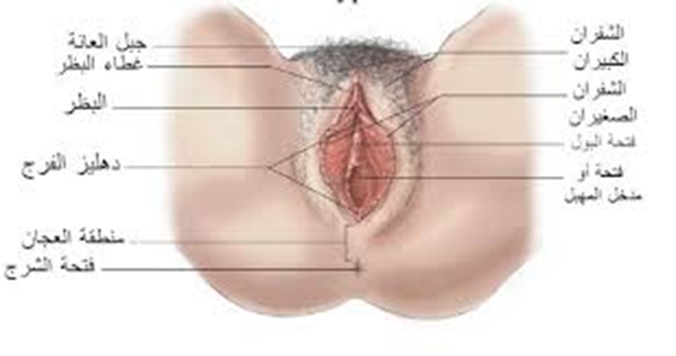
Anatomy of the External Reproductive System
1. Mons Pubis (Mons Veneris):
- A layer of fat covering the pubic area, covered with skin and hair.
- Provides protection to the pubic region.
2. Labia Majora:
- The outer folds of the vulva.
- Two fatty skin folds extending from the mons pubis to the posterior commissure.
- The outer surface is covered with hair, while the inner surface is smooth, hairless, and contains sebaceous and sweat glands.
3. Labia Minora:
- The inner folds of the vulva.
- Two thin folds of modified skin containing the external openings of the urethra and vagina.
- Protects the vagina, urethra, and clitoris.
4. Clitoris:
- A small, highly sensitive organ located beneath the pubic area.
- The external tip is covered by a fold of skin known as the clitoral hood, similar to the foreskin of the penis.
- The labia minora meet at the clitoris, which is analogous to the male penis.
5. Vestibule of the Vulva:
- The area between the inner surfaces of the labia minora and the vaginal opening.
6. Hymen:
- A thin membrane located approximately 2 cm from the vaginal entrance, partially covering the vaginal opening.
- Serves as a boundary between the external and internal reproductive organs.
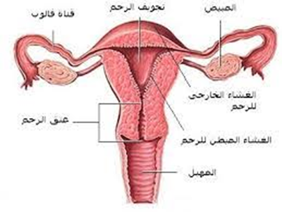
The Internal Reproductive System
The internal reproductive system consists of:
- Vagina
- Cervix
- Uterus
- Fallopian Tubes
- Ovaries
Vagina
- A muscular tube extending from the clitoris to the uterus, positioned at a 60-degree angle to the horizontal plane.
- It connects the cervix to the external reproductive organs and is located between the bladder and the intestines.
Functions:
- Serves as a passage for menstrual flow and uterine secretions.
- Acts as the birth canal during labor.
- Becomes lubricated during sexual intercourse with the help of Bartholin’s glands.
Length:
- Anterior wall: 8–9 cm
- Posterior wall: 10–11 cm
Cervix
- The cervix connects the uterus to the vagina.
- It is the lower elongated part of the uterus, measuring 2.5–3 cm.
- The cervical canal communicates with the uterine cavity superiorly and with the vagina inferiorly.
Anatomical Relations of the Cervix:
- Anteriorly: Bladder and vesicovaginal pouch.
- Posteriorly: Douglas’ pouch.
- Laterally: Broad ligament.
Uterus Anatomy
- The uterus is pear-shaped and about the size of a clenched fist. It consists of three layers: the inner endometrium, the middle myometrium, and the outer perimetrium. The endometrial layer contains highly vascularized tissue that sheds during menstruation. The strong uterine muscles expand to accommodate the growing fetus and contract during childbirth.
Measurements:
- Dimensions: 7.5 × 5.0 × 2.5 cm (length × width × depth).
- Weight: 50–60 grams (slightly larger in multiparous women).
Parts of the Uterus:
- Fundus: The rounded upper portion above the entrance of the fallopian tubes.
- Body: The main part of the uterus.
- Isthmus: The narrow region between the body and the cervix.
- Cervix: The lower elongated part of the uterus.
Position of the Uterus:
- The uterus is normally in an anteverted position (AVF), with the external os at the level of the iliac spines.
- Anteversion: The uterus tilts forward relative to the vaginal axis.
- Anteflexion: The body of the uterus bends forward over the cervix.
Anatomical Relations of the Uterus:
- Anteriorly: Bladder and vesicovaginal pouch.
- Posteriorly: Douglas’ pouch.
- Laterally: Broad ligament.
Histology of the Uterus
- Endometrium: The inner lining, containing tubular glands that undergo cyclic changes during the menstrual cycle under ovarian hormone influence.
- Myometrium: Composed of three muscle layers:
- Outer longitudinal muscle fibers.
- Middle interwoven fibers surrounding blood vessels.
- Inner circular muscle fibers.
- Perimetrium: The outermost layer, which extends anteriorly from the fundus to the isthmus, then reflects over the bladder to form the vesicovaginal pouch.
Uterine Support Structures (Ligaments)
- Broad ligament
- Round ligament
- Ovarian ligament
Fallopian Tubes
- Paired tubes measuring 10 cm in length, located in the free upper margin of the broad ligament.
- They connect the uterine horns to the peritoneal cavity, with the outer free end curving toward the ovary.
- The fallopian tubes serve as the passage for the ovum and are the site of fertilization.
- Also known as uterine tubes or oviducts.
Fertilization and Transport:
- The fallopian tube is the site of sperm-egg fertilization.
- The fertilized egg takes approximately 6–10 days to travel through the tube and implant in the uterine lining.
Parts of the Fallopian Tube:
- Interstitial part:
- Pierces the uterine wall.
- Very narrow, lacks peritoneal covering, and has a thick muscular layer.
- Isthmus:
- A straight, narrow segment with thick walls, connecting to the uterus.
- Ampulla:
- The widest segment, where fertilization usually occurs.
- Infundibulum:
- The funnel-shaped distal end, opening into the peritoneal cavity.
- Surrounded by fimbriae, with one long fimbria directed toward the ovary.
Functions of the Fallopian Tubes:
- Captures the ovum during ovulation using fimbriae.
- Transports the ovum via peristaltic movements and ciliary action.
- Produces secretions necessary for sperm activation and ovum nourishment.
Anatomical Relations:
- Superiorly: Intestinal structures.
- Inferiorly: Broad ligament and its contents.
- Medially: Uterine horns.
- Laterally: Pelvic walls.
- The ovaries are positioned posterior and inferior to the fallopian tubes.
Ovaries
- The female gonads, responsible for producing and releasing eggs (ova) monthly.
- A female is born with approximately 400,000 immature eggs (follicles).
- Over her lifetime, she will ovulate about 400–500 mature eggs.
- Ovarian follicles produce estrogen and progesterone, which regulate the menstrual cycle and prepare the uterus for implantation.
Anatomy of the Ovaries:
- Almond-shaped, located in the ovarian fossa on the pelvic wall.
- Dimensions: 3 × 2 × 1 cm.
- Surface: Whitish-pink, wrinkled due to ovulatory activity.
Anatomical Relations:
- Medially: Fallopian tubes.
- Laterally: Pelvic wall.
- Superiorly & Anteriorly: Small intestine.
- Inferiorly: Ovarian fossa, where the ureter and internal iliac vessels pass.
Pelvic Floor Muscles
- Comprises all tissues between the pelvic cavity and the external genital region.
Components:
- Pelvic peritoneum
- Extraperitoneal fat and connective tissue
- Levator ani and coccygeus muscles
- Triangular ligament (urogenital diaphragm)
- Perineal muscles
- Subcutaneous fat and fascia
