
Book
Cervical cancer
- Annexes
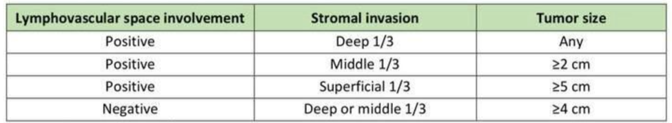
Table 5. Intermediate Risk-factors for Cervical Cancer (2).
|
The staging of cervical tumours is by the Fe´deration Internationale de Gyne´cologie et d’Obste´trique (FIGO) and TNM classification (Union for International Cancer Control), (2). |
||
|
TNM clinical classification |
||
|
TNM categories |
FIGO stages |
Definition |
|
T – Primary Tumour |
|
|
|
TX |
|
Primary tumour cannot be assessed |
|
T0 |
|
No evidence of primary tumour |
|
Tis |
|
Carcinoma in situ (preinvasive carcinoma) |
|
T1 |
I |
Tumour confined to the cervix |
|
T1a |
IA |
Invasive carcinoma diagnosed only by microscopy. Stromal invasion with a maximal depth of |
|
|
|
5.0 mm measured from the base of the epithelium and a horizontal spread of 7.0 mm or less |
|
IA1 |
Measured stromal invasion 3.0 mm or less in depth and 7.0 mm or less in horizontal spread |
|
|
IA2 |
Measured stromal invasion more than 3.0 mm and not more than 5.0 mm with a horizontal |
|
|
|
|
spread of 7.0 mm or less |
|
T1b |
IB |
Clinically visible lesion confined to the cervix or microscopic lesion greater than T1a/IA2 |
|
IB1 |
Clinically visible lesion 4.0 cm or less in greatest dimension |
|
|
IB2 |
Clinically visible lesion more than 4.0 cm in greatest dimension |
|
|
II |
Tumour invades beyond uterus but not to pelvic wall or to lower third of vagina |
|
|
IIA |
Tumour without parametrial invasion |
|
|
T2a1 |
IIA1 |
Clinically visible lesion 4.0 cm or less in greatest dimension |
|
IIA2 |
Clinically visible lesion more than 4.0 cm in greatest dimension |
|
|
T2b |
IIB |
Tumour with parametrial invasion |
|
T3 |
III |
Tumour involves lower third of vagina, or extends to pelvic wall, or causes hydronephrosis or |
|
|
|
non-functioning kidney |
|
T3a |
IIIA |
Tumour involves lower third of vagina |
|
T3b |
IIIB |
Tumour extends to pelvic wall, or causes hydronephrosis or non-functioning kidney |
|
T4 |
IVA |
Tumour invades mucosa of the bladder or rectum, or extends beyond true pelvise |
|
N – Regional Lymph Nodes |
|
|
|
NX |
|
Regional lymph nodes cannot be assessed |
|
N0 |
|
No regional lymph node metastasis |
|
N1 |
|
Regional lymph node metastasis |
|
M – Distant Metastasis |
|
|
|
M0 |
|
No distant metastasis |
|
M1 |
|
Distant metastasis (includes inguinal lymph nodes and intraperitoneal disease). It excludes |
|
|
|
metastasis to vagina, pelvic serosa, and adnexa |
|
James D, Brierley JD, Gospodarowicz MK et al. (eds). TNM Classification of Malignant Tumours, 8th edition. Oxford, UK: John Wiley & Sons, Inc. 2016.
|
||
