
كتاب
Pigeon Diseases
- Pigeon Pox Virus infection
Epidemiology:
The disease is a highly contagious viral infection affecting poultry, distinguished by the presence of lesions on unfeathered skin areas and/or diphtheritic lesions on the mucous membranes of the upper digestive and respiratory tracts. The causative agent belongs to the Poxvirus family (Poxviridae) DNA virus.
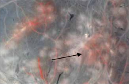
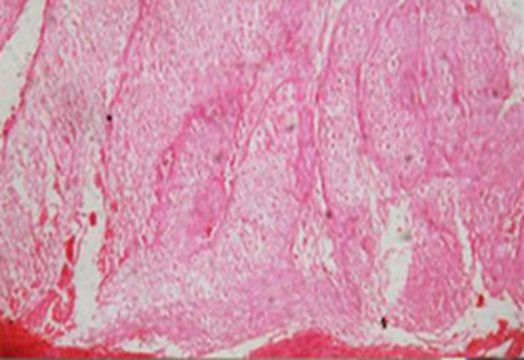
The virus replicates on the chorioallantoic membrane (CAM) of embryonated chicken eggs (ECE), resulting in the formation of grayish focal areas of cellular proliferation, known as pock lesions.
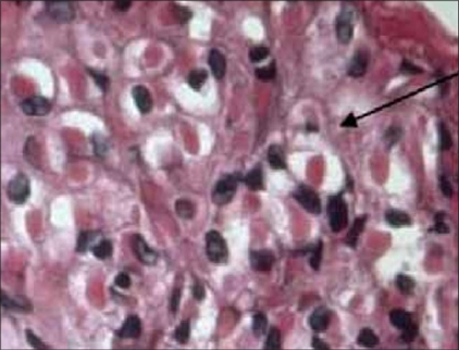
Histopathological examination reveals the presence of round to oval intracytoplasmic inclusion bodies, called ( Bollinger bodies).
Transmission: of the virus primarily occurs through wound infections due to skin abrasions, direct contact with infected birds, and mechanically via vectors such as mosquitoes. Additionally, airborne transmission may contribute to the spread of infection. Understanding these epidemiological aspects is crucial for controlling infection.
Clinical signs:
I.Skin (cutaneous or dry) form:
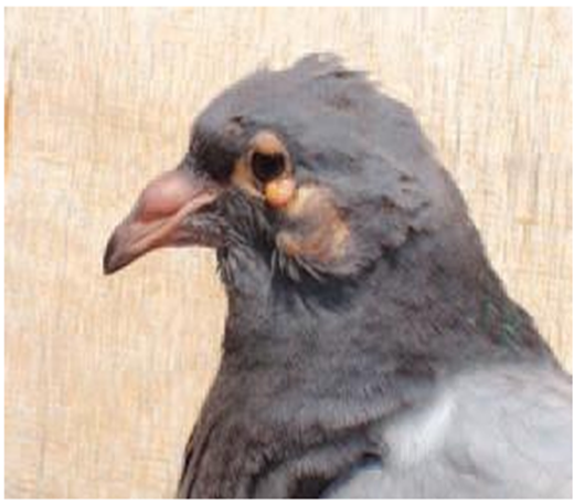
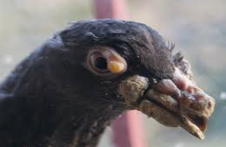
- Wart like nodules (grayish blister white spots) on the un-feathered parts of the bird’s body (around eyes, base of the peak, legs, under the wings and around the vent).
II. Mucous membrane (wet, diphtheritic) form:
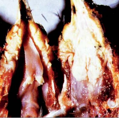
- Diphtheritic membrane (pustule like nodules) on the upper digestive, upper respiratory tract and nasal cavity.
- Yellow pustules changed to caseous necrotic material forming what is called pseudo membrane (diphtheritic membrane).
III. Mixed form:
- Cutaneous lesion and diphtheritic lesion in the same time.
|
Pigeon pox (cutaneous form) |
Diphtheroid form of pigeon pox, yellowish membranes in the oral cavity |
|
Pigeon pox (cutaneous form) |
pocks lesions were produced by field strain of fowl pox virus on CAM |
|
Cutaneous pock lesion of pigeon pox showing vacuolated prickle cells and intracytoplasmic eosinophilic inclusions. |
Chorioallantoic membrane showing intracytoplasmic inclusion bodies (×100).( called Bollinger bodies |
Diagnosis:
- The lesions are suggestive.
- Histopathological sections from the lesions revealed presence of intracytoplasmic inclusion bodies (Bollingers bodies).
- Inoculation of the suspected material on CAM of ECE gives pock lesions after 5-7 days ten confirm by histopathology.
- Identification by IGPT, NT passive HA and ELISA test.
Prevention and Control:
1. Sanitation and Management: sanitation practices and sound flock management to inhibit disease spread.
2. Vaccination:
- Apply a live attenuated virus vaccine, specifically the pigeon pox vaccine (PPV), for chickens, turkeys, and pigeons, propagated in tissue culture (TC).
- Vaccination techniques include wing web sticking or stabbing for chickens and pigeons, and brushing of feather follicles in the upper thigh or breast muscles.
- Emergency Vaccination:
1. Initiate vaccination as soon as clinical signs appear during an outbreak.
2. Utilize the live virus (pigeon pox vaccine), as it typically induces milder post-vaccinal reactions compared to the fowl pox vaccine.
3. Administer the vaccine intradermally via the wing web or feather follicle methods.
- Ectoparasite and Insect Control: Eradicate ectoparasites and insects to reduce potential disease vectors.
- Avoidance of Trauma: Address and eliminate sources of abrasions and wounds, such as cannibalism and mechanical trauma.
- Mosquito Control: Eradicate standing water to eliminate primary mosquito vectors. Isolate or cull infected birds to remove sources of the virus.
- Decontaminate feeders, waterers, bird baths, and cages using a 10% bleach solution.
Treatment:
1. Remove lesions.
2. Cleanse or treat affected areas with a tincture of iodine mixed with glycerin (1:4).
3. Administer antibiotics to prevent or treat secondary bacterial infections.
4. Provide multi-vitamins, particularly vitamin A for epithelial repair and vitamin E to enhance resistance.
5. Stimulate appetite by offering wet mash.
