
كتاب
Infertility in she camel
- D. Congenital abnormalities of the reproductive tract
The most common congenital disorders include:
o Ovarian dysgenesis/hypoplasia
o Segmental aplasia of the uterus
o Uterus didelphys
o Vaginal aplasia
o Persistent hymen
o Vulvar atresia.
Segmental aplasia of the reproductive tract
It may occur at the level of the uterine tube (formation of hydrosalpinx) or the level of the uterine horn (uterus unicornis). The affected females will often present with normal cycles and ovulation but fail to achieve or maintain pregnancy.
Diagnosis: achieved by ultrasonography and confirmed by hysteroscopy or laparoscopy.
Uterus didelphys
It is a rare congenital anomaly of the female reproductive tract characterized by a divided uterine cervix and body (Fig. 14, 15). It occurs due to abnormal development of the paramesonephric (Müllerian) duct (Mahdy and Nasr Eldeen, 2024).
Complete double cervix: It is caused by the persistence of the median walls of the Müllerian ducts along the whole length of the cervix, resulting in two cervices and one uterine body.
Incomplete double cervix: It is caused by the persistence of the median walls of the Müllerian ducts at the posterior part of the cervix, resulting in one cervical canal cranially and two cervical canals caudally.
Double external uterine orifices: It is caused by the persistence of the median walls of the Müllerian ducts at the external uterine orifice, resulting in one cervix with a band of tissue at the external os.
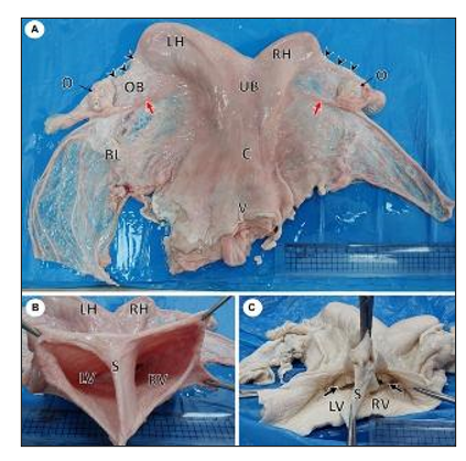
Figure 14. Gross photographs of female reproductive system of a she-camel show: (A) Apparently normal reproductive system consisted of right and left ovaries (O), each ovary was located inside an ovarian bursa (OB), the ovary attached to the uterine horn by the roundligament (red arrow), fallopian tubes (arrowheads), right (RH) and left (LH) horns, uterine body (UB), cervix (C), and vagina (V). Note the broad ligament of the uterus (BL). (B) Per vaginal view shows a completely divided vagina into the left (LV) and right (RV) vagina by a median septum (S). (C) The dorsal wall of the vagina was opened, showing each vagina had its external os (arrow) (Mahdy and Nasr Eldeen, 2024).
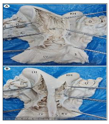
Figure 15. Gross photographs showing a dorsal view of uterus didelphys: (A) An incision was performed in the left horn (LH), body (UB), cervix (C), and vagina (LV). Note the complete septum extending from the fundus to the vagina. (B) Incised right (RH) and left (LH) horns show each horn connected to a separate uterine body that had its own internal and external os (Mahdy and Nasr Eldeen, 2024).
The abnormal condition of the vagina and vulva include: aplasia, persistent hymen, vulvar atresia, and vaginal constriction
Diagnosis
- History of rejecting the male
- Persistent straining or pain during mating or excessive vulvar swelling after mating.
- Transrectal ultrasonography generally demonstrates accumulation of fluid or mucus in the vagina/uterus (hydrometra, mucometra).
- Examination of the external genitalia and vaginal examination.
- Digital palpation is often sufficient to determine an occlusion at the level of the vestibulum.
- Cystitis and vestibular inflammation, or complete adhesion is a long-term result of vaginal inflammation.
- Abnormalities of the vulva are usually seen in small camels, which lead to accumulation of urine in the uterus, a condition known as urometra.
