كتاب
Affections Of Specific Parts and Organs of Food Animals
- III. Affections of the Liver
A- Pathological affections
1. Degeneration:
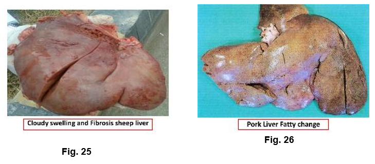
Animal suffers from acute infectious disease, the liver is the first organ undergoes macroscopic changes. These changes are manifested by cloudy swelling and or pathological fatty change.
Organ: Condemn the affected organ.
Carcass: Total condemnation of carcass showed systemic reactions.
(Fever, septicemia, pyaemia). Normal carcass approved.
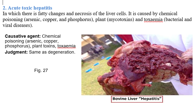
2. Acute toxic hepatitis
In which there is fatty changes and necrosis of the liver cells. It is caused by chemical poisoning (arsenic, copper, and phosphorus), plant (mycotoxins) and toxaemia (bacterial and viral diseases).
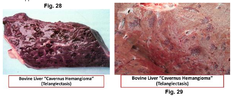
3. Cavernous-haemangioma “Telengiectasis’ ‘Plum pudding liver’
It is a common affection in imported animals. The cause is not well known but feeding factor and Sphaerophorus necrophorus may be concerned. On the other hand the lesion may have neurogenic origin caused by stretching of the spinal nerves, which may cause vasodilatation of the blood vessels of the liver. It is more common in intensive fed animals. Bluish black areas beneath the capsule and throughout the parenchyma characterize it. Lesions consist of dilated blood capillaries filled with blood.
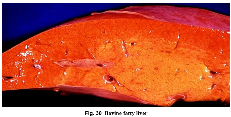
4. Fatty liver
Fatty liver is due to the mobilization of lipids from exterahepatic sources and from increased synthesis of lipids in the liver itself. The condition commonly seen in well-nourished lambs and very fatty aged cows. Affected liver is larger and heavier than normal. The liver edges are rounded, yellow or yellowish brown in color & soft in consistency.
Affected liver is fit for food, but the changes in color and consistency may make it unmarketable.
5. Liver Enlargement
Causative agent: Fatty infiltration, cirrhosis, abscess, tumor, plant poisoning.
Judgment: Case-dependent.
6. Rupture of Liver
Causative agent: Trauma, tumors, cysts, fascioliasis
Judgment: Condemn affected organ.
7. Adenoma
Causative agent: Benign tumor.
Judgment: Condemn organ and its lymph nodes.
8. Leukemia
The liver enlarged with proliferation of its interlobular connective tissue.
. Causative agent: Neoplastic proliferation.
Judgment: Total condemnation.
9. White liver disease
The condition occurs in sheep where the liver is enlarged and shows fatty changes. It is usually associated with cobalt /vitamin B12 deficiency, pregnancy toxemia in ewes and poor nutrition of cows in late pregnancy.
Causative agent: Cobalt/vitamin B12 deficiency.
Judgment: Condemn affected liver.
10. Black Liver
Causative agent: It is due to presence of the pigment lipofuscin from feeding on leaves of the mulga tree.
Judgment: Condemn affected liver.
11. Saw dust liver
The liver showed minute yellowish necrotic foci scattered throughout the substance and surface of the liver of young fatty cattle. It believed to be either early abscesses or the result of vit. E deficiency. Affected liver must be condemned.
B- Bacterial affections of the liver:
1.Tuberculosis
Tuberculosis of cattle and pig is common, while that of sheep and camel is rare. Congenital TB occurs in calves less than 2 weeks old.
Causative agent: Mycobacterium spp.
Judgment:
Localized: Condemnation of liver and its lymph nodes
Localized and emaciation: Total condemnation
Generalized: Total condemnation
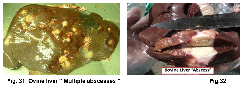
2.Liver Abscess
Liver abscess may occurs in young cattle due to umbilical infection while in older one, liver abscess may occur due to pyaemia, penetration of foreign body from reticulum or fasciola encysted metacercaria with pyaemic microorganisms from gastrointestinal tract.
Judgment: Small: Remove abscess / Multiple:
Condemn the liver.
Carcass: Total condemnation if there is systemic disturbance; otherwise,
bacteriological examination of the affected carcass.
3. Focal necrosis ‘Bacterial necrosis’
Occur in sheep and cattle but rare in pig and camel. It is caused by Seropherous necropherous which inhabitant intestinal tract of herbivores. The microorganisms penetrate the intestinal wall to reach the liver through portal vein. In acute cases the liver enlarged with formation of numerous necrotic foci and fever, while in chronic form necrotic foci with coagulated material.
Judgment: Localized: Condemn affected part /
Heavy: Condemn the liver.
Carcass: Approved if normal, total condemnation if there is fever or jaundice.
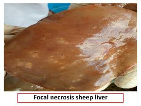
Fig.33
C-Parasitic affections of the liver:
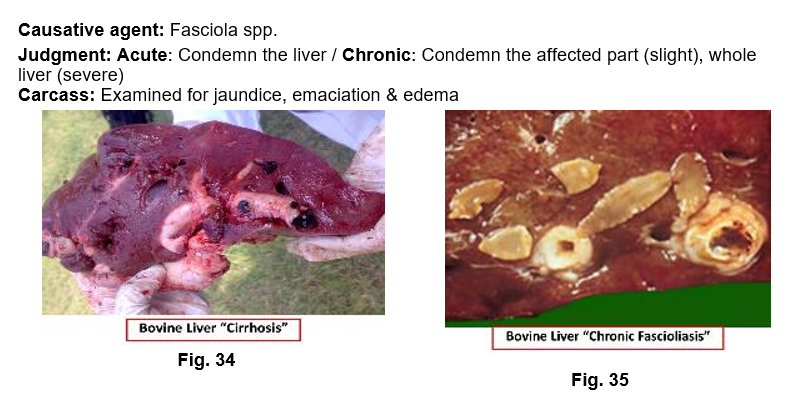
1. Fascioliasis
It is a common affection in the liver of cattle and sheep.
In acute stage, the liver is congested and swollen with petechial hemorrhages under the capsule. In chronic stage: cirrhosis and formation of connective tissue in the wall of the bile ducts and the surrounding liver tissues. In cattle the bile ducts become calcareous or pipes due to deposition of calcium salts. The flukes fail to reach the bile ducts & become encapsulated in the liver parenchyma. In sheep, there is no calcium precipitation but the liver irregularly lobulated and distorted due to connective tissue proliferation. The bile ducts thickened, dilated and have bluish color & does not undergo calcareous infiltration.
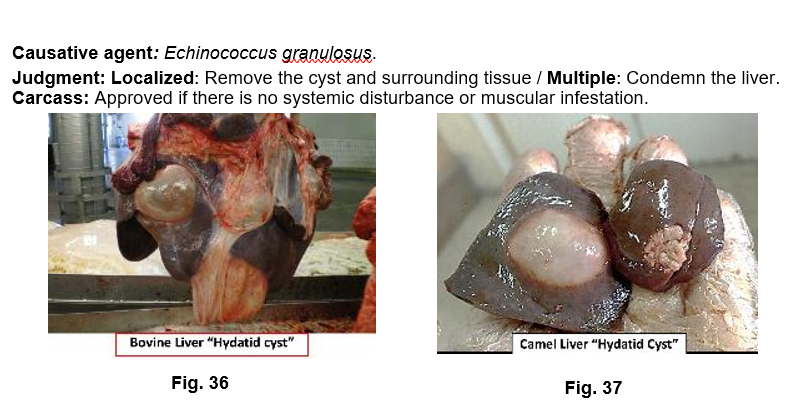
2.Hydatid Cyst
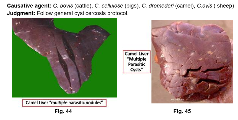
It is the larval stage of Echinococcus granulosus, which inhabit small intestine of carnivore chiefly dog. It is a common affection in the liver of camel.
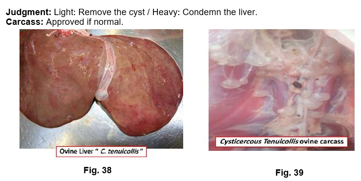
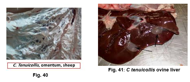
3.Cysticercus tenuicollis
It is the larval stage of Taenia hydatigena of dog. Usually present under the capsule of liver, mostly in sheep & pig, rarely in cattle and camel.
4.Linguatula larvae
Yellowish or greenish nodule under the liver capsule. It could be identified by microscopic examination to identify the typical hooks of the parasite.
Causative agent: Linguatula serrate.
Judgment: Condemn the affected part.
5.Milk Spots
It affects liver of pig and caused by the migration of Ascaris summ larvae. Milk spots are focal interstitial hepatitis and found on both surface of the liver. It is consists of white opaque center from which radiate interlobular septa. This affection may be confused with lesions caused by avian TB. In such condition grey flat like areas are found on the liver surface. Malchoire's palpation test is used to differentiate between lesion of avian TB and milk spots. The test includes, palpation of the upper part of the small intestine to manipulate Ascaris or not. Smear stained with Zeil Nelson to detect acid-fast bacilli of TB.
Judgment: Affected parts should be condemned in light cases, while the whole liver to be condemned in heavy affected infestation.

6.Coccidiosis
It is caused by Eimeria stiedea and affect rabbit liver. Appear in the form of irregular white spots on liver parenchyma.
Judgment: Condemn the liver& carcass condemned if there is emaciation.
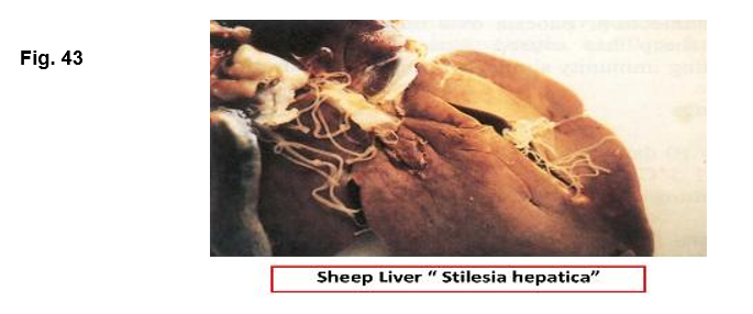
7. Stilesia hepatica
It is a cestode resembles Monzia. It affects the bile duct of sheep, goats, cattle and wild ruminants. The intermediate host is Oribatid mites. The bile duct may be occluded or form a sac-like dilatation filled with worms. Affected liver may show slight cirrhosis and the wall of the bile duct is thickened.
Judgment: Condemn the affected liver.
7. Cysticercosis