
Book
Affections Of Specific Parts and Organs of Food Animals
- IV. Affections of the Lungs
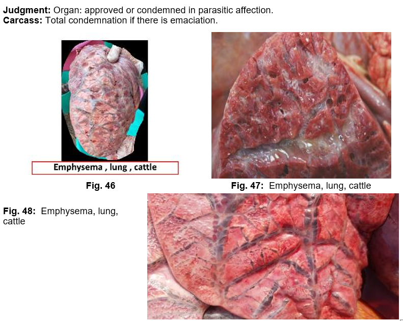
1. Interstitial or interlobular emphysema
It is a common affection in the lung of aged animals, accompanied parasitic pneumonia, and also in young animals severely affected with Dictycolus viviparous. Rupture of air alveoli results in escape of air bubbles into interstitial tissue. Emphysema may be extending to mediastinal tissue and subcutaneous tissue of thorax and abdomen.
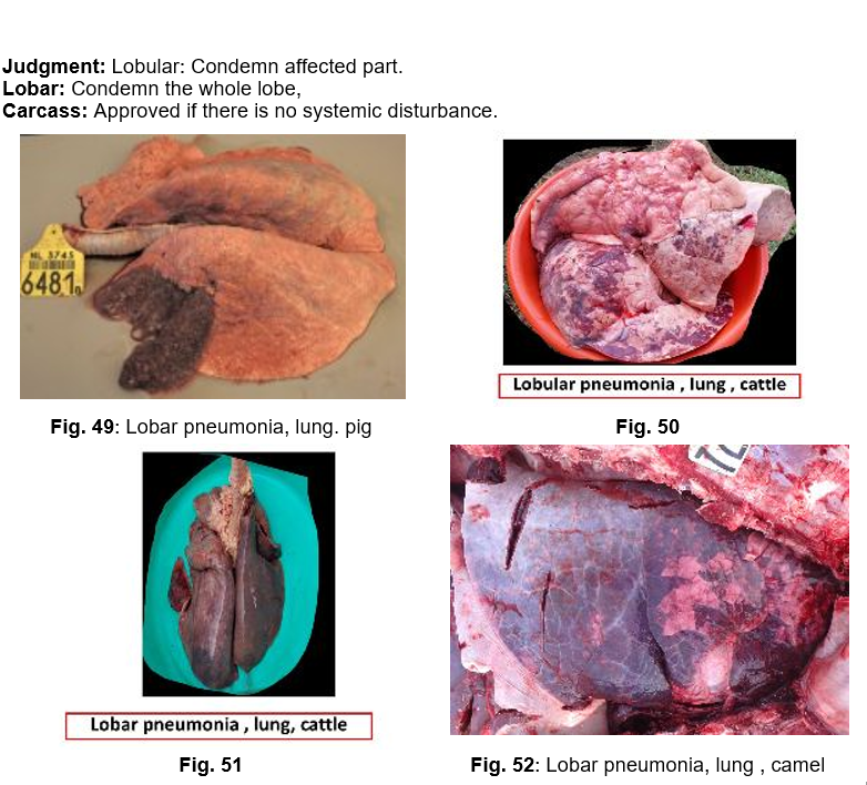
2. Pneumonia
The cause may be virus, bacteria, parasite, fungi, bad weather, inhalation of drugs or penetration of foreign bodies from reticulum. Pneumonia commonly seen in connection with specific diseases such as: bovine TB, shipping fever, enzootic pneumonia of calves and pigs, swine fever, extensive parasitic infestation of respiratory tract e.g., Ascaris. Fungi as Candida albicans and Mucor rarely give rise to pneumonia. In broncho-pneumonia (Lobular pneumonia), small pneumonic foci are intermixed with healthy tissue, while in lobar pneumonia the whole lobe or its greater part is in one stage of pneumonia.
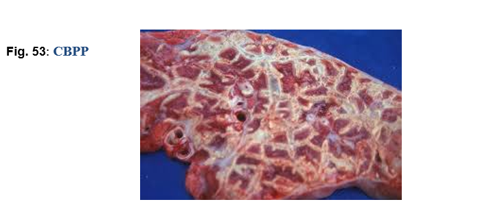
3. Contagious bovine pleuropneumonia CBPP
Contagious bovine pleuropneumonia is stages of hepatization, which surrounded by connective tissue. On cut section give marble appearance. Pneumonic foci undergo necrosis and become firm, dry and encapsulated to form sequesteraum.
Causative agent: Mycoplasma mycoides.
Judgment: Condemn the affected lung.
Carcass totally condemned if there is fever.
4. Pneumomycosis
Pneumomycosis is a fungal affection of lungs due to Aspergillus or Mucor, most common in young fowl, ducks and geese (brooder pneumonia) and occasionally seen in lambs, pig and cattle. It is characterized by diffuse hepatization of the lungs with numerous grey or greenish spots scattered throughout the lungs with a thin deposits of mould in the bronchi.
Judgment: The affected lung must be condemned, and also the carcass in case of septic
or gangrenous reactions.
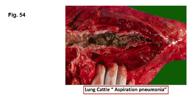
5. Contamination of trachea and lungs
Trachea and lungs may be contaminated with ingesta or blood.
Judgment: The affected trachea and lungs are condemned.
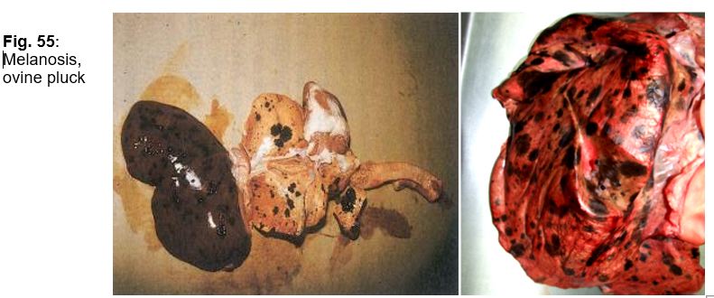
6. Melanosis
Black pigment on the lung tissue and bronchial lymph node.
Causative agent: Pigmentation disorder.
Judgment: Condemn lungs and its bronchial lymph nodes.
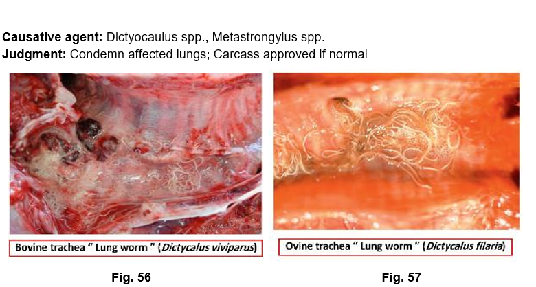
7. Parasitic
A.Parasitic Nematodes
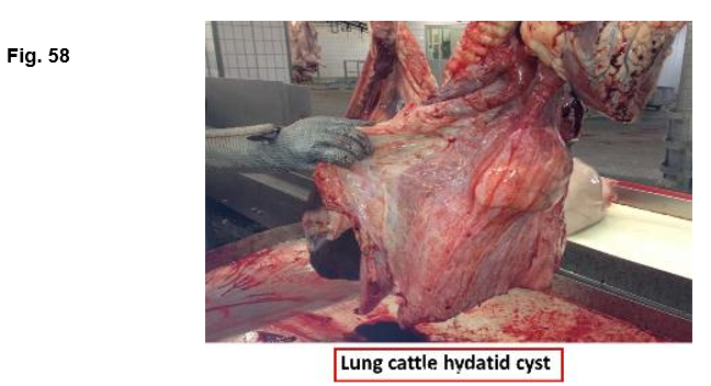
B. Hydatid Cyst
Causative agent: Larval stage of Echinococcus granulosus,
Judgment: Condemn affected part (light), condemn whole lung (heavy); Carcass approved if there is no emaciation, edema and or, muscular infestation.
C-Immature form of Fasciola.
Judgment: Condemnation of affected part of the organ (small number, or
Condemnation of lungs (large number).The carcass approved if there is no emaciation.
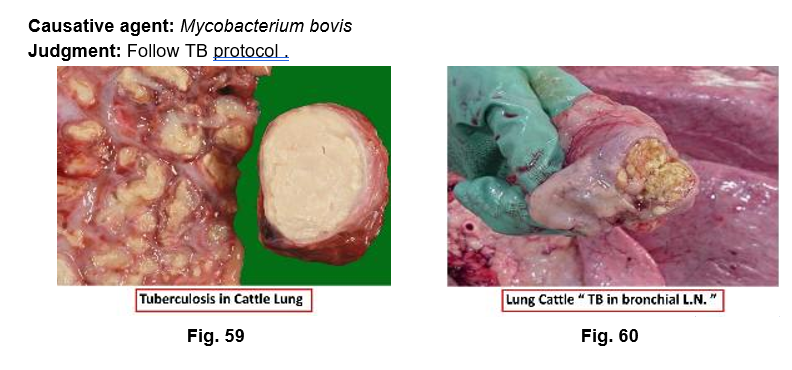
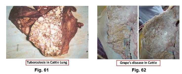
8. Tuberculosis
· Miliary TB.
· Acute early generalization.
· Acute late generalization.
· Caseous pneumonia.
· Acute acino-nodular TB.
· Chronic acino-nodular TB.
· Lung cavitation.
· Pearls or grapes disease.
7. Anthracosis
Causative agent: Bluish-black pigment of lung and its lymph nodes due to inhalation of coal dust.
Judgment: Condemn affected lungs and its lymph nodes.
8. Pulmonary Edema
Causative agent: Occurs in congestive heart failure, atypical interstitial pneumonia
(Fusarium) and organophosphate poisoning.
Judgment: Case-dependent.
9. Pulmonary Abscess
Causative agent: Single or small multiple abscesses may originate from emboli from other organs as in cases of: septic meteritis, septic mastitis, omphalophlebitis and /or penetration of foreign body. It may also occur as a part of primary diseases as: TB. actinobacillosis and aspergillosis.
Judgment: Condemn affected lungs; total condemnation if there is systemic disturbance.
10. Neoplasms
Causative agent: Lymphosarcoma, adenoma, melanoma
Judgment: Condemn affected lung or total condemnation in malignant tumors
