
كتاب
Topogaraphical, clinical and therapeutic ophthalmology
- OCULAR ANATOMY
The eyeball consists of three layers:
1- External fibrous tunic: It is the outer protective layer and consists of a transparent portion (cornea) and opaque portion (sclera).
2- Middle vascular tunic: This layer provides the nourishment for the eyeball and consists of iris, ciliary body and choroid.
3- Internal nervous tunic: This layer is represented by the retina which is really an expansion of the optic nerve.
Adnexal structures of the eyeball:
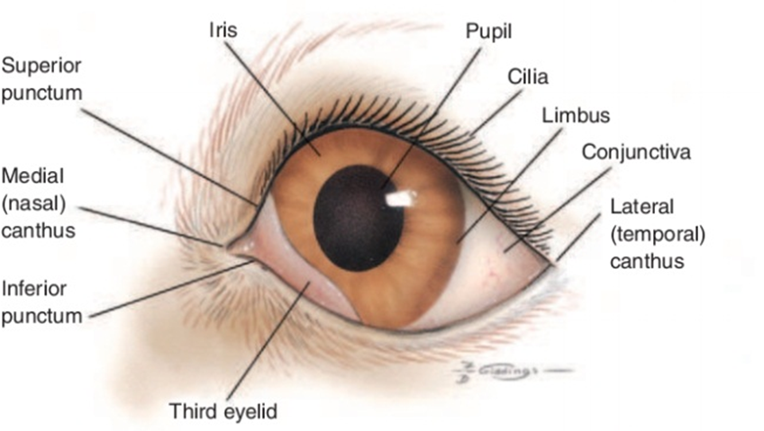
Fig.1: Frontal view of the external structures of the canine eye. Budras KD, et al. (2002):
Structures within the eyeball :
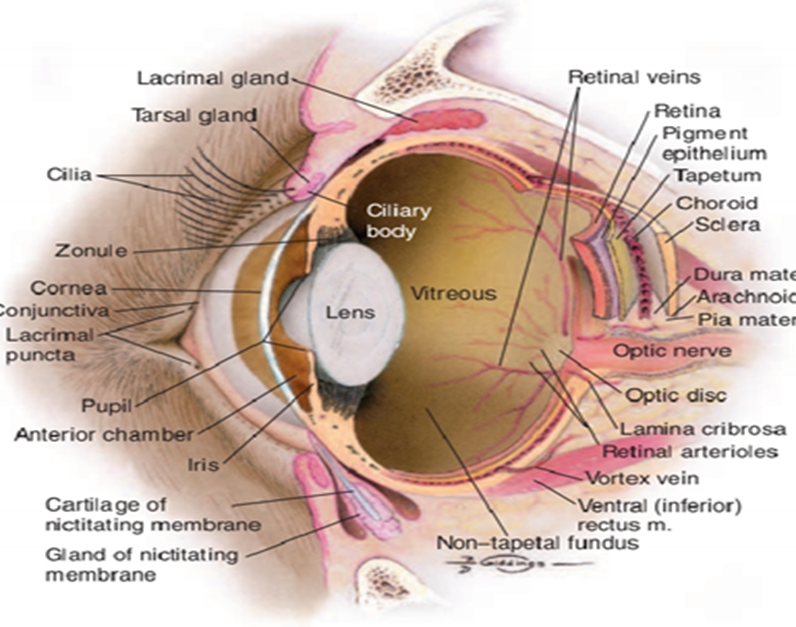
Fig.2: Internal structures of the canine eye. (Budras et al., 2002).
Arterial Supply
The major arterial supply of the eye is from the external ophthalmic artery, a branch of the internal maxillary artery, which arises from the external carotid artery The contribution from the internal carotid artery is small, unlike the situation in primates, and is via an internal ophthalmic artery, which arises from the circle of Willis. The internal ophthalmic artery enters the orbit through the optic canal with the optic nerve. From the external ophthalmic artery, numerous short posterior ciliary arteries arise and penetrate the sclera around the optic nerve head. These arteries supply the retina and choroid.
Venous Drainage
The retina is drained by the retinal veins and venules, which run from the peripheral retina toward the optic nerve head, The venous circle drains posteriorly through the sclera via the posterior ciliary veins to a dilation in the orbital vein, the superior (dorsal) ophthalmic vein.
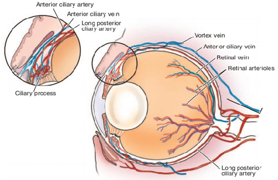
Fig.3: Vascular supply of the canine eye. (Remington , 2005).
Nerve supply:
1- Optic nerve (second cranial nerve)
The optic nerve consists of ganglion cells, whose cell bodies lie in the ganglion cell layer of the retina.
2- Oculomotor nerve(Cranial Nerve III)
The nucleus of the oculomotor nerve lies in the brainstem and has several components serving different extraocular muscles
3- Trochlear nerve (Cranial Nerve IV)
It passes through the fissure with the oculomotor nerve and the ophthalmic branch of the trigeminal nerve. The trochlear nerve innervates the dorsal oblique muscle only.
4- Trigeminal Nerve (Cranial Nerve V)
The nerve has both motor and sensory roots.
5- Abducens nerve (Cranial Nerve VI)
Supply the retractor bulbi and lateral rectus muscles.
6- Facial Nerve (Cranial Nerve VII)
The mixed facial nerve contains somatic motor and parasympathetic fibers, innervating the orbicularis oculi and retractor anguli muscles and the lacrimal gland.
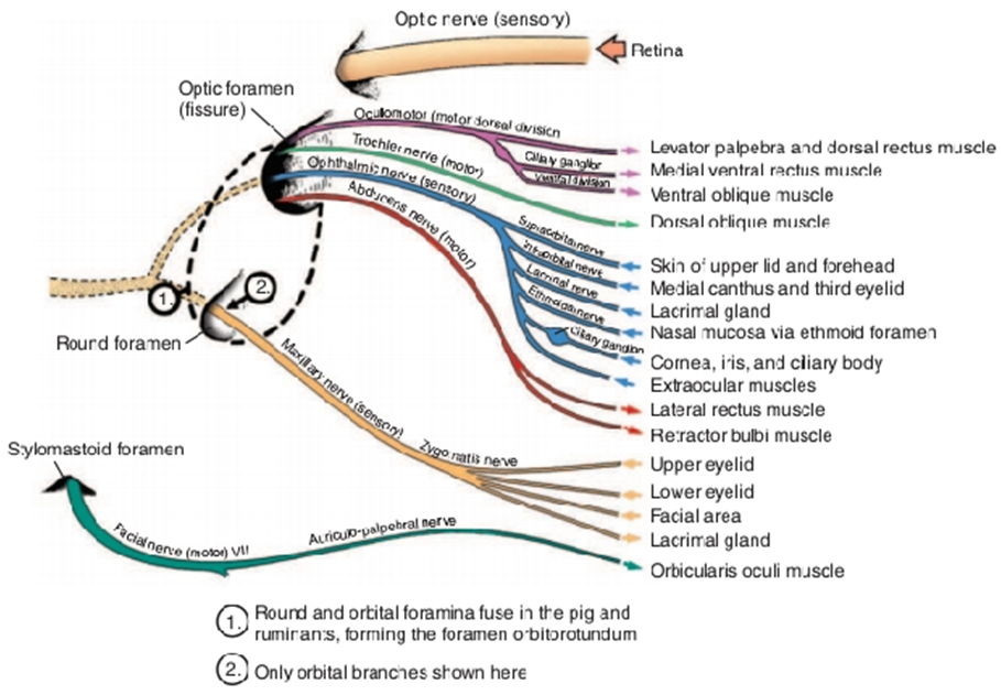
Fig.4: Nerve supply to the eye. (Evans, 1993).
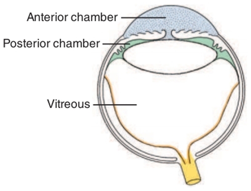
Fig.5: The chambers of the eye. The aqueous compartment is subdivided into two chambers by the iris diaphragm. The anterior chamber is anterior to the plane of the iris and pupil (blue), whereas the posterior chamber (green) is posterior to the iris-pupil plane but anterior to the vitreous (white). The retina and optic nerve are in yellow. (Budras ,2002).
