
Book
Topogaraphical, clinical and therapeutic ophthalmology
- OPHTHALMIC THERAPEUTIC AGENTS
1. Cleansing solutions
Sterile eye wash is used for removal of purulent exudates, foreign bodies, and irritants from the eyelids and conjunctival sac.
- Normal saline solution (NaCl solution 0.9%).
- B.S.S. (Balanced salt solution) and boric acid solution 2%.
2- Astringents and Cauterants
Identification and removal of the cause or more modern medical and surgical approaches usually achieve a better result in a more controlled manner, and astringents and cauterants are no longer recommended for ocular use.
- Astringents are locally acting protein precipitants.
- Cauterants are severe protein-precipitating agents that cause local tissue destruction.
Astringents as
- Zinc sulphate.
- Silver nitrate solution (1%).
- Copper sulphate .
- Yellow mercuric oxide (golden eye ointment).
.Cauterants as
- Carbolic acid (phenol.
- Tincture of iodine.
- Silver nitrate sticks
4. Mydriatics
They are drugs that dilate the pupil by stimulate the contraction of the iris dilator muscle or relax the iris sphincter muscle. they are used To make it easier to examine the retina and internal structures of the eye, treat inflammatory conditions like iritis and relieve spasms of the ciliary muscle , they can cause blurred vision, photophobia (light sensitivity), and dry eyes.
Example
• Atropine.
• Tropicamide (1% solution).
• Scopolamine (0.3 - 0.5%) solution.
• Epinephrine (Adrenalin).
• Phenylepherine.
5. Miotics
Miotics are drugs that constrict the pupil by stimulate the contraction of the iris sphincter muscle or cause the relaxation of the iris dilator muscle they are used to treat open-angle glaucoma by increasing the outflow of aqueous humor, help with presbyopia (age-related farsightedness) and to reverse the effects of mydriatics. Side effects can include eye discomfort and headache.
Example
- Pilocarpine 1-4%.
- Carbachol 1%.
- Eserine 0.25%.
6. LOCAL ANESTHETICS
a- Topical (local) anesthetics are used for ocular examinations and minor manipulative and surgical procedures, but never for therapeutic purposes. The effect on corneal sensation of a single drop of proparacaine or 2 drops separated by 1 minute.
b- Injectable analgesics
xylocaine (lidocaine) 1 - 2%, procaine (Novocaine) 1%, carbocaine (Mepivacaine HCL) 1-3%, marcaine (Bupivacaine HCL) 0.25–0.75% and duranest (Etidocaine HCL) 1%.
7. Antibiotics
The factors must be considered in the selection of an antibiotic
• The offending organism and its sensitivity.
• Location of the organism.
• Penetration of available drugs to that site.
• Pharmacokinetics of the available drugs
• Spectrum of activity of available drugs
•Toxicity of available drugs
The antibiotics used commonly in veterinary ophthalmology
- Penicillins
a large family of natural and synthetic derivatives of 6-aminopenicillanic acid that range considerably in stability, solubility, spectrum of activity, ocular penetration, and resistance to β-lactamase.
- Cephalosporins
They are generally similar to the penicillins in mechanism of action and pharmacology but are less susceptible to staphylococcal b-lactamases. A type of b-lactamase (cephalosporinase) produced by some Gram-negative organisms may inactivate them, also very useful for bacterial blepharitis. Cefazolin is the antibiotic of choice for perioperative antimicrobial prophylaxis in small animal surgery.
- Chloramphenicol
Is a broad-spectrum bacteriostatic antibiotic effective against a wide range of Gram-positive and Gram-negative organisms, Rickettsia, spirochetes, and Chlamydophila spp. However, Pseudomonas aeruginosa is often resistant. Because of its lipid solubility, Chloramphenicol may be administered orally, intramuscularly, subcutaneously, intravenously, subconjunctivally, or topically. Because absorption after oral administration results in high blood concentrations, this is the route of choice for infections in the posterior globe and orbit.
- Neomycin
Useful bactericidal agent for ocular use and is active against Gram-positive and Gram-negative bacteria, including Staphylococcus aureus and highly effective against Proteus vulgaris.
- Gentamicin
a topical agent of first choice for bacterial prophylaxis, relatively narrow against Gram-negative organisms, the value of gentamicin for treatment of more resistant organisms strains of S. aureus, Pseudomonas spp., E. coli, Aerobacter, Klebsiella spp., and Proteus spp. Topical application does not result in high intraocular concentrations, and although some drug enters the eye after subconjunctival or intravenous injection, vitreous penetration is poor regardless of route of administration. Long-term systemic therapy is limited by ototoxicity and nephrotoxicity. Gentamicin causes cataract and severe retinal degeneration when injected intraocularly.
- Tobramycin
Is effective againstβ-lactamase–producing staphylococci, resistance to tobramycin is less frequent, probably because of its more recent introduction. It is also ototoxic and nephrotoxic when given systemically but may be administered topically. or by administered via subconjunctival injection.
- Tetracyclines
broad-spectrum bacteriostatic antibiotics; however, Staphylococcus, Pseudomonas, and Proteus spp. are usually resistant. Tetracyclines are useful in treatment of infections with Chlamydophila and Mycoplasma spp. in cats. Systemic administration to dogs affected with periocular staining from pigments in the tears results in a decrease in staining, tetracycline preparations are effective for treatment of Moraxella bovis infection in cattle.
7.Antiviral drugs (DNA-synthesis inhibitors)
The use of antiviral drugs in veterinary ophthalmology is restricted to treatment of herpetic keratoconjunctivitis due to feline herpesvirus (FHV-1) in cats or, occasionally, equine herpesvirus (EHV-2) in horses.
Agents commonly used for treatment of patients with viral infection
- Idoxuridine
Idoxuridine is of the constituents of nucleic acids, which it replaces during DNA synthesis, thereby inhibiting viral replication. Can be compounded as a 0.1% solution or 0.5% ointment. It penetrates the intact cornea poorly after topical application but is generally well tolerated by cats. It must be applied at least 5 times daily.
- Vidarabine
Vidarabine interferes with viral DNA synthesis, and is moderately active against FHV-1 replication in vitro. It is usually well tolerated when applied topically as an ointment.
- Acyclovir and Valacyclovir
Acyclovir is widely available as a systemic drug, also available in a topical (ophthalmic) preparation. The efficacy of acyclovir against FHV-1 is low; cats receiving acyclovir sometimes show toxic adverse effects.
8.Antifungal agents
Important ophthalmic fungal infections may be considered in the following three categories
a- Infections of the eyelids and surrounding skin.
b- Intraocular infection (usually endophthalmitis) associated with penetrating foreign bodies or systemic mycoses, such as Cryptococcosis, Blastomycosis, Histoplasmosis, and Coccidioidomycosis.
c- Mycotic keratitis following corneal penetration or ulceration.
Agents commonly used for treatment of patients with fungal endophthalmitis orb keratitis are
- Natamycin
Natamycin is antifungal agent available as a 5% ophthalmic suspension, which is viscous but will pass through ocular lavage systems in the horse without causing obstruction. It is effective against a broad variety of fungi, including Candida, Aspergillus, Cephalosporium, Fusarium, and Penicillium spp.
- Azoles
Itraconazole, ketoconazole, fluconazole, voriconazole, clotrimazole, and miconazole are members of the azole group.They are especially useful for the treatment of systemic and ocular Cryptococcus spp. and Coccidioides immitis infections. Side effects in dogs include inappetence, pruritus, alopecia, and reversible lightening of the hair coat. In cats, anorexia, fever, depression, and diarrhea may occur. Long-term therapy, up to 6 months or longer, may be necessary because the drugs are fungistatic. Most azoles do not cross the blood-ocular barriers well.
10.Anti-inflammatory
I- Corticosteroids
The most useful and powerful drugs as anti-inflammatory agent. The following are good general rules to help govern ophthalmic use of corticosteroids:
a- Corticosteroids must not be used topically or subconjunctivally when fluorescein indicates a corneal epithelial defect.
b- Every “red” eye should be stained with fluorescein and its IOP should be measured before indiscriminate therapy with corticosteroids is initiated.
c- For non-ulcerative corneal disease or intraocular disease, a penetrating topical corticosteroid such as prednisolone or dexamethasone must be administered. Hydrocortisone does not penetrate the cornea.
d- For inflammatory disorders of the eyelids, posterior segment, optic nerve, or orbit, corticosteroids must be administered systemically, not topically.
Uses of corticosteroids:
1- Immune-mediated ocular disorders (seasonal allergic conjunctivitis, drug and contact allergies, chronic superficial keratitis or “pannus,” eosinophilic keratoconjunctivitis, episcleritis, some cases of keratoconjunctivitis sicca, lensinduced uveitis, uveodermatologic (VKH-like) syndrome, etc.)
2- Traumatic conditions resulting in severe inflammation (proptosis of the globe, contusion with hyphema).
3- Anterior uveitis
4- Postoperative immunomodulation (e.g., after corneal transplant or cataract extraction)
5- Reduction of postoperative swelling and inflammation after cryosurgery (e.g., cyclocryotherapy or cryoepilation for distichiasis or eyelid tumors)
Methods of corticosteroid application and indications are
a) Topical.
b) subconjunctival injections.
c) Systemic administration.
Corticosteroids are contraindicated in the following conditions
• Epithelial lesions of the cornea.
• Viral, mycotic and bacterial infections.
• Glaucoma.
II- Non-steroidal anti-inflammatory drugs (NSAIDs)
Such as acetylsalicylic acid 10-25 mg/kg 3 times daily.
I- Antihistamines
Antihistamine solution is very effective topically Premedication with systemic antihistamine 20–30min before intraocular surgery as
- Sodium cromoglycate.
- Olopatadine.
- Lodoxamide.
12.Carbonic anhydrase inhibitors
The enzyme carbonic anhydrase is present in ciliary body epithelium, where it is responsible, in part, for aqueous humor production. The enzyme carbonic anhydrase inhibitors decreased intraocular pressure. Carbonic anhydrase inhibitors as
- Acetazolamide (Diamox) 0.5 – 2.5 mg/ kg. b.w.
- Methazolamide (Neptazane) 1 - 2 mg/kg b.w.
13- Antiparasitic Agents
Ivermectin is now used, almost to the exclusion of all older parasiticides, for treatment of parasitism such as Habronemiasis and onchocerciasis in horses and ocular filariasis in small animals.
14.Osmotic diuretics
Osmotic drugs are applied topically to clear or reduce corneal edema ,osmotic diuretics used in acute glaucoma, prior to intraocular surgery, traumatic proptosis of the globe, and clearing of corneal edema as
- Mannitol (Osmitrol).
- glycerol (Osmoglyn).
- urea (ureaphil) .
- Topical NaCl 5% ointment is used in corneal edema.
15.Vitamins
Various vitamins have been advocated for their supposed therapeutic efficacy in the treatment of ocular disorders of animals. In the absence of a specific vitamin deficiency (e.g., vitamin A deficiency causing nyctalopia in cattle or conjunctivitis in turtles) there usually is little to be gained from such local therapy. The exception is systemic and ocular signs of warfarin Poisoning which are treated with vitamin K and its analogues, Vitamin C and Riboflavin (B2).
16. Enzymes
- Fibrinolysin is used to remove clotted blood from the anterior chamber in traumatic hyphema.
- Alpha chymotrypsin (Alphapsin) is useful for absorption of intraocular debris in cases of hypopyon.
- Alpha chymotrypsin is a proteolytic enzyme used for removal of the lens by intracapsular extraction in cases of cataract.
17.Ophthalmic stains
Ophthalmic stains are used commonly as diagnostic aids in diseases of anterior and posterior segment and nasolacrimal system.
a) Fluorescein dyes: Fluorescein is available as solution of 0.5-2.0%. Filter paper strips impregnated with fluorescein may be placed in the conjunctival sac until moistened by tears. It is readily soluble in water produces a bright green fluorescent color. It is used in the following conditions
• An indicator dye for corneal epithelial defects.
• Detection of the patency of the nasolacrimal duct.
• Detection of the lesions of the retinal and uveal vasculature.
b) Rose Bengal: It stains devitalized cells and their nuclei of the cornea and conjunctiva.
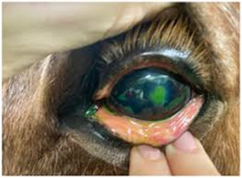
Fig.34: Corneal ulcer in horse note the Fluorescein dyes stained the dead tissue not the life tissue.
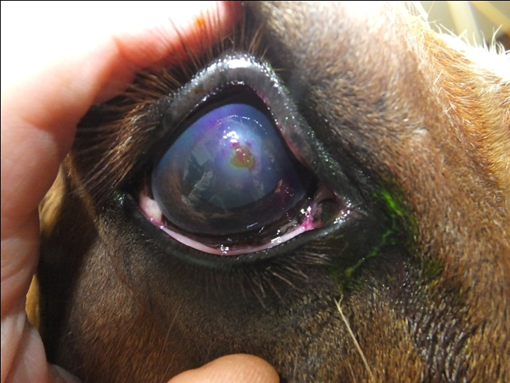
Fig.35: Corneal ulcer in horse note the Rose Bengal dyes stained the life tissue not the dead tissue.
18. Artificial tears
Artificial tear preparations are used when the normal tear quality or quantity is altered or when loss of tears is increased due to evaporation, or, in some cases, primary corneal pathology. These agents are lacrimomimetic and are not to be confused with lacrimogenic agents, such as cyclosporine. The production of endogenous tears is always preferred over the replacement of tears with “artificial” tears.
Indications for tear replacement preparations are as follows
1- For treatment of keratoconjunctivitis sicca (“dry eye”).
2- For treatment of exposure keratitis (e.g., facial nerve paralysis, buphthalmos, breed-associated lagophthalmos).
3- In patients with abnormal tear film breakup time (qualitative tear film disturbances)
4- During and after general anesthesia to prevent corneoconjunctival desiccation
5- As a lubricant, refractive/electroconductive, and cushioning solution during gonioscopy and electroretinography
6- As a diluent for compounding of some ophthalmic solutions
7- In patients with primary corneal disease, such as feline corneal sequestration and canine superficial punctate keratitis.
19. Vasoconstrictors and decongestants
Vasoconstrictor and decongestant eye drops that work by narrowing the blood vessels in the eye which activate alpha-adrenergic receptors in the small blood vessels (arterioles) of the eye's outer layer (conjunctiva) to reduce redness and congestion. These drops are effective for temporary relief of symptoms like redness and irritation but can lead to a "rebound" effect, making the eyes redder if used too frequently or over the long term
Example
- Ephedrine 0.15%.
- Phenylephrine 0.15%.
20. Corneal dehydrating agents:
Corneal dehydrating agents for corneal edema and to preserve corneal tissue for transplantation. Hypertonic drops work by drawing water out of the cornea, while preservation agents are used to dry and store donor tissue, often maintaining its properties for later use.
For treatment of corneal edema
- Hypertonic saline 2-5%: Typically used as an ophthalmic solution or ointment, 5% sodium chloride draws water from the cornea to reduce swelling.
- Glycerin: A highly concentrated solution that can rapidly dehydrate the cornea, but it is often unsuitable for routine use due to burning and photophobia.
For preserving corneal tissue
- Silica gel: A common agent for preserving corneal tissue, it is particularly good at maintaining optical transmittance.
- Silicone oil: A novel agent being explored for its stability, low toxicity, and high hydrophobicity, which may be useful for corneal tissue storage.
- Polyethylene glycol (PEG): Used to dehydrate anterior corneal grafts to a normal thickness and restore transparency before procedures
