
Book
Indigestion in ruminant
- Ruminal parakeratosis
Definition
Ruminal parakeratosis is a nutrition-related lesion of the rumen epithelium characterized by excessive keratin accumulation (hyperkeratosis/parakeratosis), thickened/hardened and often clumped papillae, reduced absorptive function and variable sloughing/erosion of the epithelium. It’s most commonly seen when ruminants receive high-concentrate/low-effective-fiber rations or experience prolonged ruminal pH depression (acidosis).
hardening of the rumen's papillae (small, finger-like projections) in ruminants like cattle. This keratinization can hinder VFA absorption, affecting feed efficiency and weight gain. It can also be associated with zinc deficiencies or a diet low in vitamin A.
Pathogenesis (how it happens)
· Excess rapidly fermentable carbohydrate → increased VFA (esp. butyrate) production and repeated/ prolonged ruminal pH depression.
· The altered chemical environment changes epithelial cell proliferation and differentiation: keratin layers accumulate (parakeratosis), cell adhesion is compromised, and papillae become short, thick, clumped or even slough. This impairs short-chain fatty acid (SCFA) absorption and the epithelial barrier.
Risk factors
· Abrupt transition to high-grain rations (feedlot finishing, early concentrate starters in calves).
·Low effective fiber or poor particle size (insufficient rumination).
· Heat-treated or pelleted forages (can predispose), and prolonged ruminal milk accumulation in bottle-fed calves.
· Zinc or vitamin A deficiency
Clinical signs
· The diseases is often subclinical
· Reduced feed efficiency, lower weight gain or milk production.
· In more severe cases: anorexia, rumen dysfunction (hypomotility), paste-like rumen contents, secondary rumenitis or ulcers, and increased risk of systemic effects from translocated bacteria/toxins.
· Gross and histologic lesions (what you’ll see) Gross: papillae thickened, clumped, occasionally short/atrophic; surface may be rough, scaly or show erosions/ulcers. (See macroscopic image in the carousel.)
· Histology: retained nucleated keratin layers (parakeratosis), hyperplasia of keratinocytes, variable spongiosis/vacuolar change, inflammatory cell infiltration and sloughing of the stratum corneum. (See histology images in the carousel.)
Diagnosis
· History of diet (high concentrates, abrupt change).
· Clinical exam (rumen dysfunction, production loss).
· Rumen fluid analysis: low pH supports SARA/acidotic states but pH alone doesn’t prove parakeratosis.
· Rumenoscopy or rumenotomy: direct visualization of papillae changes.
· Biopsy/histopathology of rumen papillae is definitive (shows parakeratosis/hyperkeratosis).
Treatment & control
· Dietary correction is mainstay: increase physically effective fiber, reduce rapidly fermentable carbohydrates, and gradually adapt animals when shifting rations. Restore rumen function and avoid abrupt changes. Merck Veterinary Manual +1
· Buffers/alkalinizing agents (e.g., oral bicarbonate) may be used to correct acute pH drops, but long-term control relies on ration formulation and management. Merck Veterinary Manual
· Feed additives: ionophores (where permitted) and rumen modifiers can reduce SARA risk in some systems — use according to regulations and veterinary guidance. Frontiers
· Severe erosions/ulcers: treat as rumenitis (antibiotics if secondary infection suspected, anti-inflammatories) under veterinary guidance.
· Prognosis Variable. With diet correction, ruminal epithelium can recover but full structural/functional recovery may take weeks to months; chronic thickening or scarring can lead to lasting reductions in absorptive efficiency. Early identification and correction improve outcomes.
· Practical management checklist (for herd use) Evaluate TMR particle size and effective fiber (shredded long hay, straw buffers).
· Review feeding schedule and ensure gradual transitions (step-up grain adaptation).
· Monitor rumen health: rumen pH spot checks, fecal consistency, milk fat depressions or drop in gain.
· Consider ration reformulation with a ruminant nutritionist; use ionophores only if appropriate.
· If individual animals show signs, perform rumen fluid analysis and consider rumenoscopy/biopsy.
PM lesions
Macroscopic rumen lesions / thickened papillae — from a slaughterhouse study.
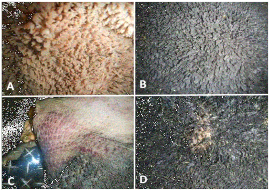
Pm lesion of ruminal parakeratosis. A/ normal rumen mucosa. B/ thickening and atrophy of the rumen papillae. C/ ruminal hemorrhages. D/E erosions, ulcers and ruminal scars (LUNA-MENDEZ et al., 2020)
