
Book
UPPER AND LOWER EYELIDS AFFECTION
- II-PROMINENT NASAL FOLDS
Unusually prominent of the nasal folds in same species like Pekingese, pugs, English bulldogs, Boston terriers, and similar brachycephalic breeds.
If nasal folds are causing keratitis, they should be removed either partially or totally, in partial removing (only the medial portion of the fold is removed, where it touches the cornea, resulting in less alteration from the breed “norm” .
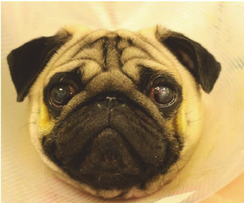
Fig.5: A 2 year old male Pug diagnosed and undergoing treatment for a corneal ulcer in his left eye (Rowena et al, 2015).
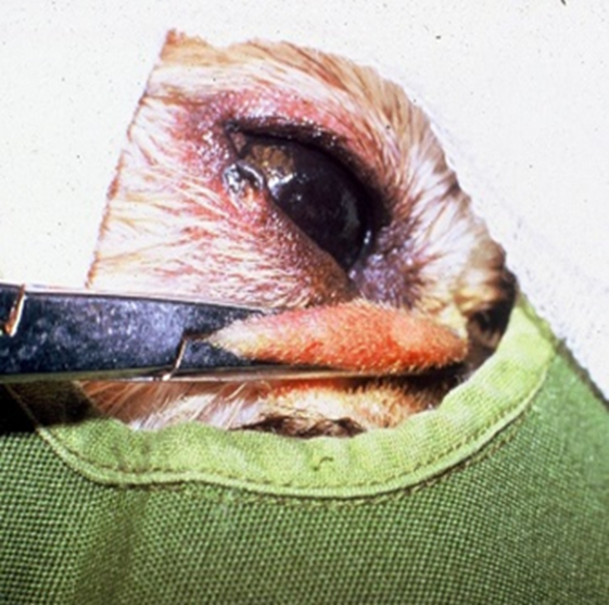
Fig.6: Crushing of the nasal fold before its excision.
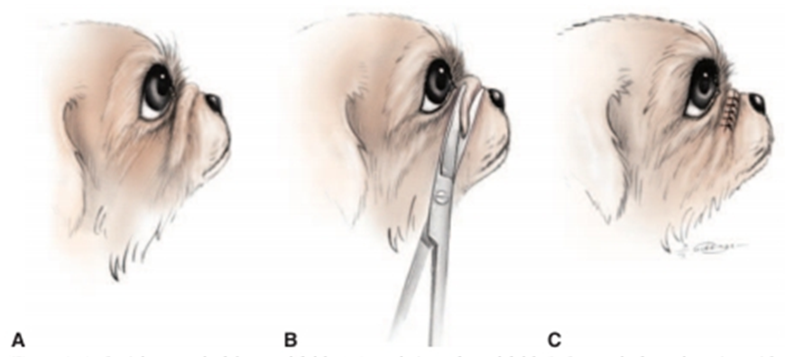
Fig.7: Partial removal of the nasal fold. (Redrawn, 2000).
A- Lateral view of nasal fold.
B- Removal of nasal portion with curved scissors.
C- The sutured wound, the knots are placed on the anterior side
of the incision in order to limit corneal contact.
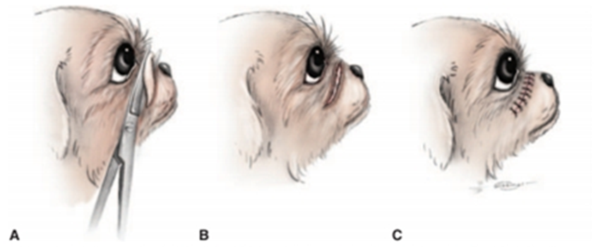
Fig.8: Total removal of the nasal fold (Redrawn, 2000).
A- Removal of the fold, starting laterally.
B- The fold removed.
C- The fold sutured. The knots are placed on the anterior side of the incision in order to reduce the chance of corneal contact.
