
Book
UPPER AND LOWER EYELIDS AFFECTION
- III- DISORDERS OF THE CILIA
Normally positioned cilia emerge from the dermal side of the eyelid margin. Cilia
disorders can be bilateral or unilateral and can affect upper or lower eyelids.
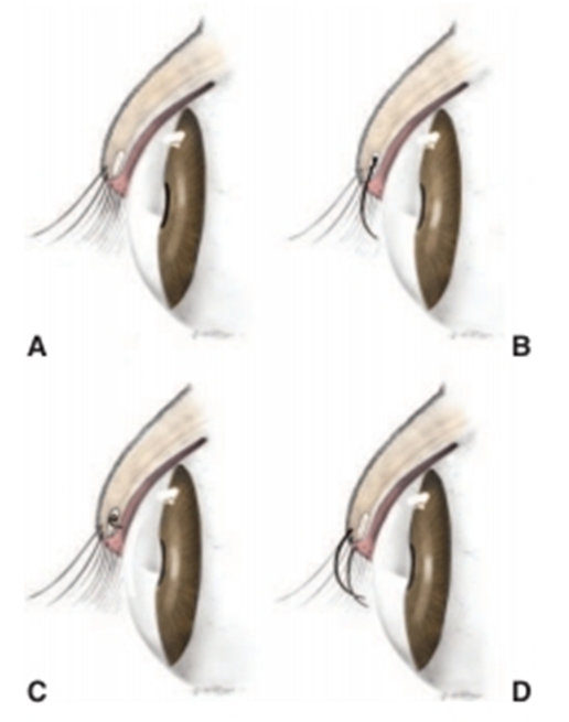
Fig.9: A- Normal eyelid (David et al, 2008).
B- Distichiasis. Cilia emerge from the meibomian gland orifice.
C-Ectopic cilium. The cilium arises from the meibomian gland but emerges
through the palpebral conjunctiva.
A- Trichiasis. Normal cilia or hairs arising from a normal location reach the cornea due to altered facial or eyelid conformation.
Clinical Signs of Cilia Disorders
1- Epiphora: Excess tearing and staining of facial hairs is usually present despite patency of the nasolacrimal apparatus.Purulent discharge is unusual except with corneal ulceration.
2- Blepharospasm: Pain associated with constant irritation and sometimes corneal ulceration is evident as blepharospasm.
3- Chronic conjunctival hyperemia:
4- Corneal ulceration
The three common disorders in which aberrant cilia or hair cause corneoconjunctival irritation are as follows
A- Distichiasis
It means presence of a second abnormal row of eyelashes. The condition is congenital. Without clinical evidence of irritation, they are considered insignificant.
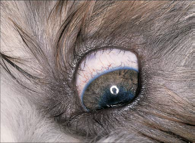
Fig.10:Marked distichia of both lids, but only visualized
well in the photo of the upper lid of a Shih TzuSymptoms
1. Blepharospasm, conjunctivitis.
2. keratitis and corneal ulceration.
3. Presence of abnormal cilia.
Treatment
1. Manual epilation: by rounded tip epilation forceps.
2. Removal of a V-shaped segment of the tarso-conjunctiva containing distichia.
3. “Eyelid-splitting” and partial tarsal plate excision techniques as postoperative cicatricial entropion, scarring of the eyelid margin, and destruction of the meibomian glands.
4. Cryoepilation or electroepilation
Simple epilation by forceps without adjunctive cryotherapy or application of an electric current is a temporary measure because the cilia regrow within 3 to 4 weeks.
Electroepilation
- For treating a small number of follicles but tends to be less reliable.
- By supplies direct current (1 to 5 mA) to the offending meibomian gland, destroying it by electrolysis.
- A fine needle (25 or 26 gauge) can be used as the applying electrode. It is passed alongside the cilium and into the follicle with use of adequate magnification. Current is applied for 20 to 30 seconds. Easy removal of the cilium, which often adheres to the epilation needle, indicates follicle destruction.
Cryoepilation:
- For treating a large numbers of follicles.
- takes advantage of the selective susceptibility of hair follicles to cold. A nitrous oxide or liquid nitrogen cryoprobe is applied to the conjunctiva overlying the meibomian glands that contain the offending cilia. The ice ball is observed as it advances over the line of gland openings on the eyelid margin. Two rapid freeze–slow thaw cycles are used. Following thawing of the second freeze, a systemically administered corticosteroid or nonsteroidal antiinflammatory drug (NSAID). An oral NSAID may be continued postoperatively for analgesia. An antibiotic-steroid ophthalmic ointment .
A- Ectopic cilia
Ectopic cilia mean single or multiple cilia arise from the meibomian glands emerging from the conjunctival surface of the eyelid, where they cause marked corneal irritation and usually ulceration. The condition is congenital and observed after several months to several years of age. They are visible only with the illumination and magnification provided by a slit-lamp or operating microscope.
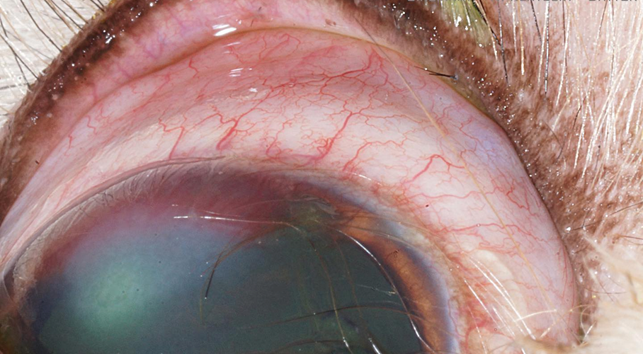
Fig.11: ectopic cilia in upper eye lid of cat (David et al, 2008).
Symptoms
1. Presence of ectopic cilia at the palpebral conjunctiva 2-6 mm from the lid margin.
2. Blepharospasm.
3. epiphora and conjunctivitis especially at the bulbar conjunctiva.
4. Vascularization of adjacent parts of the cornea accompanied by an elongated superficial corneal ulcer.
Treatment
- Electro-epilation of ectopic cilia.
- Surgical removal of the cilia with part of tarso-conjunctiva containing the follicle.
- resection of the affected cilium and meibomian gland under magnification.
- An elliptical Desmarres chalazion clamp with screw lock is placed around the cilium for hemostasis, and the eyelid is everted. A block of tissue containing the offending follicle and meibomian gland is removed with a small (No. 65 Beaver) scalpel blade, leaving the eyelid margin intact. No sutures are placed. Digital pressure for several minutes is sufficient to control hemorrhage.A topical broad-spectrum antibiotic ointment is applied three times daily for 5 to 7 days.
A- Trichiasis
Cilia or adjacent skin hairs arising from a normal location are misdirected so that they touch the cornea which irritates the globe and/or conjunctiva. This may be a primary condition (congenital) but is also a consequence of nasal folds, eyelid coloboma, eyelid agenesis, and entropion.
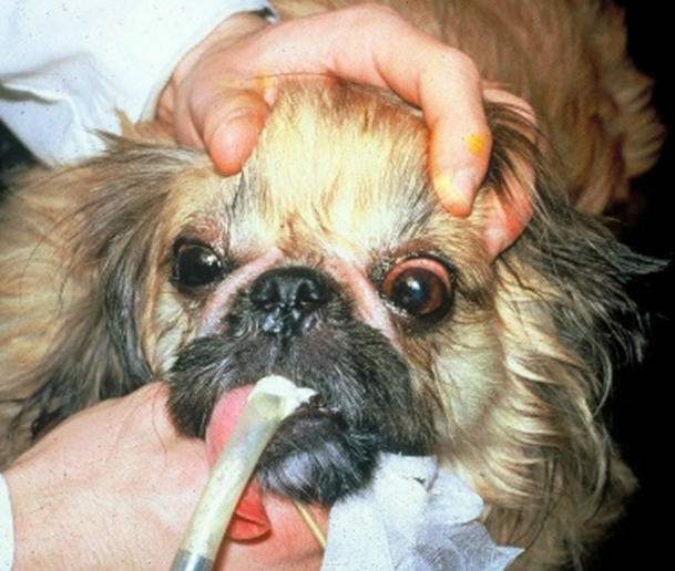
Fig12:Trichiasis due to prominent nasal folds in a Pekingese dog
Symptoms:
1. Epiphora: Excess tearing and staining of the medial canthal skin is present.
2. Blepharospasm: Pain associated with constant irritation of the cornea and conjunctiva resulting in blepharospasm and rubbing of the eye.
3. Chronic conjunctival erythema: The conjunctival blood vessels are engorged with blood.
4. Corneal ulceration: Ulcers caused by cilia are usually shallow and eccentrically placed on the cornea corresponding to the position of cilia.
5. Presence of abnormally directed cilia towards the eyeball.
Treatment
Depending on location of the offending hairs, is treated with any of the following:
1. Regular trimming of the periocular hairs.
2. Forceps epilation
3. Electro-epilation
4. Cryoepilation of the offending hairs. Especially at the medial canthus when there are a large number of hairs on the inner surface of the canthus and on the medial caruncle.
5. Surgical correction of the deformity causing the trichiasis (e.g., entropion, nasal folds, eyelid coloboma).
6. Surgical removal of dermoids
