
Book
UPPER AND LOWER EYELIDS AFFECTION
- IX- Eyelid tumors
Eyelid tumors are relatively common in all domestic species. the most common eyelid neoplasms vary by species, that the disease is virus induced. It has a “wart” like appearance or can also be ulcerative and granulomatous.
Dog: meibomian adenoma, papilloma, histiocytoma, melanoma .
• Cat: squamous cell carcinoma
• Horse: squamous cell carcinoma, equine sarcoid, melanoma
• Cattle: squamous cell carcinoma
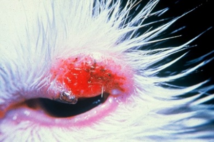
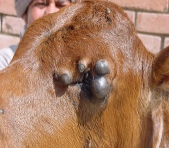
Fig.26: Basal cell carcinoma at the lower eyelid in a cat. Fig.27: Fibropapilloma at the eyelid in a cow calf.
A- Squamous Cell Carcinoma
Squamous cell carcinoma may involve the eyelids of all species especially common in poorly pigmented areas of the eyelids in horses, cattle, and cats. The tumor is associated with exposure to ultraviolet light.
The pathogenesis of eyelid squamous cell carcinoma is may metastasize to regional lymph nodes and eventually to the lungs.
Clinical signs
Chronic purulent ocular discharge, which may be temporarily or partially responsive to antibiotics. Periocular excoriation, chronic conjunctivitis, and encrusted or hemorrhagic lesions of the eyelids are common.
Diagnosis
Is confirmed with cytologic assessment of scrapings or by biopsy.
B- Meibomian Adenoma
- The most common eyelid tumor in dogs, occurring most frequently after middle age.
- The tumors originate in the meibomian glands, and although many grow rapidly and sometimes appear histologically malignant, they are usually clinically benign.
- appear as proliferative masses appearing from the meibomian gland orifice .
- Enlargement of the gland itself is often visible, especially when viewed from the conjunctival surface, be associated with glandular enlargement due to retained secretions and associated lipogranuloma formation (chalazion), may cause frictional keratoconjunctivitis and should be removed as soon as the diagnosis is made.
- Simple removal of the section of the mass protruding from the eyelid margin is inadequate, because the tumor arises from the meibomian gland and will recur if not totally removed.
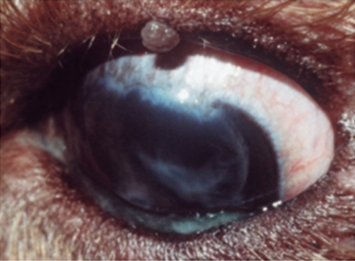
Fig.28: Upper eyelid meibomian adenoma in a dog(David et al, 2008).
A- Equine Sarcoid
- Equine sarcoid or fibrosarcoma is the second most common tumor of the equine eyelid,may be present elsewhere on the face and body.
- Sarcoids are nodular masses usually beneath intact skin and firmly adherent to the overlying dermis and subcutaneous tissues.
- Occasionally they can be ulcerated and must be distinguished from the eyelid lesions of habronemiasisin endemic areas.
- complete surgical excision of sarcoids is difficult and recurrence is likely. If surgical excision is used, a wide surrounding margin of normal tissue should be removed with reconstruction via a preplanned method.
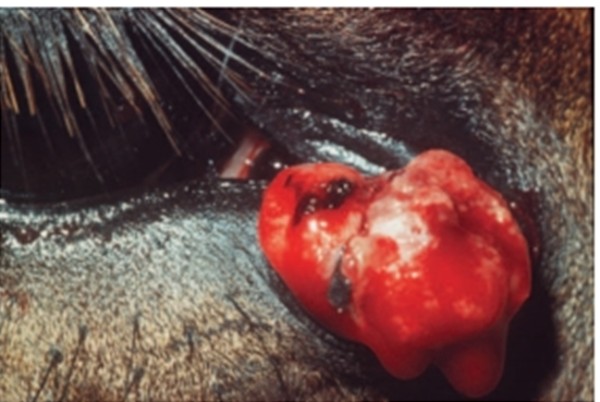
Fig.29: Equine sarcoid at the medial canthus of the right eye(David et al, 2008).
A- Viral Papillomatosis
- Viral papillomas of the eyelids and conjunctiva occur in young dogs and cattle as part of oral or generalized papillomatosis.
- When continually moistened by tears, the tumors are grayish white and soft. The disease is often self limiting.
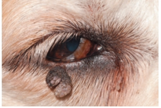
Fig.30: Lower eyelid papilloma in a dog (David et al, 2008).
A- Histiocytoma
Histiocytic masses involving the eyelids occur occasionally in dogs. may occur as single or multiple eyelid lesions
The tumors vary from benign lesions that regress spontaneously to tumors that are metastatic from or to the eyelids and therefore may involve regional lymph nodes or
other organ systems. Histiocytomas involving the eyelid typically appear as
tan to pink, somewhat alopecic masses that may grow relatively
quickly and may ulcerate quickly .
All eyelid tumors are regarded as malignant until proved otherwise by biopsy examination.
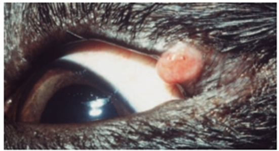
Fig.31: Solitary histiocytoma of the upper eyelid of a young dog (David et al, 2008).
Treatment
The best treatment is combination therapy of two or more of the following methods:
1- Surgical excision
- For smaller tumors involving one third or less of the eyelid margin, a simple, full-thickness wedge resection with two-layer closure.
- For more than one third of the eyelid length must be removed, total excision is usually not possible without some sort of blepharoplastic procedure, and referral may be indicated. One of the more straightforward blepharoplastic procedures is the H-plasty or full-thickness advancement flap.
2- Cryotherapy
Cryotherapy is particularly useful for eyelid lesions, as most eyelid tissues (including the nasolacrimal apparatus) are relatively resistant to the effects of cryotherapy and the lid retains function after cryonecrosis sufficient to produce tumor cell death. Cryotherapy is particularly useful for small or early tumors, especially in elderly or debilitated patients.
3- Radiation therapy
Chemotherapy