
كتاب
Metabolic disorders in Ruminant, Fat Cow Syndrome ,Fatty liver syndrome
- Diagnosis
1- Clinical History and Risk Assessment
Key indicators:
- Overconditioned cow prepartum
- Postpartum drop in dry matter intake (DMI)
- Concurrent metabolic disorders
2- Laboratory Findings
- Elevated NEFA prepartum (> 0.3 mEq/L)
- High NEFA postpartum (> 0.6 mEq/L)
- Elevated β‑hydroxybutyrate (BHBA)
- Elevated liver enzymes (AST, ALT)
- Low blood glucose level
- Bilirubinemia
- Increased non-protein nitrogen levels
3- Liver Biopsy
The gold standard for diagnosis:
· 10% fat: mild
- 10–30% fat: moderate
· 30% fat: severe fatty liver
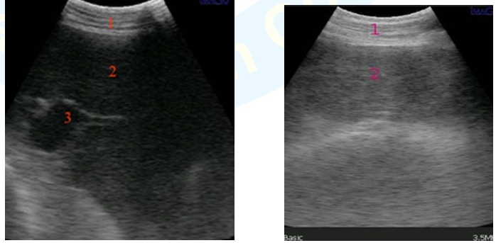
4-. Ultrasonography
Fatty infiltration appears as a hyperechoic, heterogeneous liver.
|
Hepatic ultrasonograms in control healthy cows showing normal gray echogenicity 1. Abdominal wall 2. Liver parenchyma 3. Portal vein. (Imaged through 11th ICS by convex transducer 3.5 MHz Imago). (Ghanem et al., 2016) |
Hepatic ultrasonogram in ketotic cows 1. Abdominal wall 2. Fatty infiltration (increased echogenicity, blurring of hepatic blood vessels) (Imaged through 11th ICS by convex transducer 3.5 MHz Imago) (Ghanem et al., 2016) |
