
كتاب
LACRIMAL SYSTEM
- Obstruction of the nasolacrimal duct
In cats blockage of the puncta or nasolacrimal ducts are common sequela of presumed herpetic
keratoconjunctivitis and upper respiratory tract infections. Similar may be seen in any species and frequently accompanies symblepharon.
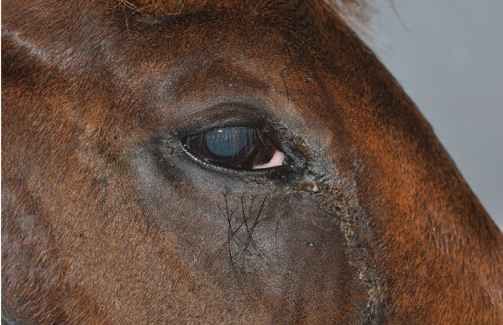
Fig.10: Duct obstruction in a horse. Note the mucoid discharge at the medial canthus.
Symptoms:
1- Epiphora.
2- moisture and tear staining of the medial canthal skin and sometimes serous or seromucoid conjunctivitis.
Treatment:
In congenital atresia of a segment of the duct, Conjunctivorhinostomy or Conjunctivobuccostomy is the treatment of choice. In cases of acquired obstruction flushing of the duct is performed 2-3 times daily for several days with ophthalmic solutions containing antibiotics and corticosteroids.
|
Fig.11:Conjunctivorhinostomy. A, The conjunctiva is removed from the inferior nasal area overlying the lacrimal bone. B, A communication is made from the conjunctival sac to the nasal cavity with a Steinmann orthopaedic pin. The pin is directed toward the contralateral external nares but is advanced only until it enters the nasal cavity. A stent of plastic tubing is sutured in place (David et al.,2008).
|
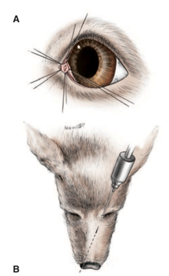
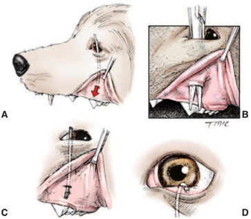
Fig.12:Conjunctivobuccostomy. A, Direction of the final drainage canal. B, A canal is made from the inferior conjunctival fornix to the oral cavity with straight hemostats. C, A tube is passed and sutured to the oral mucosa. D, The upper end of the tube is sutured to the skin in the region of the nasal canthus so as not to rub on the cornea. The tube is left in place for a minimum of 2 months. (Lavach ,1985)
