
كتاب
Predict, Prevent and Manage Severe Hyperbilirubinemia in Term and Late Preterm Newborns
- Appendix
Appendix 1: Phototherapy
Introduction:
Clinical trials have validated the efficacy of phototherapy in reducing excessive unconjugated hyperbilirubinemia, and its implementation has drastically curtailed the use of exchange transfusions. (3) The initiation and duration of phototherapy is defined by a specific range of total bilirubin values based on an infant’s postnatal age and the potential risk for bilirubin neurotoxicity. (3) Clinical response to phototherapy depends on the efficacy of the phototherapy device as well as the balance between an infant’s rates of bilirubin production and elimination. The active agent in phototherapy is light delivered in measurable doses, which makes phototherapy conceptually similar to pharmacotherapy.
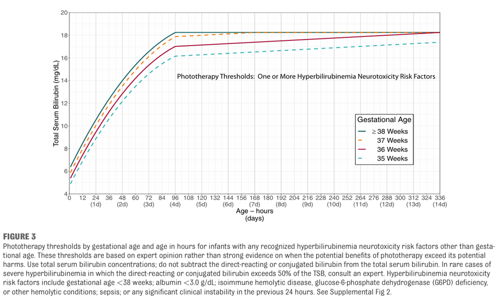
When to start phototherapy (6):
· If TsB at/above PHOTOTHERAPY THRESHOLD
· If TsB less than 3 mg/dL below PHOTOTHERAPY THRESHOLD but with signs/symptoms of ACUTE HEMOLYSIS.
· Provider may start phototherapy if TsB < 3 mg/dL below threshold in order to prevent readmission.
Figure-4: Phototherapy thresholds for babies with no neurotoxicity risk factors (6)
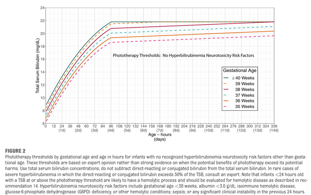
Figure-5: Phototherapy thresholds for babies with neurotoxicity risk factors (6)
Commercial light sources:
A wide selection of commercial phototherapy devices is available in Egypt.
Phototherapy devices can be categorized according to their light source as follows:
· Fluorescent-tube devices that emit different colors (cool white daylight, blue [B], special blue [BB], turquoise, and green) and are straight (F20 T12, 60 cm, 20 W), U-shaped, or spiral-shaped;
· Metal halide bulbs, used in spotlights and incubator lights;
· Light-emitting diodes (LEDs) used with fiber-optic light guides in pads, blankets, or spotlights;
· High-intensity LEDs, used as over- and under-the-body devices. (5)
Standards for phototherapy devices:
Factors to consider in prescribing and implementing phototherapy are:
· Emission range of the light source,
· Light intensity (irradiance):
- Standard PT units deliver 8–10 W/ cm2 per nm.
- Intensive PT requires 30 W/cm2 per nm, if the level of bilirubin is approaching exchange transfusion only INTENTSIVE phototherapy will be effective.
· Exposed (“treatable”) body surface area illuminated
· Reduction in total bilirubin concentration. (5)
Table-12: Factors That Affect the Dose and Efficacy of Phototherapy (PT): (5)
PT: phototherapy; LE: light-emitting. *Olympic Bili Bassinet (Olympic Medical, Seattle, WA)
|
Table-13: Practice considerations for optimal administration of phototherapy: (5)
|
Checklist |
Recommendation |
Implementation |
|
|
Light source (nm) |
Wavelength spectrum in 460- 490-nm blue-green light region |
Know the spectral output of the light source. |
|
|
Light irradiance (W·cm2·nm1) |
Use optimal irradiance: 30 W·cm2·nm1 within the 460- to 490-nm waveband |
Ensure uniformity over the light footprint area. |
|
|
Body surface area (cm2) |
Expose maximal skin area |
Reduce blocking of light. |
|
|
Timeliness of implementation |
Urgent or “crash-cart” intervention for excessive hyperbilirubinemia |
May conduct procedures while infant is on phototherapy. |
|
|
Continuity of therapy |
Briefly interrupt for feeding, parental bonding, nursing care |
After confirmation of adequate bilirubin concentration decrease. |
|
|
Efficacy of intervention |
Periodically measure rate of response in bilirubin load reduction |
Degree of total serum/plasma bilirubin concentration decrease. |
|
|
Duration of therapy |
Discontinue at desired bilirubin threshold; be aware of possible rebound increase |
Serial bilirubin measurements based on rate of decrease. |
|
What decline in the Serum Bilirubin can you expect?
· With extremely high bilirubin (more than 30 mg/dL), and intensive phototherapy, a decline of as much as 10 mg/dL can occur within few hours, and a decrease of at least 0.5 to 1 mg/dL/ hour can be expected in the first 4 to 8 hours.
· On average, for infants >35 weeks’ gestation readmitted for phototherapy, intensive phototherapy can produce a decrement of 30% to 40% in the initial bilirubin level by 24 hours after initiation of phototherapy.
· The most significant decline will occur in the first 4 to 6 hours.
· With standard phototherapy systems, a decrease of 6- 20% of the initial bilirubin level can be expected in the first 24 hours.
Safety and protective measures:
· A clinician skilled in newborn care should assess the neonate’s clinical status during phototherapy to ensure adequate hydration, nutrition, and temperature control. Clinical improvement or progression of jaundice should also be assessed, including signs suggestive of early bilirubin encephalopathy (ABE).
· The distance of the phototherapy device from the infant should be safely minimized.
· It is important to note that the intensity of light decreases at the outer perimeter of the light footprint and to recognize the effects of physical factors that could impede or obstruct light exposure to have maximum exposure.
· Temperature should be monitored during phototherapy and a neutral thermal environment ensured.
· Baby should be monitored for adequate hydration by daily weighing and assessment of wet nappies.
· Phototherapy does not use ultraviolet light and exposure to lights is mostly harmless. Devices must comply with general safety standards.
· The genitalia should be protected from light during phototherapy.
· Give baby eye protection and routine eye care.
Other clinical considerations include:
· Phototherapy is contraindicated in infants with congenital porphyria or those treated with photosensitizing drugs.
· Prolonged phototherapy is associated with increased oxidant stress and lipid peroxidation and riboflavin deficiency.
When should phototherapy be stopped?
· There is no standard for discontinuing phototherapy.
· For infants who are readmitted after their birth hospitalization (usually for TSB levels of 18 mg/dL or higher), phototherapy may be discontinued when the serum bilirubin level falls below 13 to 14 mg/dL.
· Do not delay discharge to observe the infant for rebound.
· If phototherapy is used for infants with hemolytic diseases or is initiated early and discontinued before the infant is 3 to 4 days old, a follow-up bilirubin measurement within 24 hours after discharge is recommended.
Intermittent Versus Continuous Phototherapy:
· NO rationale for intermittent phototherapy exists.
· If the infant’s bilirubin level is approaching the exchange transfusion zone, intensive phototherapy should be administered continuously until a satisfactory decline in the serum bilirubin level occurs or exchange transfusion is initiated.
· Phototherapy may be interrupted for breastfeeding or brief parental visits as long as the level of bilirubin is not approaching the exchange zone.
Hydration:
· No evidence that excessive fluid administration affects the serum bilirubin concentration.
· If a baby with high bilirubin levels is also mildly dehydrated. This may need supplemental fluid intake to correct the dehydration.
· Encourage frequent breastfeeding but if baby is dehydrated despite frequent breastfeeding, supplement with a milk-based formula, because it inhibits the enterohepatic circulation of bilirubin and should help to lower the serum bilirubin level.
· Routine intravenous fluid or other supplementation (e.g., with dextrose water) of term and near-term infants receiving phototherapy is NOT required or indicated unless there is evidence of dehydration or escalation of care is required. (6)
Safety and state of the art use:
PHOTOTHERAPY IS A DRUG like Pharmacotherapy.(20)
In order to provide effective PT and improve outcomes, the following rules should be followed:
· Use photons as a drug when prescribing phototherapy.
· Use a narrow‐spectrum blue light (450 nm).
· Measure irradiance for specific wavelength.
· Use the least irradiance dose to achieve an efficient decrease in bilirubin load.
· Use phototherapy for the shortest possible time and only when indicated. (71)
Complications:
· Phototherapy in hospital separates mother and infant. Eye patching is disturbing to parents.
· Diarrhoea, intestinal hypermotility.
· Temperature instability.
· Phototherapy rash.
· Possible long term effects:
- Teratogenic effects, one study reported that phototherapy was associated with DNA damage. However, there is no evidence that this effect on DNA at a microscopic level can lead to long-term adverse effects in phototherapy-treated babies.
- Melanocytic nevus development.
· Infants with cholestatic jaundice may develop a dark, grayish-brown discoloration of the skin, serum, and urine (the bronze infant syndrome). This syndrome generally has had few deleterious consequences, and if there is a need for phototherapy, the presence of direct hyperbilirubinemia should not be considered a contraindication to its use. This is particularly important in sick neonates.
· Rarely, purpura and bullous eruptions have been described in infants with severe cholestatic jaundice receiving phototherapy and severe blistering and photosensitivity during phototherapy have occurred in infants with congenital erythropoietic porphyria.
· Congenital porphyria or a family history of porphyria is an absolute contraindication to the use of phototherapy, as is the concomitant use of drugs or agents that are photosensitizers.
Home phototherapy:
· Home phototherapy should not be used unless there is quality assurance regarding the phototherapy device, the implementation and the monitoring of bilirubin at home. Very close supervision of medical practitioners by a regulator of the process is required to avoid malpractice. Since that is currently not available, it is not recommended to have home phototherapy until these rules are fulfilled.
Escalation of care:
The intensive care that some infants with elevated or rapid increasing bilirubin concentration require to prevent the need for an exchange and possibly prevent kernicterus. (6)
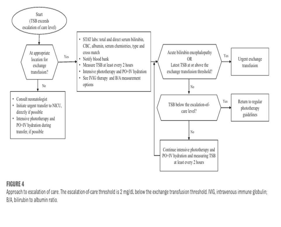
Figure-6: Approach to escalation of care
Appendix 2: Exchange Transfusion
· Exchange transfusion is an intensive invasive procedure and should not be performed in a Level 1 NICU.
· “Exchange transfusion must take place in an intensive care setting with intensive physiological and biochemical monitoring, carried out by staff trained in the procedure following written informed parental consent”, cited by British Committee for Standards in Hematology. (72)
· Exchange transfusion is usually carried out by repeatedly exchanging small samples (5-10 ml/kg) of blood through an umbilical venous catheter and replacing with same volumes by compatible blood.
· Neonatal exchange transfusion is becoming an infrequent procedure because severe Rh hemolytic disease of the newborn is becoming increasingly rare due to:
- Maternal anti-D therapy and intrauterine transfusion of affected fetuses.
- Maximizing phototherapy use prevents the need for exchange transfusion, even at very high level of TSB.
- Intravenous immunoglobulin (IVIG) acts by preventing the destruction of sensitized erythrocytes.
Benefits of Exchange Transfusion in severely jaundiced babies:
· Rapidly lowers the serum bilirubin level and reduce the risk of brain damage and kernicterus.
· Corrects anemia and increase the oxygen carrying capacity of the infant’s blood.
· Removes damaged and antibody coated red cells.
Indication for exchange transfusion in severe hyperbilirubinemia:
· Any baby showing signs of intermediate or advanced stages of acute bilirubin encephalopathy (e.g., hypertonia, arching, retrocollis, opisthotonus, high pitched cry, or recurrent apnea) irrespective of bilirubin level.
· If the TSB is at or above the exchange transfusion threshold.
· If intensive phototherapy is failing to lower TSB and it is approaching exchange level
· If the bilirubin /albumin ratio is:
- ≥8.0 If the gestational age is ≥38 weeks’ gestation and there are no hyperbilirubinemia neuro toxicity risk factors,
- ≥7.2 If the gestational age is ≥38 weeks’ gestation and there is at least 1 hyperbilirubinemia neurotoxicity risk factor,
- ≥7.2 If the gestational age is 35 through 37 weeks’ gestation with no hyperbilirubinemia neurotoxicity risk factor,
- 6.8 If the gestational age is 35 through 37 weeks’ gestation and at least 1 hyperbilirubinemia neurotoxicity risk factor. (62)
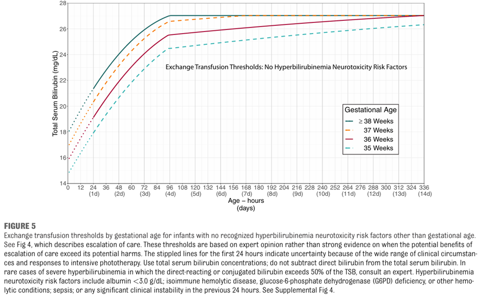
Figure-7: Exchange transfusion threshold by gestational age for infants with no recognized hyperbilirubinemia neurotoxicity risk factors
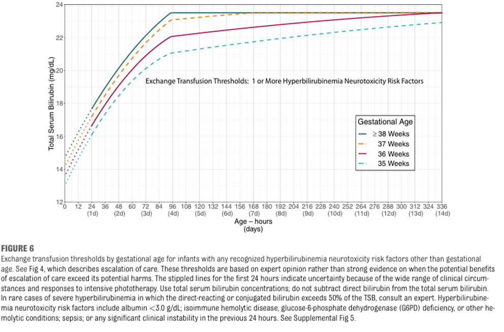
Figure-8: Exchange transfusion threshold by gestational age for infants with any recognized hyperbilirubinemia neurotoxicity risk factors(6)
Table-14: B/A Ratio at which Exchange Transfusion should be considered: (6)
· The bilirubin/ albumin ratio can be used with TSB level to help determine need for exchange transfusion.
· The bilirubin/ albumin ratio threshold for exchange transfusion, measured as TSB (measured in mg/ dL) divided by serum albumin (measured in g/dL), varies by gestational age and risk.
· An exchange transfusion may be considered if the bilirubin albumin ratio is:
- ≥8.0 if the gestational age is ≥38 weeks’ gestation and there are no hyperbilirubinemia neuro toxicity risk factors
- ≥7. 2 if the gestational age is ≥ 38 weeks’ gestation and there is at least 1 hyperbilirubinemia neurotoxicity risk factors
- ≥7.2 if the gestational age is 35 through 37 weeks’ gestation with no hyperbilirubinemia neurotoxicity risk factors
- ≥6.8 if the gestational age is 35 through 37 weeks’ gestation and at least 1 hyperbilirubinemia neurotoxicity risk factors.
Use of IVIG: (6)
Intravenous immunoglobulin (IVIG) 0.5-1 g/kg over 2 hours:
Provide to infants with isoimmune hemolytic disease (i.e. positive DAT) whose TSB reaches or exceeds the escalation of care threshold. This dose can be repeated in 12 hours.
Table-15: Procedures for Exchange Transfusion:
|
Steps |
Preparation for exchange transfusion |
|
1 |
Parent/care giver Information and consent Informed parental consent about the procedure and risks of exchange transfusion should be obtained and documented wherever possible. This should be a written consent. |
|
2 |
Order the blood : Which blood group to use? · Cross-matched washed packed red blood cells mixed with thawed adult fresh-frozen plasma to a hematocrit approximating 40% is preferred for exchange transfusions. The additional albumin-containing fresh frozen plasma that infants receive by keeping the hematocrit close to 40% will augment bilirubin removal. (72-74). · Use blood group O packed RBCs (with low titer hemolysins) Rh negative or Rh identical with the neonate suspended in AB plasma for exchange transfusion. · If not found then: if - Rh isoimmunization: use Rh negative and blood group O or blood group of the baby suspended in AB plasma. Cross matched with the baby’s and mother’s blood. - ABO incompatibility: Rh compatible and blood group O (not that of the baby), suspended in AB plasma, cross matched with baby’s and mother’s blood. - Other conditions: baby’s group and Rh type, cross matched with the baby’s and mother’s blood. Use a double-volume exchange transfusion: · Do not perform a single volume exchange. · A double volume exchange transfusion will exchange approximately 80% of the infant’s blood volume:(2 x 80 mls / Kg = 160 / per Kg = double volume blood exchange) Red Blood Cell Requirements: · 5 days old or less. · Negative for any antigens to which the mother or baby has antibodies. · Irradiated – use within 24 hours. Irradiation is a MUST if the infant has had a previous intrauterine transfusion and is preferable for all exchange transfusions if no delay will occur. · CMV negative. · Plasma reduced with HCT 40%. · It should be warmed to 37oc immediately before transfusion by a blood warmer. (Not to be transfused straight from 40C) |
|
3 |
Gain intravenous access: · Central via the umbilical vein. · Or a combination of central and peripheral lines. · Any central line should have its position confirmed by X-ray prior to procedure and checked for patency. |
|
4 |
Prepare equipment: Make a check list with the following before starting exchange: · Continuous cardio-respiratory monitoring equipment, including SpO2, and temperature should be available and resuscitation equipment. · Blood sugar monitoring available. · Exchange Transfusion tray. · Blood: do not collect blood until ready to begin exchange. · Protective clothing - Sterile gowns, gloves and masks. · Blood warmer and extension set. · Blood giving set with appropriate 170-200-micron filter. · Stand for blood. · Laboratory bottles = Full blood count x 2, urea and electrolytes x 2 including calcium and glucose. Blood culture x 2, capillary gas for pH x 2, (x 2 = pre and post specimens required ). · 2 ml, 5 ml, 10 ml and 60 ml syringes and needles. · Ventilator and intubation equipment, also suction equipment. (available if needed) · Sharps bin box. · High stools for doctor and nurse as the procedure up to 3 hours. · Timing device for timing infusion and withdrawal of aliquots. |
|
5 |
Ensure a neonatal screening blood spot has been taken prior to procedure. |
|
6 |
A minimum of 2 staff are required for the procedure (senior doctor and trained nurse). |
|
Steps |
Management prior to exchange transfusion |
|
1 |
· Nurse the infant in the radiant overhead heater if available, in the intensive care nursery with continuation of phototherapy. |
|
2 |
· Monitor heart rate, respiratory rate, oxygen saturation, blood pressure and temperature, a baseline set of observations including general color and tone and blood sugar should be documented before the procedure starts. |
|
3 |
· Stop enteral feeds one hour before the procedure, leaving the nasogastric tube on free drainage, (to reduce the risk of necrotizing enterocolitis). |
|
4 |
· Recheck TSB and check blood sugar (and effect of treatment) prior to picking up blood. |
|
5 |
· Set up the blood warmer. Using aseptic non touch technique when appropriate. |
|
6 |
· Collect and check the blood with another trained member of staff. · Warm the blood in a towel if warmer is not available. · Do not place blood on servo heater. Prime tubing. |
|
7 |
· Establish volume of aliquots prior to commencement · Aliquots usually tolerated for exchange transfusion: Ø <1500 grams: 5 ml Ø 1500-2490 grams :10 ml Ø 2500 grams – 3500 : 15 ml |
|
8 |
Bloods tests, blood sugar and a starting set of observations are recorded |
|
Steps |
Management during Exchange Transfusion |
|
1 |
Define responsibility: · Senior doctor (person A) is responsible for infusing and withdrawing an equal amount of blood. · Person B is responsible for the accurate recording of blood input and withdrawal on the Exchange Transfusion Fluid Chart (Table-17). |
|
2 |
· One member of staff (person A) flushes the input line with sodium chloride, connects the blood giving set, and connects the first syringe to the withdrawal line. Start by withdrawal of aliquot send to lab, and continue to infuse and withdraw equal amounts so that an equal rate is maintained. · Person B records this. ·
Small aliquots exchanged at a slower rate are usually better
tolerated by infants with cardiovascular instability. The time for each
in/out should be around 4-10 minutes (withdrawal and infusion of blood should
be performed at the same rate). In and
out aliquots are repeated sequentially until desired volume for exchange · A timer may be used for guiding speed of aliquots. |
|
3 |
· Whilst the exchange transfusion is in progress, the volume of blood exchanged in and out is recorded in the fluid balance chart with the Time. |
|
4 |
· The first aliquot of blood removed should be sent for estimation of full blood count, (FBC), differential, urea and electrolytes (U&Es), liver function tests, split bilirubin, Ca, hematocrit and glucose. · In severe cases consider: - coagulation screen, glucose 6-phosphate dehydrogenase (G6PD). |
|
5 |
· This process of administration and withdrawal continues until the agreed volume of blood has been exchanged or the condition of the baby determines cessation. · At the end of the procedure the infant’s fluid balance should be in deficit of one aliquot. |
|
6 |
· The last withdrawn blood should be sent for blood sugar, urea and electrolytes, bilirubin - total + direct, FBC, hematocrit, magnesium, calcium, blood gases and consider coagulation screen. |
|
7 |
· Continuously observation is required for Any signs of deterioration including cyanosis, pallor, abdominal distension, vomiting and blood in stools - Record heart rate, respirations, temperature, oxygen saturation and blood pressure every 15 minutes, any changes in these should be reported immediately. - Signs of hypocalcaemia - Tachycardia, irritability, seizures. - Monitor capillary blood glucose every 30 minutes. - All lines should be monitored for extravasation, blanching, erythema, leaking and edema. |
|
8 |
· Aim for procedure should take 2 hours (no longer than 3 hours). · On completion of the exchange transfusion, the lines are again flushed with NaCl. |
|
9 |
· Disposal of equipment –in accordance with local policy. |
|
10 |
· Documentation – ALL the procedure should be documented including: - All transfusion records, timing, accurate fluid balance, observation, samples taken and their results. - Also any untoward reaction, corrective measures taken and outcomes. |
|
11 |
· The baby should be left in a comfortable position and parents informed that the procedure has been completed and the condition of the baby. |
|
12 |
· Stop the procedure immediately and review if there are any concerns or untoward reactions. |
|
13 |
· Do NOT routinely administer intravenous calcium. (23) |
|
Steps |
Management following exchange transfusion |
|
1 |
· Continue intensive phototherapy after exchange, checking serum bilirubin as required. |
|
2 |
· Take blood pressure at completion of procedure. · Continue to monitor vital signs hourly for 6 hours or as the condition of the baby indicates. · After this time, and if stable and within normal limits routine observations may be recommenced. |
|
3 |
· Measure TSB level within 2 hours and manage according to threshold graphs (Figures-7 & 8). · Continue to monitor CBC and reticulocytes. |
|
4 |
· Monitor the blood sugar 1, 2 and 4 hours after exchange regularly according to condition for rebound hypoglycemia. |
|
5 |
· Observe catheter sites for bleeding/signs of infection. |
|
6 |
· Observe urine for blood, observe stools for blood. |
|
7 |
· Feeding: - Observe for abdominal distension, and for signs of feed intolerance, gastric aspirate and vomiting. - Commence feeds on medical advice, but not before 3 hours post-exchange transfusion. |
|
8 |
· Neonatal screening must not be taken until 4 days after the exchange as blood transfusion will invalidate all the tests. |
|
9 |
· All infants should receive regular audiology testing as they are at risk of hearing loss due to the severely raised serum bilirubin levels. |
Risks/Complications of exchange transfusion
There is a 5% risk of complication (3)
Exchange Transfusion Reaction:
· Tachycardia or bradycardia.
· Respiratory Distress.
· Dramatic changes in blood pressure.
· Temperature irregularity.
· Rash.
· Cyanosis or pallor.
· Hypoglycemia
· Hypocalcaemia (treatment see below)
· Cardiac Arrhythmias.
· Heart failure (fluid overload).
Other risk/complications:
· Administration of incorrect blood product.
· Volume overload.
· Hemodynamic instability.
· Acid base instability.
· Apnea.
· Electrolyte imbalances (hypocalcaemia, hypoglycemia, hypomagnesaemia).
· Convulsions.
· Hypotension (volume depletion).
· Embolism (air/thrombus).
· Graft versus host reaction.
· Infection.
· Necrotizing enterocolitis.
· Temperature instability.
· Thrombocytopenia.
· Thrombosis.
· Intraventricular hemorrhage.
Table-16: Exchange Transfusion Fluid Chart: (24)
|
Name |
|
|||||||
|
DOB |
|
|||||||
|
Hospital no |
|
|||||||
|
Aliquot Number |
Time |
IN |
OUT |
Total Balance (in/out) |
||||
|
aliquot mls |
Total mls |
aliquot mls |
Total mls |
|||||
|
1 |
|
|
|
|
|
|
|
|
|
2 |
|
|
|
|
|
|
|
|
|
3 |
|
|
|
|
|
|
|
|
|
4 |
|
|
|
|
|
|
|
|
|
5 |
|
|
|
|
|
|
|
|
|
6 |
|
|
|
|
|
|
|
|
|
7 |
|
|
|
|
|
|
|
|
|
8 |
|
|
|
|
|
|
|
|
|
9 |
|
|
|
|
|
|
|
|
|
10 |
|
|
|
|
|
|
|
|
|
11 |
|
|
|
|
|
|
|
|
|
12 |
|
|
|
|
|
|
|
|
|
13 |
|
|
|
|
|
|
|
|
|
14 |
|
|
|
|
|
|
|
|
|
15 |
|
|
|
|
|
|
|
|
|
16 |
|
|
|
|
|
|
|
|
|
17 |
|
|
|
|
|
|
|
|
|
18 |
|
|
|
|
|
|
|
|
|
19 |
|
|
|
|
|
|
|
|
|
20 |
|
|
|
|
|
|
|
|
|
21 |
|
|
|
|
|
|
|
|
|
22 |
|
|
|
|
|
|
|
|
|
23 |
|
|
|
|
|
|
|
|
|
24 |
|
|
|
|
|
|
|
|
|
25 |
|
|
|
|
|
|
|
|