
Book
Most common fish viral diseases
- Koi Herpesvirus (KHV) disease or Carp Interstitial Nephritis and Gill Necrosis Virus (CNGV)
Definition:
It is an acute highly contagious viral disease affecting common carp and koi characterized clinically by high mortalities, signs of hypoxia, enophthalmia and hemorrhagic spots on the body surface and erratic swimming behavior.
Etiology:
The etiological agent of Koi Herpesvirus (KHV) or Carp Interstitial Nephritis and Gill Necrosis Virus (CNGV), is a third Cyprinid herpesvirus-3 (CyHV-3) a spherical to pleomorphic envelope with linear double-stranded DNA (Gotesman et al., 2013).
Mode of transmission:
Horizontal transmission through cohabitation with infected fish, CyHV-3 may penetrate via the skin, and the gill (Costes et al. 2009). Ingesting CyHV-3-positive material offers an alternative route for CyHV-3 to enter via the pharyngeal periodontal mucosa, where virus proliferates extensively in the gut (Fournier et al. 2012).
Susceptible host:
All varieties and subspecies of common carp and common carp hybrids
Clinical signs:
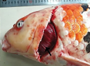
Skin had patches of pale coloration or reddening, excessive or reduced mucous secretion and sandpaper-like skin texture, enophthalmia (sunken eyes), hemorrhagic spots on the skin and base of the fins, and fin erosion, Signs of hypoxia, loss of equilibrium and disorientation,
PM lesions:
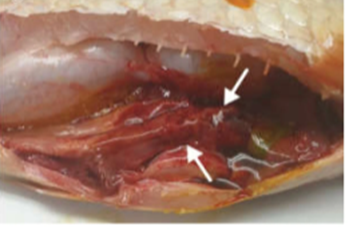
Gills had excess mucus, paleness and mottled with red and grayish-white patches, adhesion of abdominal cavity, enlarged kidney and liver, and petechial hemorrhages on the internal organs.
|
|
|
Histopathological lesions:
Necrosis in primary and secondary lamellae, fusion of secondary lamellae and adhesion of gill filaments, and hyperplasia and hypertrophy of branchial epithelium.
Necrosis of Kidneys hematopoietic tissues, severe interstitial nephritis with heavy infiltration with inflammatory cells and destruction of renal tubules.
Inflammation and necrosis in liver and spleen
Intranuclear inclusions in the cells of gills and kidneys.
Signet ring’ appearance of branchial epithelial cells (chromatin margins to the edges of the nucleus creating a distinct visual ring.)