Book
Management of Steroid Sensitive Nephrotic Syndrome
- Implementation Tools and Considerations
To improve healthcare provision, quality, safety, and patient outcome, evidence-based recommendations must not only be developed, but also disseminated and implemented at national and local levels and integrated into clinical practice.
Dissemination involves educating related healthcare providers to improve their awareness, knowledge and understanding of the guideline’s recommendations. It is one part of implementation, which involved translation of evidence-based guidelines into real life practice with improvement of health outcomes for the patients.
Implementation requires an evidence-based strategy involving professional groups and stakeholders and should consider the local cultural and socioeconomic conditions. Cost-effectiveness of implementation programs should be assessed.
Specific steps need to be followed before clinical practice recommendations can be integrated into local clinical practice, particularly in low resource settings.
Steps of implementing diagnosis, treatment, and prevention strategies into the Egyptian health system:
1. Develop a multidisciplinary working group.
2. Assess the status of nutritional care delivery, care gaps and current needs.
3. Select the material to be implemented, agree on the main goals, identify the key recommendations for diagnosis, treatment and prevention and adapt them to the local context or environment.
4. Identify barriers to, and facilitators of implementation.
5. Select an implementation framework and its component strategies.
6. Develop a step-by-step implementation plan:
· Select the target populations and evaluate the outcome.
· Identify the local resources to support the implementation.
· Set timelines.
· Distribute the tasks to the members.
· Evaluate the outcomes.
7. Continuously review the progress and results to determine if the strategy requires modification.
Guideline implementation strategies will focus on the following: -
1. For Practitioners
· Educational meetings: conferences, lectures, workshops, grand rounds, seminars, and symposia.
· Educational materials: printed or electronic information (software).
· Web-based education: computer-based educational activities.
· A trained person meets with providers in their practice setting to provide information with the intention of changing the provider’s practice. The information may include feedback on the performance of the provider(s).
· Reminders: the provision of information verbally, on papers or on a computer screen to prompt a health professional to recall information or to perform or avoid a particular action related to patient care.
· Optimize professional-patient interactions, through mass media campaigns, reminders, and education materials.
· Practice tools: tools designed to facilitate behavioral/practice changes, e.g., flow charts.
2. For Patients and care givers
· Patient education materials (Arabic booklet): Printed/electronic information aimed at the patient/consumer, family, caregivers, etc.
· Reminders: the provision of information verbally, on papers or electronically to remind a patient/consumer to perform a particular health-related behavior.
· Mass media campaigns.
3. For Nurses
· Educational meetings: lectures, workshops or traineeships, seminars, and symposia.
· Educational materials: printed.
· A trained person meets with nurses in their practice setting to provide information with the intention of changing the provider’s practice.
· Reminders: the provision of information verbally, on paper or on a computer screen to prompt them to recall information or to perform or avoid a particular action related to patient care.
· Practice tools: tools designed to facilitate behavioral/practice changes.
4. For Stakeholders
Plans have been made to contact with all the health sectors in Egypt including all sectors of the Ministry of Health and Population, National Nutrition Institute, University Hospitals, Ministry of Interior, Ministry of Defense, Non-Governmental Organizations, Private sector, and all Health Care Facilities.
· Information and communication technology: Electronic decision support, order sets, care maps, electronic health records, office-based personal digital assistants, etc.
· Any summary of clinical provision of health care over a specified period may include recommendations for clinical action. The information is obtained from medical records, databases, or observations by patients. Summary may be targeted at the individual practitioner or the organization.
· Administrative policies and procedures.
· Formularies: Drug safety programs, electronic medication administration records.
5. Other activities to assist the implementation of the adapted guideline’s recommendations include:
· International initiative: Dissemination of the presented adapted CPG internationally via sending the final adapted CPG to the Guidelines International Network (GIN) Adaptation Working Group and contacting the CPG developers.
· Gantt chart has been designed to manage the dissemination and implementation stages for the adapted CPG over an accurate time frame (Appendix).
Evidence to Decision Tables: (if any)
Guideline Implementation Tools
Educational materials based on this Adapted CPG for treatment of CAP in children have been made available in several forms including:
1. Manual for physician for diagnosis and algorithm for management of acute malnutrition
3. Arabic Educational materials for nurses and mothers
IPNA 2020 & KDIGO 2021 Tables
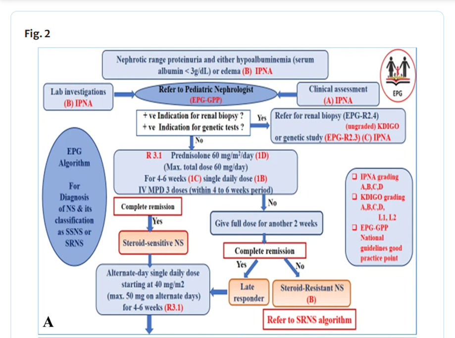
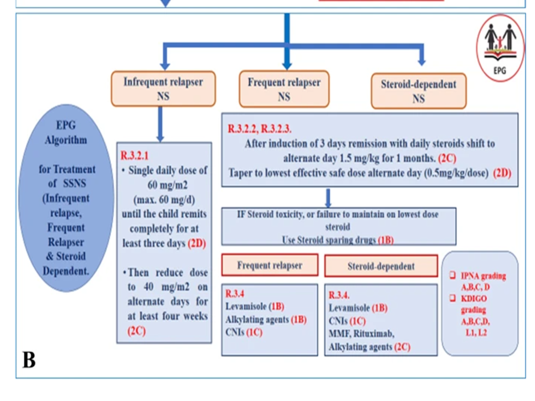
Table (1): Definitions related to Nephrotic Syndrome in Children.
(IPNA 2020 and KDIGO 2021)
|
Term |
Definitions |
|
Nephrotic-range proteinuria
|
UPCR ≥ 200 mg/mmol (2 mg/mg) in first morning void or 24 h urine sample ≥ 1000 mg/m2/day corresponding to 3+ or 4+ by urine dipstick. |
|
Nephrotic syndrome |
Nephrotic-range proteinuria and either hypoalbuminemia (serum albumin < 30 g/l) or edema when serum albumin level is not available. |
|
Complete remission
|
UPCR (based on first morning void or 24 h urine sample) ≤ 20 mg/mmol (0.2 mg/mg) or negative or trace dipstick on three or more consecutive occasions. |
|
Partial remission
|
UPCR (based on first morning void or 24 h urine sample) > 20 but < 200 mg/mmol and, if available, serum albumin ≥ 30 g/l. |
|
Relapse |
Relapse Recurrence of nephrotic-range proteinuria. * In children, relapse is commonly assessed by urine dipstick and is thus defined as dipstick ≥ 3+ on 3 consecutive days, or UPCR ≥ 200 mg/mmol (2 mg/mg) on a first morning urine sample, with or without reappearance of edema in a child who had previously achieved partial or complete remission. |
|
Confirmation Period
|
Time period between 4 and 6 weeks from PDN initiation during which response to further oral PDN and/or pulses of iv MPDN and RAASi are ascertained in patients achieving only partial remission at 4 weeks. * A patient achieving complete remission at 6 weeks is defined as a late responder. * A patient not achieving complete remission at 6 weeks although he had achieved partial remission at 4 weeks is defined as SRNS. |
|
SSNS |
Complete remission within 4 weeks of prednisone or prednisolone (PDN) at standard dose (60 mg/m2/day or 2 mg/kg/day, maximum 60 mg/day). |
|
Infrequent relapsing NS |
˂ 2 relapses per 6 months or ˂ 4 relapses per 12 months.
|
|
Frequent relapsing NS |
≤ 2 relapses per 6 months or ≤ 4 relapses per 12 months.
|
|
Steroid dependent NS |
Relapses during therapy with prednisone or prednisolone (either at full dose or during tapering) or within 15 days of prednisone or prednisolone discontinuation. |
|
SRNS |
Lack of complete remission within 4 weeks of treatment with PDN at standard dose. |
|
Late Responder NS |
Complete remission at 6 weeks.
|
|
CNI-resistant SRNS |
Absence of at least partial remission after 6 months of treatment with a CNI at adequate doses and/or levels. |
|
Multi-drug-resistant SRNS |
Absence of complete remission after 12 months of treatment with 2 mechanistically distinct steroid-sparing agents at standard doses (see text). |
|
Secondary steroid resistance |
Children with initial steroid-sensitivity who in subsequent relapses develop SRNS. |
|
|
|
|
Table abbreviations |
|
|
UPCR urine protein/creatinine ratio, SSNS steroid sensitive nephrotic syndrome, SRNS steroid-resistant nephrotic syndrome, PDN prednisolone or prednisone, MPDN methylprednisolone, RAASi renin-angiotensin-aldosterone system, CNI calcineurin inhibitor |
|
Reproduced with permission from IPNA 2020 and KIDIGO 2021.
Table 2: Initial workup and follow-up for a child with steroid-resistant nephrotic syndrome (IPNA 2020).
|
Table 2 Pediatr Nephrol (2020) 35:1529–1561 |
||
|
Investigations |
Initial work up |
Follow-up mentoring |
|
Clinical Evaluation |
|
|
|
Patient history – Including results of dipstick assessments at home, physical activity, fever episodes, pain, abdominal discomfort, swelling, fatigue, school attendance, adherence to medication, menstrual cycle in female adolescents - Search for risk factors for secondary causes As appropriate (sickle cell disease, HIV, SLE, HepB, malaria, parvovirus B19) - Check for tuberculosis in endemic areas before starting immunosuppressant drugs |
✓
✓ ✓ |
Every 3 months
As appropriate As appropriate |
|
Physical examination - Assessing fluid status including signs of edema (e.g., ascites, pericardial & pleural effusions), tetany, lymphadenopathy - Drug toxicity (e.g., eyes, skin) Every 3 months - - Skeletal status - Extrarenal features, e.g., dysmorphic features ambiguous genitalia - Full neurological examination & standardized assessment of cognitive status - Pubertal status: Tanner stage, testicular volume in boys ( in patients aged > 10 years)
- Vital parameters: blood pressure
- Anthropometry Growth chart: height/length, weight, Head circumference < 2 years Calculation of BMI and annual height velocity - Vaccination status Check and complete, especially for encapsulated bacteria— Pneumococcal, Meningococcal, Hemophilus Influenza, and Varicella-Zoster. - Family history. - Renal and extrarenal manifestations. - Consanguinity |
✓ ✓ ✓ ✓ ✓
|
As appropriate
Every 3 months Every 3 months Every 3 months As appropriate
Every 12 months or as appropriate
Every 12 months
Every 3 months; yearly 24h ambulatory BP monitoring hypertension, if feasible.
Every 3 months (monthly in infants) Every 12 month or as appropriate Every 12 month or as appropriate |
|
Biochemistry |
|
|
|
Urine Spot urine (first morning void) or 24 h urine: protein/creatinine Urinalysis including hematuria Spot urine: calcium/creatinine ratio, low molecular weight proteinuria (e.g., α1-microglobulin/creatinine ratio
Blood Complete blood count (CBC) Creatinine, BUN, or urea Electrolytes (including ionized calcium, potassium* and albumin corrected albumin if available) Serum albumin, total protein Blood gas analysis (HCO3) C-reactive protein Estimated GFRb ALP, PTH, 25(OH) vitamin D Lipid profile (LDL- and HDL-cholesterol, triglycerides)
Baseline coagulation tests (prothrombine time (INR), aPTT, fibrinogen, ATIII), detailed thrombophilic screening in patients with reported previous thrombotic events, central venous lines, persistent nephrotic range proteinuria and/or increased familial history for thrombotic events.
Thyroid function (T3, FT4, TSH)
Immunoglobulin G
Glucose/fasting glucose HbA1c C3, antinuclear antibodies ds-DNA, ENA, ANCA HBs-Ag, anti-HCV-IgG, syphilis, and HIV tests Vaccination status including blood titer tests |
✓ ✓ Conditional ✓
✓ ✓ ✓
✓ ✓ ✓
✓
✓
✓
✓
Conditional |
Essential Every 3 months (more frequently until remission) Every 6–12
Essential Every 3 months (more frequently until remission) and in CKD stage 4–5) Every day or every other day when using high dose diuretics
As required (clinical decision) Every 3 months (more frequently in CKD stage 4). Every 12 months (more frequently in patients with CKD stages( 3- 5) Every 12 months or as appropriate At diagnosis and then as appropriate, e.g., in case of relapses. Every 12 months or as appropriate especially in patients with prolonged proteinuria. In case of recurrent infections
Every 6 months or as appro Every 12 months or as appro As appropriate As appropriate Before prednisolone & as app Yearly or as appropriate |
|
Urine Spot urine (first morning void) or 24 h urine: protein/creatinine Urinalysis including hematuria Spot urine: calcium/creatinine ratio, low molecular weight proteinuria (e.g., α1-microglobulin/creatinine ratio
Blood Complete blood count (CBC) Creatinine, BUN, or urea Electrolytes (including ionized calcium, potassium* and albumin corrected albumin if available) Serum albumin, total protein Blood gas analysis (HCO3) C-reactive protein Estimated GFRb ALP, PTH, 25(OH) vitamin D Lipid profile (LDL- and HDL-cholesterol, triglycerides)
Baseline coagulation tests (prothrombine time (INR), aPTT, fibrinogen, ATIII), detailed thrombophilic screening in patients with reported previous thrombotic events, central venous lines, persistent nephrotic range proteinuria and/or increased familial history for thrombotic events.
Thyroid function (T3, FT4, TSH)
Immunoglobulin G
Glucose/fasting glucose HbA1c C3, antinuclear antibodies ds-DNA, ENA, ANCA HBs-Ag, anti-HCV-IgG, syphilis, and HIV tests Vaccination status including blood titer tests |
✓ ✓ Conditional ✓
✓ ✓ ✓
✓ ✓ ✓
✓
✓
✓
✓
Conditional |
Essential Every 3 months (more frequently until remission) Every 6–12
Essential Every 3 months (more frequently until remission) and in CKD stage 4–5) Every day or every other day when using high dose diuretics
As required (clinical decision) Every 3 months (more frequently in CKD stage 4). Every 12 months (more frequently in patients with CKD stages( 3- 5) Every 12 months or as appropriate At diagnosis and then as appropriate, e.g., in case of relapses. Every 12 months or as appropriate especially in patients with prolonged proteinuria. In case of recurrent infections
Every 6 months or as appro Every 12 months or as appro As appropriate As appropriate Before prednisolone & as app Yearly or as appropriate |
|
Genetics |
|
|
|
Next-generation sequencing (NGS)/Whole Exome Sequencing (WES) |
✓ |
Extended screening for patients with SRNS depending on new findings (Table 3); whole exome sequencing if indicated transplantation, if not previously performed. |
|
Drug-specific monitoring
|
|
|
|
CsA: and Tacrolimus: Drug trough levels
|
Weekly during titration (for 4 weeks) |
Thereafter every 3 months or as appropriate
|
|
MMF: mycophenolic acid kinetic (2 h) c
Rituximab – CD19 B cell count: baseline
Statins: creatinine kinase (CK) – If on statins, every 6 months Prolonged glucocorticoid therapy Conditional
Ophthalmological examination for cataract and intraocular pressure Bone mineral density by lumbar DEXA |
AUC after 4 weeks of treatment.
1 month after the first dose (nadir) every 6 months Conditional |
Thereafter every 6–12 months or as appropriate.
Every 1–3 months until B cell recovery
every 6 months
Conditional |
|
Imaging |
|
|
|
Renal ultrasound: renal echogenicity and size of kidneys
|
✓
|
At presentation (mandatory prerenal biopsy)
|
|
Ultrasound of abdomen & pleural space (ascites, effusions, thrombosis)
Cardiac ultrasound (left ventricular mass, effusions)
Chest X-ray
X-ray of the left wrist (bone age assessment in children aged > 5 years, mineralization) |
✓
✓
✓
✓ |
as appropriate
Every 12 months in hypertensive patients or in case of severe edema Optional If indicated
Every 12 months or as appropriate |
|
Histopathological Renal Biopsy |
|
|
|
|
✓
|
See text: at diagnosis, and subsequently if indicated: in case of unexplained drop in eGFR, unexplained increase in proteinuria, to rule out and/or to monitor CNI nephrotoxicity during prolonged (< 2 years) treatment. |
|
Dietary assessment |
|
|
|
Dietician review and advice by a dietician regarding salt, potassium, caloric and protein intake
|
✓ |
Every 3 months (more frequently in infants, malnourished patients, and patients with CKD stage 4–5) |
|
Assessment for extrarenal involvement |
|
|
|
Depending on underlying disease and clinically evident extrarenal features: - Brain MRI (e.g., microcephaly, psychomotor delay, mental retardation, myoclonic epilepsy, tremor, ataxia, hypotonia) Interdisciplinary evaluation by Ophthalmology (e.g., microcoria, cataract, glaucoma, optic atrophy, keratoconus, macular spots, lenticonus, nystagmus). Cardiology (e.g., congenital heart defects) |
✓
✓
✓ |
If indicated
If indicated |
Reproduced with permission from IPNA 2020.
Table 3- Steroid sparing therapy in SSNS (KDIGO 2021)
Table 3- Steroid sparing therapy in SSNS (KDIGO 2021)

Reproduced with permission from KDIGO 2021.